Showing posts with label Artificial Life. Show all posts

Thursday, 20 May 2010

Artificial Life: First Self-replicating Synthetic Bacterial Cell Created

A fully synthesized genome transforms one species of bacterium into another
Thursday, May 20, 2010

Mycoplasma mycoides JCVI-syn1.0. Credit: J. Craig Venter Institute.

Researchers at the J. Craig Venter Institute (JCVI), a not-for-profit genomic research organization, published results today describing the successful construction of the first self-replicating, synthetic bacterial cell. The team synthesized the 1.08 million base pair chromosome of a modified Mycoplasma mycoides genome. The synthetic cell is called Mycoplasma mycoides JCVI-syn1.0 and is the proof of principle that genomes can be designed in the computer, chemically made in the laboratory and transplanted into a recipient cell to produce a new self-replicating cell controlled only by the synthetic genome.

This research will be published by Daniel Gibson et al in the May 20th edition of Science Express and will appear in an upcoming print issue of Science.

“For nearly 15 years Ham Smith, Clyde Hutchison and the rest of our team have been working toward this publication today – the successful completion of our work to construct a bacterial cell that is fully controlled by a synthetic genome,” said J. Craig Venter, Ph.D., founder and president, JCVI and senior author on the paper.

“We have been consumed by this research, but we have also been equally focused on addressing the societal implications of what we believe will be one of the most powerful technologies and industrial drivers for societal good. We look forward to continued review and dialogue about the important applications of this work to ensure that it is used for the benefit of all.”

According to Dr. Smith:

“With this first synthetic bacterial cell and the new tools and technologies we developed to successfully complete this project, we now have the means to dissect the genetic instruction set of a bacterial cell to see and understand how it really works."

To complete this final stage in the nearly 15 year process to construct and boot up a synthetic cell, JCVI scientists began with the accurate, digitized genome of the bacterium, M. mycoides. The team designed 1,078 specific cassettes of DNA that were 1,080 base pairs long. These cassettes were designed so that the ends of each DNA cassette overlapped each of its neighbours by 80bp. The cassettes were made according to JCVI’s specifications by the DNA synthesis company, Blue Heron Biotechnology.

The JCVI team employed a three stage process using their previously described yeast assembly system to build the genome using the 1,078 cassettes. The first stage involved taking 10 cassettes of DNA at a time to build 110, 10,000 bp segments. In the second stage, these 10,000 bp segments are taken 10 at a time to produce eleven, 100,000 bp segments. In the final step, all 11, 100 kb segments were assembled into the complete synthetic genome in yeast cells and grown as a yeast artificial chromosome.

The complete synthetic M. mycoides genome was isolated from the yeast cell and transplanted into Mycoplasma capricolum recipient cells that have had the genes for its restriction enzyme removed. The synthetic genome DNA was transcribed into messenger RNA, which in turn was translated into new proteins. The M. capricolum genome was either destroyed by M. mycoides restriction enzymes or was lost during cell replication. After two days viable M. mycoides cells, which contained only synthetic DNA, were clearly visible on Petri dishes containing bacterial growth medium.

The initial synthesis of the synthetic genome did not result in any viable cells so the JCVI team developed an error correction method to test that each cassette they constructed was biologically functional. They did this by using a combination of 100 kb natural and synthetic segments of DNA to produce semi-synthetic genomes. This approach allowed for the testing of each synthetic segment in combination with 10 natural segments for their capacity to be transplanted and form new cells. Ten out of 11 synthetic fragments resulted in viable cells; therefore the team narrowed the issue down to a single 100 kb cassette. DNA sequencing revealed that a single base pair deletion in an essential gene was responsible for the unsuccessful transplants. Once this one base pair error was corrected, the first viable synthetic cell was produced.

Dr. Gibson stated:

“To produce a synthetic cell, our group had to learn how to sequence, synthesize, and transplant genomes. Many hurdles had to be overcome, but we are now able to combine all of these steps to produce synthetic cells in the laboratory.”

“We can now begin working on our ultimate objective of synthesizing a minimal cell containing only the genes necessary to sustain life in its simplest form. This will help us better understand how cells work,” he added.

This publication represents the construction of the largest synthetic molecule of a defined structure; the genome is almost double the size of the previous Mycoplasma genitalium synthesis. With this successful proof of principle, the group will now work on creating a minimal genome, which has been a goal since 1995. They will do this by whittling away at the synthetic genome and repeating transplantation experiments until no more genes can be disrupted and the genome is as small as possible. This minimal cell will be a platform for analyzing the function of every essential gene in a cell.

According to Dr. Hutchison:

“To me the most remarkable thing about our synthetic cell is that its genome was designed in the computer and brought to life through chemical synthesis, without using any pieces of natural DNA. This involved developing many new and useful methods along the way. We have assembled an amazing group of scientists that have made this possible.”

As in the team’s 2008 publication in which they described the successful synthesis of the M. genitalium genome, they designed and inserted into the genome what they called watermarks. These are specifically designed segments of DNA that use the “alphabet” of genes and proteins that enable the researcher to spell out words and phrases. The watermarks are an essential means to prove that the genome is synthetic and not native, and to identify the laboratory of origin. Encoded in the watermarks is a new DNA code for writing words, sentences and numbers. In addition to the new code there is a web address to send emails to if you can successfully decode the new code, the names of 46 authors and other key contributors and three quotations: "To live, To err, To fall, To triumph, To recreate life out of life." – James Joyce; "See things not as they are, but as they might be.” – A quote from the book, “American Prometheus”; "What I cannot build, I cannot understand." – Richard Feynman. 

The JCVI scientists envision that the knowledge gained by constructing this first self-replicating synthetic cell, coupled with decreasing costs for DNA synthesis, will give rise to wider use of this powerful technology. This will undoubtedly lead to the development of many important applications and products including biofuels, vaccines, pharmaceuticals, clean water and food products. The group continues to drive and support ethical discussion and review to ensure a positive outcome for society.

Funding for this research came from Synthetic Genomics Inc., a company co-founded by Drs. Venter and Smith.

Background
The research published today was made possible by previous breakthroughs at JCVI. In 2007 the team published results from the transplantation of the native M. mycoides genome into the M. capricolum cell which resulted in the M. capricolum cell being transformed into M. mycoides. This work established the notion that DNA is the software of life and that DNA dictates the cell phenotype.

In 2008 the same team reported on the construction of the first synthetic bacterial genome by assembling DNA fragments made from the four chemicals of life — ACGT. The final assembly of DNA fragments into the whole genome was performed in yeast by making use of the yeast genetic systems. However, when the team attempted to transplant the synthetic bacterial genome out of yeast and into a recipient bacterial cell, viable transplants could not be recovered.

Ethical Considerations
Since the beginning of the quest to understand and build a synthetic genome, Dr. Venter and his team have been concerned with the societal issues surrounding the work. In 1995 while the team was doing the research on the minimal genome, the work underwent significant ethical review by a panel of experts at the University of Pennsylvania (Cho et al, Science December 1999:Vol. 286. no. 5447, pp. 2087 – 2090). The bioethical group's independent deliberations, published at the same time as the scientific minimal genome research, resulted in a unanimous decision that there were no strong ethical reasons why the work should not continue as long as the scientists involved continued to engage public discussion.

Dr. Venter and the team at JCVI continue to work with bioethicists, outside policy groups, legislative members and staff, and the public to encourage discussion and understanding about the societal implications of their work and the field of synthetic genomics generally. As such, the JCVI’s policy team, along with the Center for Strategic & International Studies (CSIS), and the Massachusetts Institute of Technology (MIT), were funded by a grant from the Alfred P. Sloan Foundation for a 20-month study that explored the risks and benefits of this emerging technology, as well as possible safeguards to prevent abuse, including bioterrorism. After several workshops and public sessions the group published a report in October 2007 outlining options for the field and its researchers.

Most recently in December of 2008, JCVI received funding from the Alfred P. Sloan Foundation to examine ethical and societal concerns that are associated with the developing science of synthetic genomics. The ongoing research is intended to inform the scientific community as well as educate our policymakers and journalists so that they may engage in informed discussions on the topic.

About the J. Craig Venter Institute
The JCVI is a not-for-profit research institute in Rockville, MD and La Jolla, CA dedicated to the advancement of the science of genomics; the understanding of its implications for society; and communication of those results to the scientific community, the public, and policymakers. Founded by J. Craig Venter, Ph.D., the JCVI is home to approximately 400 scientists and staff with expertise in human and evolutionary biology, genetics, bioinformatics/informatics, information technology, high-throughput DNA sequencing, genomic and environmental policy research, and public education in science and science policy. The legacy organizations of the JCVI are: The Institute for Genomic Research (TIGR), The Center for the Advancement of Genomics (TCAG), the Institute for Biological Energy Alternatives (IBEA), the Joint Technology Center (JTC), and the J. Craig Venter Science Foundation. The JCVI is a 501 (c) (3) organization.

Reference:
Creation of a Bacterial Cell Controlled by a Chemically Synthesized Genome
Daniel G. Gibson, John I. Glass, Carole Lartigue, Vladimir N. Noskov, Ray-Yuan Chuang, Mikkel A. Algire, Gwynedd A. Benders, Michael G. Montague, Li Ma, Monzia M. Moodie, Chuck Merryman, Sanjay Vashee, Radha Krishnakumar, Nacyra Assad-Garcia, Cynthia Andrews-Pfannkoch, Evgeniya A. Denisova, Lei Young, Zhi-Qing Qi, Thomas H. Segall-Shapiro, Christopher H. Calvey, Prashanth P. Parmar, Clyde A. Hutchison III, Hamilton O. Smith, and J. Craig Venter
Science, Published online May 20 2010; DOI: 10.1126/science.1190719
.........

ZenMaster

For more on stem cells and cloning, go to CellNEWS at
http://cellnews-blog.blogspot.com/

Sunday, 8 March 2009

Assembling Cells Into Artificial 3-D Micro-tissues, Including a Tiny Gland

Hybridized DNA is glue that sticks these cells together to resemble real tissue Sunday, 08 March 2009 As synthetic biologists cram more and more genes into microbes to make genetically engineered organisms produce ever more complex drugs and chemicals, two University of California chemists have gone a step further. A 3-D reconstruction using deconvolution fluorescence microscopy of a single multicellular structure encapsulated in agarose gel. Cells are stained different colours according to the oligonucleotide sequence attached to their surfaces. Credit: Bertozzi lab, UC Berkeley.They have assembled different types of genetically engineered cells into synthetic micro-tissues that can perform functions such as secreting and responding to hormones, promising more complex biological capabilities than a single cell alone could produce. "This is like another level of hierarchical complexity for synthetic biology," said co-author Carolyn Bertozzi, University of California at Berkeley professor of chemistry and of molecular and cell biology and director of the Molecular Foundry at Lawrence Berkeley National Laboratory. "People used to think of the cell as the fundamental unit. But the truth is that there are collections of cells that can do things that no individual cell could ever be programmed to do. We are trying to achieve the properties of organs now, though not yet organisms." While the synthetic tissues today comprise only a handful of cells, they could eventually be scaled up to make artificial organs that could help scientists understand the interactions among cells in the body and might some day substitute for human organs. "We are really taking this into the third dimension now, which for me is particularly exciting," said first author Zev J. Gartner, a former UC Berkeley post-doctoral fellow who recently joined the UC San Francisco faculty as an assistant professor of pharmaceutical chemistry. "We are not simply linking cells together, we are linking them together in 3-D arrangements, which introduces a whole new level of cellular behaviour which you would never see in 2-D environments." Gartner and Bertozzi, the T.Z. and Irmgard Chu Distinguished Professor at UC Berkeley and a Howard Hughes Medical Institute investigator, report on their assembly of three-dimensional micro-tissues this week in the online early edition of the journal Proceedings of the National Academy of Sciences. One type of cell that needs other cells to make it work properly is the stem cell, Bertozzi noted. Theoretically, using Gartner and Bertozzi's chemical technique, it should be possible to assemble stem cells with their helper cells into a functioning tissue that would make stem cells easier to study outside the body. "In principal, we might be able to build a stem cell niche from scratch using our techniques, and then study those very well defined structures in controlled environments," Bertozzi said. Bertozzi noted that most of the body's organs are a collection of many cell types that need to be in actual physical contact to operate properly. The pancreas, for example, is a collection of specialized cells, including insulin-secreting beta cells, that "sense glucose from the environment and respond by producing insulin. A complex feedback regulatory loop goes into all of this, and you need more than one cell type to achieve such regulation." "If you really want to understand the way these cells behave in an organism, especially a human, you would like to recapitulate that environment as closely as possible in vitro," Gartner said. "We are trying to do that, with the aim that the rules we learn may help us control them better." Gartner and Bertozzi assembled three types of cultured cells into onion-like layers by using two established technologies: DNA hybridization and Staudinger chemistry. DNA hybridization is like a "programmable glue," she said, that can stick cells together because of the highly precise nature of binding between complementary DNA strands: One strand of the DNA helix binds only to its complementary strand and nothing else. By putting a short DNA strand on the surface of one cell and its complementary strand on another cell, the researchers assure that the two lock together exclusively. To get these specific DNA strands onto the cells, they used chemical reactions that do not interfere with cellular chemistry but nevertheless stick desired chemicals onto the cell surface. The technique for adding unusual but benign chemicals to cells was developed by Bertozzi more than a decade ago based on a chemical reaction called the Staudinger ligation. After proving that they could assemble cells into micro-tissues, Gartner and Bertozzi constructed a minute gland – analogous to a lymph node, for example – such that one cell type secreted interleukin-3 and thereby kept a second cell type alive. "What we did is build a little miniaturized, stripped-down system that operates on the same principle and looks like a miniaturized lymph node, an arrangement where two cells communicate with each another and one requires a signal from the other," she said. "The critical thing is that the two cells have to have a cell junction. If you just mix the cells randomly without connection, the system doesn't have the same properties." She expects that eventually, clusters could be built on clusters to make artificial organs that someday may be implanted into humans. "Our method allows the assembly of multicellular structures from the bottom up. In other words, we can control the neighbours of each individual cell in a mixed population," she said. "By this method, it may be possible to assemble tissues with more sophisticated properties." One interesting aspect of the technique is that DNA hybridization seems to be temporary, like a suture. Eventually, the cells may substitute their own cell-cell adhesion molecules for the DNA, creating a well-knit and seemingly normal, biological system. ......... ZenMaster


For more on stem cells and cloning, go to CellNEWS at http://cellnews-blog.blogspot.com/ and http://www.geocities.com/giantfideli/index.html

A New Way to Assemble Cells Into 3-D Micro-tissues

Cellular engineers make multicellular tissues from the bottom up Sunday, 08 March 2009 Scientists at the US Department of Energy's Lawrence Berkeley National Laboratory can now control how cells connect with one another in vitro and assemble themselves into three-dimensional, multicellular micro-tissues. The researchers demonstrated their method by constructing a tailor-made artificial cell-signalling system, analogous to natural cell systems that communicate via growth factors. Artificial tissues are presently used in medicine for a range of applications such as skin grafts, bone marrow transplants, or blood substitutes, as well as in basic medical and biological research. Tissue engineers try to improve upon or repair natural tissues by manipulating living cells from one or more donors, sometimes in combination with synthetic materials. Unfortunately, in this "top down" approach, the cells assemble themselves randomly, losing the 3-D organization that is key to many tissue functions.


Assembly of micro-tissues.After cell types labelled with red and green dye markers are joined (bottom), the resulting 3-D structures are purified to eliminate unreacted cells (centre). More cells can then be added to form even more complex structures (top). There is no theoretical limit to the number of different cell types that can be assembled; micro-tissues with three or four different kinds of cells should be feasible. Credit: Carolyn Bertozzi, Lawrence Berkeley National Laboratory.
"Our method allows the assembly of multicellular structures from the 'bottom up,'" says Carolyn Bertozzi, principal investigator in the research, who directs DOE's Molecular Foundry nanoscience research facility at Berkeley Lab and is a member of the Lab's Materials Sciences and Physical Biosciences Divisions. "In other words, we can control the neighbours of each individual cell in a mixed population. By this method, it may be possible to assemble tissues with more sophisticated properties." An example of a state that depends on sophisticated cellular connectivity and communication is the stem-cell niche, "which can maintain stem cells in an undifferentiated state, or in some cases guide their differentiation down a specific pathway," says Bertozzi. She is also a professor in the Departments of Chemistry and Molecular and Cell Biology at the University of California at Berkeley and a Howard Hughes Medical Institute investigator. Bertozzi and her colleague Zev Gartner, who is now an assistant professor of Pharmaceutical Chemistry at the University of California at San Francisco, based their method of assembling 3-D micro-tissues on an approach familiar to synthetic organic chemists, who build up novel molecular structures by combining one atom or functional group at a time. In the same way, Bertozzi and Gartner use synthetic methods to build micro-tissues one cell type at a time.

Controlling cellular connections The researchers enabled cells to react with other cells in a controlled way by coating the cell surfaces with DNA (not where DNA is normally found!). First they induced the cells to express artificial sugars bearing special chemical groups. Lengths of synthetic DNA, introduced into the cell-growth medium, were equipped to recognize these synthetic sugars on the cell surfaces and chemically bind to them.


How to build a micro-tissue.How to build a micro-tissue: At the bottom, cells bearing complementary single strands of DNA on their surfaces react with each other to form stable cell–cell contacts. At centre are Jurkat cells stained red or green, labelled with different, complementary DNA sequences, and combined at a ratio of 50 (red) to 1 (green). At top, the two cell types are shown joined in a 3-D multicellular structure. Credit: Carolyn Bertozzi, Lawrence Berkeley National Laboratory.
The researchers coated cell surfaces from one group with strands of single-stranded DNA only 20 bases long, and the surfaces of another group with the complementary DNA strand. When a cell from one group meets its counterpart, the single strands recognize each other and form double-stranded DNA, which binds the cells together. Bertozzi and Gartner discovered three variables that determine how cells from different groups react with one another. One is the ratio of the two kinds of cells: if both cell populations are equal, every cell finds a single partner and no complex assemblies form. But if there are, say, 50 times more cells from one group than from the other, numerous cells from the larger group will cluster around each cell from the smaller group. Another variable is the complexity of the synthetic DNA sequence. The researchers can specify the complexity – for example, from a simple repeat of two bases such as cytosine and adenine (CACACA...) on one strand, which binds to a complementary repeating sequence of thymine and guanine (TGTGTG...) on the other, up to sequences whose base order varies over the full length of the 20-base strand of synthetic DNA. The more complex the sequences they display, the longer it takes the cells to bind together. A third variable is the density of the DNA on the cell surface. By controlling how many artificial sugars the cells express, the researchers can control the DNA surface density. The greater the density, the faster the cells bind to one another. Bertozzi says more variables for controlling cell assemblies are possible. "For example, it might be possible to cluster DNA strands on specific cellular structures. Thus, distribution of DNA on the cell surface might be yet another parameter we can exploit to guide cell-cell interactions." Building complex micro-tissues one step at a time By controlling these variables to assemble small cellular structures, then separating the desired structures from unwanted ones and unreacted cells and assembling more cells on the purified collection – then repeating the steps again – the researchers can synthesize large, complex micro-tissues in much the same way a synthetic organic chemist assembles a complex molecule. Bertozzi and Gartner applied these methods to build a signalling network where one kind of cell controls the growth of a second kind of cell. They maintained the survival and replication of hematopoietic progenitor cells (a kind of stem cell for blood cells), which depend on the presence of the growth factor interleukin-3, by combining them in micro-tissues with CHO cells (Chinese hamster ovary cells) that were engineered to secrete interleukin-3. When the two cell types were randomly mixed, the stem cells did not grow. But structured micro-tissues built from the two cell types stimulated their own growth, forming a simple artificial signalling network that behaved much like the natural networks that control immune-cell expansion or tumour proliferation. "Since DNA has essentially an unlimited capacity for information storage, there is no theoretical limit on the number of different cell types we can assemble in a structure," says Bertozzi. The key is to give each cell type its own unique DNA "bar code," enabling its programmed interaction with any other specified cell type. "In practice, I think structures with three or four cell types are quite feasible. Such structures would be relevant to many biological organs." Structured micro-tissues have numerous research applications, says Bertozzi, particularly "in probing how the local cellular environment affects the behaviour of a particular cell. Also, we can study how systems of cells work together to produce complex organ functions. Examples include how T cells and B cells work together in the lymph nodes to mount an immune response against foreign antigens." Practical challenges remain, such as scaling up the production of tailored micro-tissues to quantities needed for biomedical applications. Beyond that, Bertozzi hopes to refine the present method of modifying cell-surface DNA. "As it stands, the need for unnatural sugar biosynthesis limits the kinds of cells that we can use in micro-tissue construction," she says. "There are other ways in which DNA can be conjugated to cells, independent of their sugar metabolic pathways, and we intend to explore those avenues." Reference: Programmed assembly of 3-dimensional microtissues with defined cellular connectivity Zev J. Gartner and Carolyn R. Bertozzi PNAS Early Edition, In press, 2009. ......... ZenMaster
For more on stem cells and cloning, go to CellNEWS at http://cellnews-blog.blogspot.com/ and http://www.geocities.com/giantfideli/index.html

Thursday, 9 October 2008

Artificial Cells Could Power Medical Implants

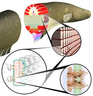
Models of eel cells suggest electrifying possibilities Thursday, 09 October 2008 Researchers at Yale University have created a blueprint for artificial cells that are more powerful and efficient than the natural cells they mimic and could one day be used to power tiny medical implants. The scientists began with the question of whether an artificial version of the electrocyte – the energy-generating cells in electric eels – could be designed as a potential power source. "The electric eel is very efficient at generating electricity," said Jian Xu, a postdoctoral associate in Yale's Department of Chemical Engineering. "It can generate more electricity than a lot of electrical devices." Xu came up with the first blueprint that shows how the electrocyte's different ion channels work together to produce the fish's electricity while he was a graduate student under former Yale assistant professor of mechanical engineering David LaVan, now at the National Institute of Standards and Technology. But the scientists didn't stop there. "We're still trying to understand how the mechanisms in these cells work," said LaVan. "But we asked ourselves: 'Do we know enough to sit down and start thinking about how to build these things’ — and to optimize that design? Nobody had really done that before." Using the new blueprint as a guide, LaVan and Xu set about designing an artificial cell that could replicate the electrocyte's energy production. "We wanted to see if nature had already optimized the power output and energy conversion efficiency of this cell," said Xu. "And we found that an artificial cell could actually outperform a natural cell, which was a very surprising result." The artificial cell LaVan and Xu modelled is capable of producing 28 percent more electricity than the eel's own electrocyte, with 31 percent more efficiency in converting the cell's chemical energy – derived from the eel's food – into electricity. Electric eels channel the output of thousands of specialized cells called electrocytes to generate electric potentials of up to 600 volts, according to biologists. The mechanism is similar to nerve cells. The arrival of a chemical signal triggers the opening of highly selective channels in a cell membrane causing sodium ions to flow in and potassium ions to flow out. The ion swap increases the voltage across the membrane, which causes even more channels to open. Past a certain point the process becomes self-perpetuating, resulting in an electric pulse travelling through the cell. The channels then close and alternate paths open to “pump” the ions back to their initial concentrations during a “resting” state.


Electric eel anatomy.Electric eel anatomy. The first detail shows stacks of electrocytes, cells linked in series (to build up voltage) and parallel (to build up current). Second detail shows an individual cell with ion channels and pumps penetrating the membrane, The Yale/NIST model represents the behaviour of several such cells. Final detail shows an individual ion channel, one of the building blocks of the model. Credit: Daniel Zukowski, Yale University
In all, according LaVan, there are at least seven different types of channels, each with several possible variables to tweak, such as their density in the membrane. Nerve cells, which move information rather than energy, can fire rapidly but with relatively little power. Electrocytes have a slower cycle, but deliver more power for longer periods. LaVan and Xu developed a complex numerical model to represent the conversion of ion concentrations to electrical impulses and tested it against previously published data on electrocytes and nerve cells to verify its accuracy. Then they considered how to optimize the system to maximize power output by changing the overall mix of channel types. Their calculations show that substantial improvements are possible. One design for an artificial cell generates more than 40 percent more energy in a single pulse than a natural electrocyte. Another would produce peak power outputs over 28 percent higher. In principle, say the authors, stacked layers of artificial cells in a cube slightly over 4 mm on a side are capable of producing continuous power output of about 300 microwatts to drive small implant devices. The individual components of such artificial cells — including a pair of artificial membranes separated by an insulated partition and ion channels that could be created by engineering proteins — already have been demonstrated by other researchers. Like the natural counterpart, the cell’s energy source would be adenosine triphosphate (ATP), synthesized from the body’s sugars and fats using tailored bacteria or mitochondria. While eels use thousands of electrocytes to produce charges of up to 600 volts, LaVan and Xu’s calculations show it would be possible to create a smaller "bio-battery" using several dozen artificial cells. The tiny bio-batteries would only need to be about 4 mm thick to produce the small voltages needed to power tiny electrical devices such as retinal implants or other prostheses. Although the engineers came up with a design, it will still be some time before the artificial cells are actually built. For one thing, they still need a power source before they could start producing electricity. LaVan speculates the cells could be powered in a way similar to their natural counterparts. It is possible, he said, that bacteria could be employed to recycle ATP – responsible for transferring energy within the cell – using glucose, a common source of chemical energy derived from food. With an energy source in place, the artificial cells could one day power medical implants and would provide a big advantage over battery-operated devices. "If it breaks, there are no toxins released into your system," said Xu. "It would be just like any other cell in your body." Reference: Designing artificial cells to harness the biological ion concentration gradient J. Xu and D.A. LaVan. Nature Nanotechnology advanced online publication 10.1038/nnano.2008.274 ......... ZenMaster
For more on stem cells and cloning, go to CellNEWS at http://cellnews-blog.blogspot.com/ and http://www.geocities.com/giantfideli/index.html

Living Cells as Nanotechnology Factories

Bacteria used as factories to make complex DNA based nanostructures Thursday, 09 October 2008 In the tiny realm of nanotechnology, scientists have used a wide variety of materials to build atomic scale structures. But just as in the construction business, nanotechnology researchers can often be limited by the amount of raw materials. Now, Biodesign Institute at Arizona State University researcher Hao Yan has avoided these pitfalls by using cells as factories to make DNA based nanostructures inside a living cell. Yan specializes in a fast-growing field within nanotechnology — commonly known as structural DNA nanotechnology — that uses the basic chemical units of DNA, abbreviated as C, T, A, or G, to self-fold into a number of different building blocks that can further self-assemble into patterned structures. "This is a good example of artificial nanostructures that can be replicated using the machineries in live cells" said Yan. "Cells are really good at making copies of double stranded DNA and we have used the cell like a copier machine to produce many, many copies of complex DNA nanostructures." DNA nanotechnologists have made some very exciting achievements during the past five to 10 years. Nevertheless, DNA nanotechnology has been limited by the need to chemically synthesize all of the material from scratch. To date, it has strictly been a test tube science, where researchers have developed many toolboxes for making different DNA nanostructures to attach and organize other molecules including nanoparticles and other biomolecules. "If you need to make a single gram of a DNA nanostructure, you need to order one gram of the starting DNA materials. Scientists have previously used chemical methods to copy branched DNA structures, and there has also been significant work in using long-stranded DNA sequences replicated from cells or phage viruses to scaffold short helper DNA sequences to form 2-D or 3-D objects," said Yan, who is a professor in the Department of Chemistry and Biochemistry at ASU. "We have always dreamed of scaling up DNA nanotechnology. One way to scale it up is to use the cellular system because simple DNA can be replicated inside the cell. We wanted to know if the cell's copy machine could tolerate single stranded DNA nanostructures that contain complicated secondary structures." Yan and his fellow researchers, Chenxiang Lin, Sherri Rinker and Yan Liu at ASU and their collaborators Ned Seeman and Xing Wang at New York University, wanted to test the nanoscale manufacturing capabilities of cells. They went back to reproducing the very first branched nanostructure made up of DNA — a cross-shaped, four-arm DNA junction. Another DNA junction structure containing a different crossover topology was also tested. To copy these branched DNA nanostructures inside a living cell, the ASU and NYU research team first shipped the cargo inside a bacterial cell. They cut and pasted the DNA necessary to make these structures into a phagemid, a virus-like particle that infects a bacterial cell. Once inside the cell, the phagemid used the cell just like a photocopier machine to reproduce millions of copies of the DNA. By theoretically starting with just a single phagemid infection, and a single millilitre of cultured cells, Yan found that the cells could churn out trillions of the DNA junction nanostructures. The DNA nanostructures produced in the cells were also found to fold correctly, just like the previously built test tube structures. According to Yan, the results proved the key existence of the DNA nanostructures during the cell's routine DNA replication and division cycles. "When a DNA nanostructure gets replicated, it does exist and can survive the complicated cellular machinery. And it looks like the cell can tolerate this kind of structure and still do its job. It's amazing," Yan said. Yan acknowledges that this is just the first step, but foresees there are many interesting DNA variations to consider next. "The fact that the natural cellular machinery can tolerate artificial DNA objects is quite intriguing, and we don't know what the limit is yet." Yan's group may be able to change and evolve DNA nanostructures and devices using the cellular system and the technology may also open up some possibilities for synthetic biology applications. "I'm very excited about the future of DNA nanotechnology, but there is a lot of work to be done. An interesting research topic to pursue is the interface of DNA nanostructures with live cells; it is full of opportunities," said Yan. The results were published in the early online edition of the Proceedings of the National Academy of Sciences. ......... ZenMaster


For more on stem cells and cloning, go to CellNEWS at http://cellnews-blog.blogspot.com/ and http://www.geocities.com/giantfideli/index.html

Saturday, 28 June 2008

Artificial Polio Virus Could Make Safe New Vaccine

‘Wimpy’ viruses could become a method to produce new and better vaccines Saturday, 28 June 2008 A team of molecular biologists and computer scientists at Stony Brook University has designed and synthesized a new class of weakened polioviruses. They used their synthesizing method with computer software to systematically re-code the poliovirus genome. In doing so, the team is the first to demonstrate that a synthetic weakened virus can immunize an animal. These results show promise in the creation of new attenuated (‘live virus’) anti-viral vaccines and are reported in the June 27 issue of Science. Six years ago, Eckard Wimmer, Ph.D., Distinguished Professor, Department of Molecular Genetics and Microbiology at Stony Brook University, and colleagues synthesized and generated poliovirus, the first artificial synthesis of any virus. Dr. Wimmer and other scientists within the Department built on that finding in their recent work. “Synthesizing the wild-type poliovirus was an essential and important first step toward our current research,” says Dr. Wimmer, noting that the new method involves impeding the synthesis of viral proteins, a new approach to developing attenuated vaccines. This type of vaccine is created by mutating the virus so it cannot cause disease. Generally, attenuated vaccines are easy to administer, inexpensive, and sometimes offer the best protection against disease. “As all viruses depend on their host’s cellular machinery to produce their proteins, targeting the synthesis of viral proteins by the host may be universally applicable to creating weakened strains of other viruses,” says Steffen Mueller, Ph.D., Senior Author and Research Assistant Professor of Molecular Genetics and Microbiology, referring to the implications of the research results. Because of the redundancy of the genetic code, there are an unimaginably large number of ways to encode any given protein. For poliovirus proteins, there are more possible encodings (10 to the 442 power) than atoms in the universe. Using a powerful computer algorithm, the team found particular re-codings of the genome predicted to weaken the virus. The researchers made hundreds of small mutations in the genome that perfectly preserved the viral proteins but changed the way those proteins were encoded by RNA (ribonucleic acid), so that pairs of amino acids were added by transfer RNAs (tRNAs) that rarely work together in normal proteins. They call the process “Synthetic Attenuated Virus Engineering,” or “SAVE.” The resulting virus contains completely authentic, wild-type poliovirus proteins. However, each of the hundreds of mutations causes a tiny defect by creating an obstacle – a genetic “speed bump” – in translating the genetic code into a protein. “Translation of this unusual genome into viral proteins was inefficient, and the most highly re-coded virus was weakened to the point where it was unable to infect cells,” says J. Robert Coleman, Lead Author and a graduate student in Molecular Genetics and Microbiology. The reduced translational efficiency of these chimeric viruses reduced their ability to cause disease. The team injected mice with the re-coded polioviruses. Most mice showed no signs of disease but did produce anti-polio antibodies. These mice were then immune against infection by the normal, fully virulent poliovirus. “Ultimately we created a wimpy poliovirus that can be customized and does not cause disease unless given at high doses,” explains Bruce Futcher, Ph.D., Co-author and Professor of Molecular Genetics and Microbiology. “These viruses are still far from suitable vaccines for humans, but there is a lot of potential for this approach,” continues Dr. Futcher. “A virus modified using ‘SAVE’ might act as a vaccine by providing immunity against the normal virus.” The inclusion of computer programming essential to developing these synthetic polioviruses featured the work of Steven Skiena, Ph.D., Professor of Computer Science. Dr. Skiena, in collaboration with his graduate student Dimitris Papamichail, developed the sequence design algorithm. “Sophisticated computer algorithms are necessary to design the hundreds of changes to sufficiently cripple the virus for our ‘death by a thousand cuts’ approach,” summarizes Dr. Skiena. “Because of the large number of changes, the weakened virus can never mutate back to wild-type.” The research team hopes this “death by a thousand cuts” virus mutation strategy can be applicable to attenuating many kinds of viruses. They are looking into applications with other viruses. References: Virus Attenuation by Genome-Scale Changes in Codon Pair Bias J. Robert Coleman, Dimitris Papamichail, Steven Skiena, Bruce Futcher, Eckard Wimmer, Steffen Mueller Science 27 June 2008, Vol. 320. no. 5884, pp. 1784 – 1787, DOI: 10.1126/science.1155761 Dangerous Virus Made from Mail-order Kits CellNEWS - Thursday, 11 July 2002 Pentagon Behind The Creation of The "Synthetic" Poliovirus CellNEWS - Friday, 12 July 2002 ......... ZenMaster


For more on stem cells and cloning, go to CellNEWS at http://cellnews-blog.blogspot.com/ and http://www.geocities.com/giantfideli/index.html

Friday, 30 May 2008

Self-assembled, Artificial Viruses for Gene Therapy

Intended for use in gene therapy, artificial viruses efficiently carry genes and drug molecules into tumour cells Friday, 30 May 2008 Viruses are true experts at importing genetic material into the cells of an infected organism. This trait is now being exploited for gene therapy, in which genes are brought into the cells of a patient to treat genetic diseases or genetic defects. Korean researchers have now made an artificial virus. As described in the journal Angewandte Chemie, they have been able to use it to transport both genes and drugs into the interior of cancer cells. Natural viruses are extremely effective at transporting genes into cells for gene therapy; their disadvantage is that they can initiate an immune response or cause cancer. Artificial viruses do not have these side effects, but are not especially effective because their size and shape are very difficult to control — but crucial to their effectiveness. A research team headed by Myongsoo Lee has now developed a new strategy that allows the artificial viruses to maintain a defined form and size. The researchers started with a ribbon-like protein structure (â-sheet) as their template. The protein ribbons organized themselves into a defined threadlike double layer that sets the shape and size. Coupled to the outside are "protein arms" that bind short RNA helices and embed them. If this RNA is made complementary to a specific gene sequence, it can very specifically block the reading of this gene. Known as small interfering RNAs (siRNA), these sequences represent a promising approach to gene therapy.


A filament-shaped artificial virus is formed by using a pre-organized supra-molecular nano-ribbon as a template. The artificial virus, which is composed of the nano-ribbon, small interfering RNAs (blue, double-helix shape), and hydrophobic guests (red), is highly efficient in delivering genes and drugs to the inside of cells. Credit: (C) Wiley-VCH 2008, http://dx.doi.org/10.1002/anie.200800266
Glucose building blocks on the surfaces of the artificial viruses should improve binding of the artificial virus to the glucose transporters on the surfaces of the target cells. These transporters are present in nearly all mammalian cells. Tumour cells have an especially large number of these transporters. Trials with a line of human cancer cells demonstrated that the artificial viruses very effectively transport an siRNA and block the target gene. In addition, the researchers were able to attach hydrophobic (water repellent) molecules — for demonstration purposes a dye — to the artificial viruses. The dye was transported into the nuclei of tumour cells. This result is particularly interesting because the nucleus is the target for many important antitumor agents. Article: Filamentous Artificial Virus from a Self-Assembled Discrete Nanoribbon Yong-beom Lim, Eunji Lee, You-Rim Yoon, Myeong Sup Lee, Myongsoo Lee Angewandte Chemie International Edition 2008, 47, No. 24, 4525– 4528, doi: 10.1002/anie.200800266 ......... ZenMaster
For more on stem cells and cloning, go to
CellNEWS at http://cellnews-blog.blogspot.com/ and http://www.geocities.com/giantfideli/index.html
Friday, 25 January 2008

Synthetic Bacterial Genome

Venter Institute Scientists Create First Synthetic Bacterial Genome Publication Represents Largest Chemically Defined Structure Synthesized in the Lab Team Completes Second Step in Three Step Process to Create Synthetic Organism Friday, 25 January 2008 A team of 17 researchers at the J. Craig Venter Institute (JCVI) has created the largest man-made DNA structure by synthesizing and assembling the 582,970 base pair genome of a bacterium, Mycoplasma genitalium JCVI-1.0. This work, published online today in the journal Science by Dan Gibson, Ph.D., et al, is the second of three key steps toward the team’s goal of creating a fully synthetic organism. In the next step, which is ongoing at the JCVI, the team will attempt to create a living bacterial cell based entirely on the synthetically made genome.

M. genitalium: Living Organism with the Smallest Genome.


The team achieved this technical feat by chemically making DNA fragments in the lab and developing new methods for the assembly and reproduction of the DNA segments. After several years of work perfecting chemical assembly, the team found they could use homologous recombination (a process that cells use to repair damage to their chromosomes) in the yeast Saccharomyces cerevisiae to rapidly build the entire bacterial chromosome from large subassemblies. “This extraordinary accomplishment is a technological marvel that was only made possible because of the unique and accomplished JCVI team,” said J. Craig Venter, Ph.D., President and Founder of JCVI. “Ham Smith, Clyde Hutchison, Dan Gibson, Gwyn Benders, and the others on this team dedicated the last several years to designing and perfecting new methods and techniques that we believe will become widely used to advance the field of synthetic genomics.” The building blocks of DNA — adenine (A), guanine (G), cytosine (C) and thiamine (T) — are not easy chemicals to artificially synthesize into chromosomes. As the strands of DNA get longer they get increasingly brittle, making them more difficult to work with. Prior to today’s publication the largest synthesized DNA contained only 32,000 base pairs. Thus, building a synthetic version of the genome of the bacteria M. genitalium genome that has more than 580,000 base pairs presented a formidable challenge. However, the JCVI team has expertise in many technical areas and a keen biological understanding of several species of mycoplasmas. “When we started this work several years ago, we knew it was going to be difficult because we were treading into unknown territory,” said Hamilton Smith, M.D., senior author on the publication. “Through dedicated teamwork we have shown that building large genomes is now feasible and scalable so that important applications such as biofuels can be developed.” Methods for Creating the Synthetic M. genitalium The process to synthesize and assemble the synthetic version of the M. genitalium chromosome began first by resequencing the native M. genitalium genome to ensure that the team was starting with an error free sequence. After obtaining this correct version of the native genome, the team specially designed fragments of chemically synthesized DNA to build 101 “cassettes” of 5,000 to 7,000 base pairs of genetic code. As a measure to differentiate the synthetic genome versus the native genome, the team created “watermarks” in the synthetic genome. These are short inserted or substituted sequences that encode information not typically found in nature. Other changes the team made to the synthetic genome included disrupting a gene to block infectivity. To obtain the cassettes the JCVI team worked primarily with the DNA synthesis company Blue Heron Technology, as well as DNA 2.0 and GENEART.


Circular map of the M. genitalium chromosome. Genes are coloured according to the functional classification of the encoded proteins. The length of each projection is proportional to the number of amino acid residues in the respective protein. The outside ring shows the products of the genes that are transcribed clockwise, and the inside ring shows those transcribed counter clockwise.
From here, the team devised a five stage assembly process where the cassettes were joined together in subassemblies to make larger and larger pieces that would eventually be combined to build the whole synthetic M. genitalium genome. In the first step, sets of four cassettes were joined to create 25 subassemblies, each about 24,000 base pairs (24kb). These 24kb fragments were cloned into the bacterium Escherichia coli to produce sufficient DNA for the next steps, and for DNA sequence validation. The next step involved combining three 24kb fragments together to create 8 assembled blocks, each about 72,000 base pairs. These 1/8th fragments of the whole genome were again cloned into E. coli for DNA production and DNA sequencing. Step three involved combining two 1/8th fragments together to produce large fragments approximately 144,000 base pairs or 1/4th of the whole genome. At this stage the team could not obtain half genome clones in E. coli, so the team experimented with yeast and found that it tolerated the large foreign DNA molecules well, and that they were able to assemble the fragments together by homologous recombination. This process was used to assemble the last cassettes, from 1/4 genome fragments to the final genome of more than 580,000 base pairs. The final chromosome was again sequenced in order to validate the complete accurate chemical structure. The synthetic M. genitalium has a molecular weight of 360,110 kilodaltons (kDa). Printed in 10 point font, the letters of the M. genitalium JCVI-1.0 genome span 147 pages.
This handout photo from the J. Craig Venter Institute shows a single molecule from the synthetic Mycoplasma genitalium bacteria over a period of 0.6 seconds. US scientists have taken a major step toward creating the first ever artificial life form by synthetically reproducing the DNA of a bacteria.
“This is an exciting advance for our team and the field. However, we continue to work toward the ultimate goal of inserting the synthetic chromosome into a cell and booting it up to create the first synthetic organism,” said Dan Gibson, lead author. The research to create the synthetic M. genitalium JCVI-1.0 was funded by Synthetic Genomics, Inc. Key Milestones in JCVI’s Synthetic Genomics Research The work described by Gibson et al. has its genesis in research by Dr. Venter and colleagues in the mid-1990s after sequencing M. genitalium and beginning work on the minimal genome project. This area of research, trying to understand the minimal genetic components necessary to sustain life, began with M. genitalium because it is a bacterium with the smallest genome that we know of that can be grown in pure culture. That work was published in the journal Science in 1995. In 2003 Drs. Venter, Smith and Hutchison made the first significant strides in the development of a synthetic genome by their work in assembling the 5,386 base pair bacteriophage ΦX174 (phi X). They did so using short, single strands of synthetically produced, commercially available DNA (known as oligonucleotides) and using an adaptation of polymerase chain reaction (PCR), known as polymerase cycle assembly (PCA), to build the phi X genome. The team produced the synthetic phi X in just 14 days. In June 2007 another major advance was achieved when JCVI researchers led by Carole Lartigue, Ph.D., announced the results of work on genome transplantation methods allowing them to transform one type of bacteria into another type dictated by the transplanted chromosome. The work was published in the journal Science, and outlined the methods and techniques used to change one bacterial species, Mycoplasma capricolum, into another, Mycoplasma mycoides Large Colony (LC), by replacing one organism’s genome with the other one’s genome. Genome transplantation was the first essential enabling step in the field of synthetic genomics as it is a key mechanism by which chemically synthesized chromosomes can be activated into viable living cells. Today’s announcement of the successful synthesis of the M. genitalium genome is the second step leading to the next experiments to transplant a fully synthetic bacterial chromosome into a living organism and “boot up” the cell. Ethical Considerations Since the beginning of the quest to understand and build a synthetic genome, Dr. Venter and his team have been concerned with the societal issues surrounding the work. In 1995 while the team was doing the research on the minimal genome, the work underwent significant ethical review by a panel of experts at the University of Pennsylvania (Cho et al, Science December 1999:Vol. 286. no. 5447, pp. 2087 – 2090). The bioethical group's independent deliberations, published at the same time as the scientific minimal genome research, resulted in a unanimous decision that there were no strong ethical reasons why the work should not continue as long as the scientists involved continued to engage public discussion. Dr. Venter and the team at JCVI continue to work with bioethicists, outside policy groups, legislative members and staff, and the public to encourage discussion and understanding about the societal implications of their work and the field of synthetic genomics generally. As such, the JCVI’s policy team, along with the Center for Strategic & International Studies (CSIS), and the Massachusetts Institute of Technology (MIT), were funded by a grant from the Alfred P. Sloan Foundation for a 20-month study that explored the risks and benefits of this emerging technology, as well as possible safeguards to prevent abuse, including bioterrorism. After several workshops and public sessions the group published a report in October 2007 outlining options for the field and its researchers. About the J. Craig Venter Institute The JCVI is a not-for-profit research institute dedicated to the advancement of the science of genomics; the understanding of its implications for society; and communication of those results to the scientific community, the public, and policymakers. Founded by J. Craig Venter, Ph.D., the JCVI is home to approximately 400 scientists and staff with expertise in human and evolutionary biology, genetics, bioinformatics/informatics, information technology, high-throughput DNA sequencing, genomic and environmental policy research, and public education in science and science policy. The legacy organizations of the JCVI are: The Institute for Genomic Research (TIGR), The Center for the Advancement of Genomics (TCAG), the Institute for Biological Energy Alternatives (IBEA), the Joint Technology Center (JTC), and the J. Craig Venter Science Foundation. The JCVI is a 501 (c)(3) organization. Publication: Complete Chemical Synthesis, Assembly, and Cloning of a Mycoplasma genitalium Genome Published Online January 24, 2008Science DOI: 10.1126/science.1151721

Links: J. Craig Venter Institute Synthetic biology for energy production Mycoplasma genitalium genome More from CellNEWS: Venter makes synthetic life... or not yet? October 09, 2007 Next Step Towards Artificial Life Whole Genome Transplantation Achieved in Mycobacterium June 28, 2007 Venter attempt at the minimalistic approach of creating ‘artificially-made’ life November24, 2002 ......... ZenMaster


For more on stem cells and cloning, go to
CellNEWS at http://www.geocities.com/giantfideli/index.html

Wednesday, 19 December 2007

The Latest About Synthetic Life

Read The Latest About Synthetic Life Synthetic DNA on the Brink of Yielding New Life Forms Washington Post - Monday, December 17, 2007 ......... ZenMaster For more on stem cells and cloning, go to CellNEWS at http://www.geocities.com/giantfideli/index.html