Monday 30 March 2009

Adult Stem Cells Increase Blood Flow after Heart Attack

Own bone marrow stem cells increased circulation within the heart Monday, 30 March 2009 Patients treated with their own bone marrow stem cells after a heart attack experienced increased circulation within the heart, a study by Emory University School of Medicine physicians has found. Principal investigator Arshed Quyyumi, MD, professor of medicine at Emory University School of Medicine, presents the results Monday at the American College of Cardiology conference in Orlando. "These results show that treatment with a patient's own bone marrow stem cells has the potential to reduce long-term complications after a heart attack," Quyyumi says. "We are encouraged by these results and are planning to conduct a more extensive study." A severe enough heart attack can lead to remodelling of the heart muscle and increased risk of heart failure and other complications. Several groups of researchers around the world have reported clinical trials in which cells from the bone marrow are used to try to restore the heart's pumping power, with mixed results. This study was one of the first to use a preparation of bone marrow cells enriched for endothelial progenitor cells, which are thought to replenish the linings of blood vessels. Emory University, Vanderbilt University, The Lindner Research Center, Cincinnati, and Texas Heart Center in Houston participated. In the clinical trial, which began in June 2006, 31 patients were treated by angioplasty and stent placement after a heart attack. Within a week after their heart attacks, 16 of the patients had bone marrow cells infused into the coronary artery where a blockage caused the heart attack. Members of this group received three different amounts of magnetically sorted bone marrow cells (5, 10 and 15 million cells). The control group received standard medication only. No significant adverse events were reported. The patients will be followed for up to five years. Doctors assessed healing and remodelling of patients' hearts with nuclear (single photon emission computed tomography - SPECT) stress testing, magnetic resonance imaging, and echocardiography three months and six months later. Patients receiving higher doses of cells had greater improvement in blood flow within the heart than those patients treated with lower doses or those receiving medication alone. "This is critical information for future study design – the more cells a patient receives, the more beneficial effect we see in the heart," Quyyumi says. Higher doses of cells also appeared to provide some benefit in cardiac function, determined by measuring the percentage of blood pumped out with each heartbeat and tissue death due to loss of adequate blood supply, but these results were not considered significant statistically. ......... ZenMaster


For more on stem cells and cloning, go to CellNEWS at http://cellnews-blog.blogspot.com/ and http://www.geocities.com/giantfideli/index.html

Adult Stem Cells May Reduce Angina in Heart Patients

Reduced pain and improved walking observed in largest CD34+ stem cell study for heart disease Monday, 30 March 2009 Preliminary data has shown the first evidence that delivering a potent form of autologous (from the patient) adult stem cells into the heart muscle of patients with severe angina may result in less pain and improved exercise tolerance. The results was presented on March 28 as a late-breaking abstract at the American College of Cardiology's 58th annual scientific session. The six-month, Phase II data were presented by principal investigator Douglas Losordo, M.D., director of the Feinberg Cardiovascular Research Institute and of the Program in Cardiovascular Regenerative Medicine at Northwestern Memorial Hospital. Baxter International Inc. sponsored the trial. "The results from this study provide the first evidence that a patient's own stem cells could actually be used as a treatment for their heart disease," said Losordo, who also is the Eileen M. Foell Professor of Heart Research at the Feinberg School. "The study provides potential hope for those patients with currently untreatable angina to be more active with less pain." "Baxter sponsored this trial in order to continue advancing the science of adult stem cell therapies for cardiovascular disease," said Hartmut J. Ehrlich, MD, vice president of global research and development for Baxter's BioScience business. "While the preliminary results from this early-stage trial seem encouraging, further studies will be necessary to evaluate the effectiveness of this adult stem cell therapy." Losordo also cautioned that the findings of the 26-site trial, while encouraging, are not yet definitive and require verification in a larger study. Northwestern Memorial Hospital was the lead site of the study. Trial design This prospective, randomized, double-blind, placebo-controlled, multi-centre study included 167 adult patients who were on maximal medical therapy and were not suitable candidates for conventional procedures to improve blood flow to the heart, such as angioplasty, stents, or coronary artery bypass surgery. All patients were given a drug to stimulate release of CD34+ adult stem cells from the bone marrow, and these cells were then collected from the bloodstream using a process called apheresis. The CD34+ cells were then separated from the other blood components for use in this investigational therapy using Baxter's ISOLEX 300i Magnetic Cell Selection System, currently approved for use with cancer patients. The CD34+ adult stem cells were injected into 10 locations in the heart muscle of patients in the treatment group. Patients in the placebo group received saline. A sophisticated electromechanical mapping technology identified where the heart muscle was alive but not functioning, because it was not receiving enough blood supply. This state is called hibernating myocardium. "Muscle hibernates because it wants to decrease energy consumption to stay alive," Losordo explained. "It's not getting enough oxygenated blood to perform normally, so it shuts down its contractile function." Results The autologous stem cell transplant is the first therapy to produce an improvement in patients with severe angina, measured by their ability to walk on a treadmill. Six months after the procedure, the autologous stem cell transplant patients were able to walk longer (average of 60 seconds) on a treadmill than the placebo group. It also took longer until they experienced angina pain on a treadmill compared to the placebo group and, when they felt pain, it went away faster with rest. In addition, they had a reduction of episodes of chest pain compared to the control group. ......... ZenMaster


For more on stem cells and cloning, go to CellNEWS at http://cellnews-blog.blogspot.com/ and http://www.geocities.com/giantfideli/index.html
Thursday 26 March 2009

New Way to Make Stem Cells

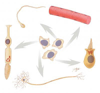
Avoids risk of inducing cancer Thursday, 26 March 2009 Stem cells (centre ones) can develop into any cell type. They are valuable as research tools and might, in the future, be used to treat a wide range of diseases. Credit: Judith Stoffer.A team of scientists has advanced stem cell research by finding a way to endow human skin cells with embryonic stem cell-like properties without inserting potentially problematic new genes into their DNA. The team was led by James A. Thomson, V.M.D., Ph.D., of the University of Wisconsin-Madison and supported in part by the National Institute of General Medical Sciences, a component of the National Institutes of Health. This is not the first time that scientists have endowed differentiated cells like skin cells with the capacity to develop into any of the roughly 220 types of cells in the body, a process known as induced pluripotency. However, it is the first time that they have done so without using viruses, which can insert potentially harmful genes into the cells' genetic material and trigger cancer. Thomson's new method imports the necessary genes on a small circle of DNA known as a plasmid. Over time, the plasmid disappears naturally from the cell population, avoiding the danger posed by using viruses. Scientists view pluripotent cells as invaluable to studies of normal and disease processes and to understanding the effects of certain drugs. In the future, doctors might be able to use such cells therapeutically to replace those affected by diseases such as Alzheimer's and Parkinson's or lost to traumatic injuries. Reference: Human Induced Pluripotent Stem Cells Free of Vector and Transgene Sequences Junying Yu, Kejin Hu, Kim Smuga-Otto, Shulan Tian, Ron Stewart, Igor I. Slukvin and James A. Thomson Science Express, March 26 2009; 10.1126/science.1172482 ......... ZenMaster


For more on stem cells and cloning, go to CellNEWS at http://cellnews-blog.blogspot.com/ and http://www.geocities.com/giantfideli/index.html

The Egg Makes Sure that Sperm Don't Get Too Old

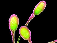
Egg cells can extend the telomeres of a fertilising sperm cell Thursday, 26 March 2009 In contrast to women, men are fertile throughout life, but research at the Sahlgrenska Academy, University of Gothenburg, Sweden, has now shown that a fertilising sperm can get help from the egg to rejuvenate. The result is an important step towards future stem cell therapy. Sperm head, false coulored.The risk of chromosomal abnormalities in the foetus is highly correlated to the age of the mother, but is nearly independent of the age of the father. One possible explanation is that egg cells have a unique ability to reset the age of a sperm. "We are the first to show that egg cells have the ability to rejuvenate other cells, and this is an important result for future stem cell research", says Associate Professor Tomas Simonsson, who leads the research group at the Sahlgrenska Academy that has made this discovery. Each time a cell divides, the genetic material at the ends of the chromosomes becomes shorter. The ends of the chromosomes, known as "telomeres", are important for the genetic stability of the cell and they act as a DNA clock that measures the age of the cell. The cell stops dividing and dies when the telomeres become too short. The discovery that the egg cell can extend the telomeres of a fertilising sperm cell is important in the development of stem cell therapy. Stem cell therapy involves replacing the cell nucleus in unfertilised egg with a nucleus from a somatic cell that has come from a patient who needs a stem cell transplantation. As soon as the cell has divided a few times, it is possible to harvest stem cells that are then allowed to mature to the cell type that the recipient needs. "The genetic stability of the transplanted cells has been a serious concern up until now, and it was feared that the lifetime of these cells would depend on the age of the cell nucleus that was transferred. Our results suggest that this is not the case", says Tomas Simonsson. About the Sahlgrenska Academy: The Sahlgrenska Academy is the faculty of health sciences at the University of Gothenburg. Education and research are conducted within the fields of pharmacy, medicine, odontology and health care sciences. About 4000 undergraduate students and 1000 postgraduate students are enrolled at Sahlgrenska Academy. The staff is about 1500 persons. 850 of them are researchers and/or teachers. ......... ZenMaster


For more on stem cells and cloning, go to CellNEWS at http://cellnews-blog.blogspot.com/ and http://www.geocities.com/giantfideli/index.html

Therapeutic Cloning Gets a Boost with New Research Findings

San Antonio and Honolulu researchers make important discoveries about point mutation rates in cloned mouse foetuses Thursday, 26 March 2009 Germ cells, the cells which give rise to a mammal's sperm or eggs, exhibit a five to ten-fold lower rate of spontaneous point mutations than adult somatic cells, which give rise to the body's remaining cell types, tissues and organs. Despite their comparatively higher mutation rates, however, adult somatic cells are used as the donor cells in a cloning process called somatic cell nuclear transfer (SCNT). This made researchers wonder if cloning by SCNT leads to progeny with more mutations than their naturally conceived counterparts. Also, would cloned foetuses receive DNA programming predisposing them to develop mutations faster than natural foetuses of the same age? Those scenarios are simply not likely, say researchers at the University of Texas at San Antonio, The University of Texas Health Science Center at San Antonio and The University of Hawaii at Honolulu's John A. Burns School of Medicine. The team, which spent more than five years analyzing mutation rates and types in cloned Big Blue® mouse foetuses recently published its findings in the online Early Edition of the Proceedings of the National Academy of Sciences in a paper titled "Epigenetic regulation of genetic integrity is reprogrammed during cloning." The paper offers the first direct demonstration that cloning does not lead to an increase in the frequency of point mutations. John McCarrey, professor of cellular and molecular biology at UTSA and the study's principal investigator, suggests a "bottleneck effect" is partially responsible for the observations his team recorded. "To create a cloned foetus by somatic cell nuclear transfer, only one adult somatic cell – one donor cell – is needed," he explains. "Because a random cell population exhibits a low mutation rate overall and only one cell from that population is used for cloning, the likelihood is remote that the cell chosen to be cloned will transfer a genetic mutation to its cloned offspring. Therefore, the bottleneck effect limits the transfer of mutations from donor cells to cloned offspring." Not only did the researchers find that SCNT does not lead to an increase in the frequency of point mutations in cloned mice, the team also found that naturally conceived foetuses and cloned foetuses that are the same age have similar rates of spontaneous mutation development. They attribute this finding to epigenetic reprogramming. It is known in the scientific community that germ cells contain an epigenome, a programmed state of the genome, which keeps mutation rates low. They suggest this type of epigenome is found in germ cells because those cells are responsible for contributing genetic information to subsequent generations. Adult somatic cells (the donor cells in SCNT) have higher mutation rates and less stringent epigenetic programming to avoid mutations than germ cells. Offspring produced from somatic cells by cloning have mutation rates similar to those in offspring produced by natural reproduction, suggesting that the epigenome of an adult somatic cell is reprogrammed during cloning to maintain the genetic integrity of that cell's progeny. Reference: Epigenetic regulation of genetic integrity is reprogrammed during cloning Patricia Murphey, Yukiko Yamazaki, C. Alex McMahan, Christi A. Walter, Ryuzo Yanagimachi, and John R. McCarrey PNAS, March 2, 2009, doi: 10.1073/pnas.0900687106 ......... ZenMaster


For more on stem cells and cloning, go to CellNEWS at http://cellnews-blog.blogspot.com/ and http://www.geocities.com/giantfideli/index.html

Wednesday 25 March 2009

Human Adult Testes Cells Can Become Embryonic-like

Human Adult Testes Cells Can Become Embryonic-like Tuesday, 24 March 2009 Using what they say is a relatively simple method, scientists at Georgetown University Medical Center have extracted stem/progenitor cells from testes and have converted them back into pluripotent embryonic-like stem cells. Researchers say that the naïve cells are now potentially capable of morphing into any cell type that a body needs, from brain neurons to pancreatic tissue. And because they produced these stem cells without the use of additional genes, the technology should be safe for human use, the researchers say in a paper published online in the journal Stem Cells and Development. "Given these advances, and with further validation, it is possible that in the not–too-distant-future, men could be cured of disease with a biopsy of their own testes," says the study's senior investigator, Martin Dym, PhD, a professor in the Department of Biochemistry and Molecular & Cellular Biology. The Georgetown researchers are among the first scientists to show that human testes stem cells can become embryonic stem-like cells, and they have done this work using testis tissue from organ donors, which they say has provided enough valuable tissue to allow them to make their discoveries. While they have published their preliminary results before, they are now disclosing a new and simpler method to isolate the testes stem/progenitor cells than has not been seen in other published procedures in humans and rodents. Being able to use adult stem cells for this type of cell-based therapy offers a number of advantages over other strategies currently being explored, says Dym. The use of embryonic stem cells is controversial because it necessitates destruction of an embryo, and pushing fully mature cells, such as skin cells, back into a stem-like state requires use of cancer genes, and has therefore been viewed as potentially risky for human treatment, he says. The idea with this approach is that men with an incurable disorder or disease could have a biopsy of their testes, which Dym says is a common procedure in patients suspected of having testicular cancer. Testes stem/progenitor cells – those cells that can go on to produce sperm – would be removed from the biopsy tissue, and grown in the laboratory with the addition of certain chemicals and growth factors. This causes the cells to revert back into an embryonic-like stem cell state, which could then be driven into chosen cell types. "We are taking adult spermatogonial stem/progenitor cells, which in the body are unipotent, capable of only making sperm, and coaxing them back to embryonic-like stem cells, which are pluripotent," Dym says. Once these new cell types are produced – several weeks after initial collection – they can be frozen and used at any point in the future, the researchers say. He and the research team conducted the study using testes donated to GUMC from four organ donors, aged 16-52 years old. "This is novel data which strengthens the argument for carrying out further research on pluripotent cells derived from human testes," Dym says. The next step, he says, is to get differentiated cells to cure disease in animal models and the researchers are now working on a project that uses testes spermatogonial stem/progenitor cells that morphed into pancreatic cells to treat diabetes in mouse models of human diabetes. ......... ZenMaster


For more on stem cells and cloning, go to CellNEWS at http://cellnews-blog.blogspot.com/ and http://www.geocities.com/giantfideli/index.html
Stem Cell Therapy May Lead to Treatment for Deafness

A new study has successfully isolated human auditory stem cells from foetal cochleae Tuesday, 24 March 2009 Deafness affects more than 250 million people worldwide. It typically involves the loss of sensory receptors, called hair cells, for their "tufts" of hair-like protrusions, and their associated neurons. The transplantation of stem cells that are capable of producing functional cell types might be a promising treatment for hearing impairment, but no human candidate cell type has been available to develop this technology. A new study led by Dr. Marcelo N. Rivolta of the University of Sheffield has successfully isolated human auditory stem cells from foetal cochleae (the auditory portion of the inner ear) and found they had the capacity to differentiate into sensory hair cells and neurons. The study is published in the April issue of STEM CELLS. The researchers painstakingly dissected and cultured cochlear cells from 9-11 week-old human foetuses. The cells were expanded and maintained in vitro for up to one year, with continued division for the first 7 to 8 months and up to 30 population doublings. This is similar to other non-embryonic stem cell populations, such as bone marrow. Gene expression analysis showed that all cell lines expressed optic markers that lead to the development of the inner ear as well as markers expressed by pluripotent embryonic stem cells, from which all tissues and organs develop. They were able to formulate conditions that allowed for the progressive differentiation into neurons and hair cells with the same functional electrophysiological characteristics as cells seen in vivo. "The results are the first in vitro renewable stem cell system derived from the human auditory organ and have the potential for a variety of applications, such as studying the development of human cochlear neurons and hair cells, as models for drug screening and helping to develop cell-based therapies for deafness," say the authors. Although the hair cell-like cells did not show the typical formation of a hair bundle, the authors suggest that future studies will aim to improve the differentiation system. They are currently working on using the knowledge gleaned from this study to optimize the differentiation of human embryonic stem cells into ear cell types. "Although considerable information has been obtained about the embryology of the ear using animal models, the lack of a human system has impaired the validation of such information," the authors note. "Access to human cells that can differentiate should allow the exploration of features unique to humans that may not be applicable to animal models," says Donald G. Phinney, co-editor of the journal. The protocol they developed to expand and isolate human foetal auditory stem cells may be able to be adapted for deriving clinical-grade cells with potential therapeutic applications. Dr Ralph Holme, director of biomedical research for Royal National Institute for Deaf and Hard of Hearing People, said: "There are currently no treatments to restore permanent hearing loss so this has the potential to make a difference to millions of deaf people." Reference: Human Fetal Auditory Stem Cells (hFASCs) Can Be Expanded In Vitro And Differentiate Into Functional Auditory Neurons And Hair Cell-Like Cells Wei Chen, Stuart L. Johnson, Walter Marcotti, Peter W. Andrews, Harry D. Moore, Marcelo N. Rivolta STEM CELLS Express March 23, 2009, 10.1002/stem.62 ......... ZenMaster


For more on stem cells and cloning, go to CellNEWS at http://cellnews-blog.blogspot.com/ and http://www.geocities.com/giantfideli/index.html

Friday 20 March 2009

Lab-grown Nerves Promote Nerve Regeneration after Injury

Lab-grown Nerves Promote Nerve Regeneration after Injury Friday, 20 March 2009 Researchers at the University of Pennsylvania School of Medicine have engineered transplantable living nerve tissue that encourages and guides regeneration in an animal model. Results were published this month in Tissue Engineering. About 300,000 Americans suffer peripheral nerve injuries every year, in many cases resulting in permanent loss of motor function, sensory function, or both. These injuries are a common consequence of trauma or surgery, but there are insufficient means for repair, according to neurosurgeons. In particular, surgeons need improved methods to coax nerve fibres known as axons to regrow across major nerve injuries to reconnect healthy targets, for instance muscle or skin. "We have created a three-dimensional neural network, a living conduit in culture, which can be transplanted en masse to an injury site," explains senior author Douglas H. Smith, MD, Professor, Department of Neurosurgery and Director of the Center for Brain Injury and Repair at Penn.. Smith and colleagues have successfully grown, transplanted, and integrated axon bundles that act as 'jumper cables' to the host tissue in order to bridge a damaged section of nerve.


This is a surviving cluster of transplanted neurons at the graft extremity (top) with axons in the centre (bottom). In both images, transplanted nerve cells are labelled green and axons are stained red. These axons are a mix of the transplanted axons and host axons, which intertwined as regeneration occurred directly across the transplanted tissue. Credit: Douglas H. Smith, MD, University of Pennsylvania.This is a surviving cluster of transplanted neurons at the graft extremity (top) with axons in the centre (bottom). In both images, transplanted nerve cells are labelled green and axons are stained red. These axons are a mix of the transplanted axons and host axons, which intertwined as regeneration occurred directly across the transplanted tissue. Credit: Douglas H. Smith, MD, University of Pennsylvania.
Previously, Smith and colleagues have "stretch-grown" axons by placing neurons from rat dorsal root ganglia (clusters of nerves just outside the spinal cord) on nutrient-filled plastic plates. Axons sprouted from the neurons on each plate and connected with neurons on the other plate. The plates were then slowly pulled apart over a series of days, aided by a precise computer-controlled motor system. These nerves were elongated to over 1 cm over seven days, after which they were embedded in a protein matrix (with growth factors), rolled into a tube, and then implanted to bridge a section of nerve that was removed in a rat. "That creates what we call a 'nervous-tissue construct'," says Smith. "We have designed a cylinder that looks similar to the longitudinal arrangement of the nerve axon bundles before it was damaged. The long bundles of axons span two populations of neurons, and these neurons can have axons growing in two directions – toward each other and into the host tissue at each side.” The constructs were transplanted to bridge an excised segment of the sciatic nerve in rats. Up to 16 weeks post-transplantation, the constructs still had their pre-transplant shape, with surviving transplanted neurons at the extremities of the constructs spanned by tracts of axons. Remarkably, the host axons appeared to use the transplanted axons as a living scaffold to regenerate across the injury. The authors found host and graft axons intertwined throughout the transplant region, suggesting a new form of axon-mediated axonal regeneration. "Regenerating axons grew across the transplant bridge and became totally intertwined with the transplanted axons," says Smith Axons throughout the transplant region showed extensive myelination, the fatty layer surrounding axons. What's more, graft neurons had extended axons beyond the margins of the transplanted region, penetrating deep into the host nerve. Remarkably, the constructs survived and integrated without the use of immunosuppressive drugs, challenging the conventional wisdom regarding immune tolerance in the peripheral nervous system. The researchers suspect that the living nerve-tissue construct encourages the survival of the supporting cells left in the nerve sheath away from the injury site. These are cells that further guide regeneration and provide the overall structure of the nerve. "This may be a new way to promote nerve regeneration where it may not have been possible before," says co-first author D. Kacy Cullen, PhD, a postdoctoral fellow in the Smith lab. "It's a race against time – if nerve regeneration happens too slowly, as may be the case for major injuries, the support cells in the extremities can degenerate, blunting complete repair. Because our living axonal constructs actually grow into the host nerve sheath, they may 'babysit' these support cells to give the host more time to regenerate." Reference: Long-Term Survival and Integration of Transplanted Engineered Nervous Tissue Constructs Promotes Peripheral Nerve Regeneration Jason H. Huang, D. Kacy Cullen, Kevin D. Browne, Robert Groff, Jun Zhang, Bryan J. Pfister, Eric L. Zager, Douglas H. Smith Tissue Engineering Part A. February 2009, doi:10.1089/ten.tea.2008.0294 ......... ZenMaster
For more on stem cells and cloning, go to CellNEWS at http://cellnews-blog.blogspot.com/ and http://www.geocities.com/giantfideli/index.html

Epigenetic Mark Guides Stem Cells toward Their Destiny

Protein that controls access to genes plays an important role in embryonic skin stem cells Friday, 20 March 2009 Not all stem cells are completely blank slates. Some, known as adult stem cells, have already partially embraced their fates, and are capable of becoming only cells of a particular type of tissue. So how do these tissue-specific stem cells restrict their fate? In research to appear in the March 20 issue of Cell, scientists at Rockefeller University have uncovered a gene control mechanism that guides the development and differentiation of epidermal skin stem cells in mouse embryos, and shown that this mechanism tempers the development of the skin barrier. The work, led by Elaine Fuchs, head of the Laboratory of Mammalian Cell Biology and Development, and first author Elena Ezhkova, a postdoc in the lab, highlights the interactions between genetic and epigenetic regulators in embryonic skin stem cells and how these interactions change as stem cells begin the process of specialization. The findings also provide insights that could lead to therapeutic advance in treating prematurely born infants who have not yet fully formed the skin. The skin is composed of two principle parts. The bottom 90 percent is known as the dermis; the outer 10 percent is the epidermis. The epidermis is composed of an inner layer (known as the basal layer) of dividing cells that gives rise to about 10 differentiated layers, the outermost of which is our body surface. "In contrast to embryonic stem cells, which can make all the cells of the body, epidermal stem cells restrict their options to making just these 10 differentiating layers," explains Fuchs, who is also Rebecca C. Lancefield Professor at Rockefeller and a Howard Hughes Medical Institute investigator. Clues as to how epidermal stem cells restrict their fate began to emerge when Ezhkova profiled the molecular makeup of the basal layer of the epidermis and compared it to the differentiating layers. As the skin stem cells began to differentiate, a complex of proteins (Polycomb repressor complex) that restrict access to genes decreased their expression while a transcription factor (AP1), which activates genes, increased its expression. Of the five major proteins that make up the Polycomb repressor complex, Ezhkova focused on an enzyme called Ezh2. It has long been known that Ezh2 prevents cells from reading segments of chromosomes by adding a small modification that acts as a molecular clamp to the gene and physically blocks transcription factors from gaining access to them. Past research had shown that Ezh2 blocked access to skin differentiation genes in embryonic stem cells, but its role in skin stem cells had never been explored in a living animal. Working with Alexander Tarakhovsky, head of the Laboratory of Lymphocyte Signalling at Rockefeller, Fuchs's team bred mice with and without Ezh2 and found that mice without it were born with thicker skin than their normal counterparts. Without Ezh2 to block access to the skin differentiation genes, the skin stem cells began to differentiate much earlier. As a result, extra skin tissue built up layer after layer. Previous research has shown that when embryonic stem cells lack Ezh2, genes for many cell differentiation pathways, not just the skin, lose their "molecular clamps" and become activated. In skin stem cells, however, these genes also lose these molecular clamps, but only the skin differentiation genes become activated. In examining why only the skin pathway is switched on when the clamp is missing, the researchers found that AP1, the transcription factor that selectively activates skin differentiation genes, is present in both basal and differentiating skin cells. Without Ezh2, AP1 could bind and start to activate these genes in the basal layer, before the genes are normally expressed. Another difference is that in embryonic stem cells, as soon as the molecular clamp is removed from muscle and neuronal genes, for example, an "activating mark" helps to switch on genes. In the skin stem cells, this mark is not present on the non-skin genes, helping to keep them silent. "Embryonic stem cells must be flexible – they produce all the cells of the animal," explains Fuchs. "As development proceeds, the resident progenitors of developing tissues become increasingly more restricted in their repertoire of differentiation programs. As the embryo develops, tissue-specific stem cells seem to remove the activating mark on those programs that will never be used, thereby switching off the wrong programs permanently. Then, to activate the right programs, the genes become dependent upon tissue-specific transcription factors." "The system is clearly more complicated than this, but the result is a series of intrinsic and extrinsic factors that control gene expression," Fuchs adds. Reference: Ezh2 Orchestrates Gene Expression for the Stepwise Differentiation of Tissue-Specific Stem Cells Elena Ezhkova, H. Amalia Pasolli, Joel S. Parker, Nicole Stokes, I-hsin Su, Gregory Hannon, Alexander Tarakhovsky and Elaine Fuchs Cell, Volume 136, Issue 6, 1122-1135, 20 March 2009, doi:10.1016/j.cell.2008.12.043 ......... ZenMaster


For more on stem cells and cloning, go to CellNEWS at http://cellnews-blog.blogspot.com/ and http://www.geocities.com/giantfideli/index.html

Wednesday 18 March 2009

Protein Key to Embryonic Stem Cell Differentiation

Burnham scientists identify protein that controls differentiation in human and mouse embryonic stem cells Wednesday, 18 March 2009 Investigators at Burnham Institute for Medical Research (Burnham) have learned that a protein called Shp2 plays a critical role in the pathways that control decisions for differentiation or self-renewal in both human embryonic stem cells (hESCs) and mouse embryonic stem cells (mESCs). The research, led by Gen-Sheng Feng, Ph.D., differs with some earlier findings that suggested hESCs and mESCs differentiate as a result of different signalling mechanisms. The discovery that Shp2 has a conserved role between mice and humans suggests an interesting common signalling mechanism between mESCs and hESCs, despite the known distinct signalling paths and biological properties between the two types of pluripotent stem cells. The study was published online in the journal PLoS ONE on March 17, 2009. Embryonic stem cells (ESCs) are pluripotent cells that can differentiate to become more than 200 different cell types. Because of their plasticity, ESCs have been suggested as potential therapies for numerous diseases and conditions, including neurodegenerative diseases, spinal cord injury and tissue damage. Development of such therapies is largely dependent on fully understanding and controlling the processes that lead to differentiation of hESCs into specialized cell types. "There are many signalling pathways that help embryonic stem cells decide their fate," said Dr. Feng. "We found that the Shp2 protein acts as a coordinator that fine-tunes the signal strength of multiple pathways and gives us a better understanding of the fundamental signalling methods that determine whether a stem cell's fate will be self-renewal or differentiation." In the study, the Feng lab created mutant Shp2 mESCs and showed that differentiation was dramatically impaired as the cells self-renewed as stem cells. The researchers also demonstrated small interfering RNAs in hESCs reduce Shp2 expression and subsequent cell differentiation. Feng and colleagues screened chemical libraries and identified a small-molecule inhibitor of Shp2 that, in small doses, partially inhibits differentiation in both mESCs and hESCs. Taken together, these results suggest a conserved role for Shp2 in ESC differentiation and self-renewal in both mice and humans. "This opens the door for new experimental reagents that will amplify the self-renewal process to create more stem cells for research and potential clinical use in the future," Dr. Feng added. "This research also suggests that comparative analysis of mouse and human embryonic stem cells will provide fundamental insight into the cellular processes that determine 'stemness,' a critical question that remains to be answered in the stem cell biology field." About Burnham Institute for Medical Research: Burnham Institute for Medical Research is dedicated to revealing the fundamental molecular causes of disease and devising the innovative therapies of tomorrow. Burnham, with operations in California and Florida, is one of the fastest-growing research institutes in the country. The Institute ranks among the top-four institutions nationally for NIH grant funding and among the top 25 organizations worldwide for its research impact. Burnham utilizes a unique, collaborative approach to medical research and has established major research programs in cancer, neurodegeneration, diabetes, infectious and inflammatory and childhood diseases. The Institute is known for its world-class capabilities in stem cell research and drug discovery technologies. Burnham is a non-profit, public benefit corporation. Reference: A Conserved Mechanism for Control of Human and Mouse Embryonic Stem Cell Pluripotency and Differentiation by Shp2 Tyrosine Phosphatase Dongmei Wu, Yuhong Pang, Yuehai Ke, Jianxiu Yu, Zhao He, Lutz Tautz, Tomas Mustelin, Sheng Ding, Ziwei Huang, Gen-Sheng Feng PLoS ONE 4(3) 2009: e4914. doi:10.1371/journal.pone.0004914 ......... ZenMaster


For more on stem cells and cloning, go to CellNEWS at http://cellnews-blog.blogspot.com/ and http://www.geocities.com/giantfideli/index.html

Tuesday 17 March 2009

Stem Cells Crucial to Diabetes Cure in Mice

Stem Cells Crucial to Diabetes Cure in Mice Tuesday, 17 March 2009 More than five years ago, Dr. Lawrence C.B. Chan and colleagues in his Baylor College of Medicine laboratory cured mice with type 1 diabetes by using a gene to induce liver cells to make insulin. "Now we know how it works," said Chan, director of the federally designed Diabetes and Endocrinology Research Center at BCM and chief of the division of endocrinology in BCM's department of medicine. "The answer is adult stem cells." A gene called neurogenin3 proved critical to inducing cells in the liver to produce insulin on a continuing basis, said Chan and Dr. Vijay Yechoor, assistant professor of medicine-endocrinology and first author of the report that appears in the current issue of the journal Developmental Cell. The research team used a disarmed virus called a vector to deliver the gene to the livers of diabetic mice by a procedure commonly known as gene therapy. "The mice responded within a week," said Yechoor. The levels of sugar in their blood plummeted to normal and stayed that way for the rest of their normal lives. The quick response generated more questions as did the length of time that the animals stayed healthy. They found that there was a two-step response. At first, the neurogenin3 gene goes into the mature liver cells and causes them to make small quantities of insulin – enough to drop sugar levels to normal, said Yechoor. "This is a transient effect," he said. "Liver cells lose the capacity to make insulin after about six weeks." However, they found that other cells that made larger quantities of insulin showed up later, clustered around the portal veins (blood vessels that carry blood from the intestines and abdominal organs to the liver). "They look similar to normal pancreatic islet cells (that make insulin normally)," said Yechoor. They found that these "islet" cells came from a small population of adult stem cells usually found near the portal vein. Only a few are needed usually because they serve as a safety net in case of liver injury. When that occurs, they quickly activate to form mature liver cells or bile duct cells. However, neurogenin3 changes their fates, directing them down a path to becoming insulin-producing islet cells located in the liver. The mature liver cell cannot make this change because its fate appears to be fixed before exposure to neurogenin3. The islet cells in the liver look similar to those made by pancreas after an injury, said Yechoor. "If we didn't use neurogenin3, none of this would happen," he said. "Neurogenin3 is necessary and sufficient to produce these changes." Chan cautioned that much more work is needed before similar results could be seen in humans. The gene therapy they undertook in the animals used a disarmed viral vector that could still have substantial toxic effects in humans. "The concept is important because we can induce normal adult stem cells to acquire a new cell fate. It might even be applicable to regenerating other organs or tissues using a different gene from other types of adult stem cells," he said. Finding a way to use the treatment in human sounds easier than it is, he said. The environment in which cells grow appears to be an important part of the cell fate determination. However, he and Yechoor plan to continue their work with the eventual goal of providing a workable treatment for people with diabetes. Reference: Neurogenin3 Is Sufficient for Transdetermination of Hepatic Progenitor Cells into Neo-Islets In Vivo but Not Transdifferentiation of Hepatocytes Vijay Yechoor, Victoria Liu, Christie Espiritu, Antoni Paul, Kazuhiro Oka, Hideto Kojima andLawrence Chan Developmental Cell, Volume 16, Issue 3, 358-373, 17 March 2009, doi:10.1016/j.devcel.2009.01.012 ......... ZenMaster


For more on stem cells and cloning, go to CellNEWS at http://cellnews-blog.blogspot.com/ and http://www.geocities.com/giantfideli/index.html

China Restrict Clinical Tests of Stem Cells Obtained by Therapeutic Cloning

Other medical procedures also restricted Tuesday, 17 March 2009 China's Ministry of Health has issued a temporary ban on clinical use of therapeutic cloning, effective from May 1, on its official Web site on Tuesday. The MoH also prohibited other techniques, like xenotransplantation of stem cells and some gene therapy trials in clinical studies. Article 58th in the guidelines stated: “…xenogeneic stem cell therapy technology, xenotransplantation gene therapy technology, human somatic cell cloning technology in medical technology may not be used in clinical suspense.” This temporary ban is issued because of major concerns on ethical issues, safety, and effectiveness, until further verification of the technologies are achieved. The restrictions are applied to a broader set of clinical techniques. They also include treatment by cloning technologies, autologous stem cell and immune cell therapy, gene therapy techniques, xenotransplantation technology, surgical treatment of central nervous system, stereotactic surgical treatment of mental illness technologies, allogeneic stem cell transplantation technology, and vaccines as therapeutic techniques. It is uncertain how these restrictions will affect new medical procedures in the long run. However, it shows a sensitivity to the complexity of these issues in the medical community in China. These new restrictions should also be seen in the light of the recently initiated medical reform program in China. "The biggest beneficiaries of medical reform are the masses of the people, the goal is to benefit everyone." China’s health minister Chen Zhu said in a recent interview with Xinhua. ......... ZenMaster


For more on stem cells and cloning, go to CellNEWS at http://cellnews-blog.blogspot.com/ and http://www.geocities.com/giantfideli/index.html

Monday 9 March 2009

Obama Reverses Bush’s Stem Cell Policy

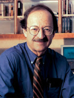
Ceremony in the White House overturns the 8-year-old Bush ban on federal support for stem cell research Monday, 09 March 2009 President Barack Obama, at a ceremony in the East Room at White House today, overturned the 8-year-old Bush ban on federal support of human embryonic stem cell research. In an Executive Order, the Bush’s ban on federal funding for embryonic stem cell research is overturned. Barack Obama at the ceremony.“Medical miracles do not happen simply by accident,” the President said. "They result from painstaking and costly research — from years of lonely trial and error, much of which never bears fruit — and from a government willing to support that work." This research should be pursued, but with strict guidelines and ethics oversight. The President would also support research on iPS cell research. "The majority of Americans — from across the political spectrum, and of all backgrounds and beliefs — have come to a consensus that we should pursue this research," said Obama. "That the potential it offers is great, and with proper guidelines and strict oversight, the perils can be avoided.” He clearly stated that cloning for human reproductive purposes would never be allowed. Obama's move is part of a broader effort to separate science and politics, sending a clear signal, that science — not political ideology — will guide his administration. Therefore, Obama signed a Presidential Memorandum on Scientific Independence from politics and beliefs. The presidential memorandum will establish greater independence for federal science policies and programs in an effort to separate science and politics and "restore scientific integrity in governmental decision-making”. "This order is an important step in advancing the cause of science in America. But let's be clear: promoting science isn't just about providing resources — it is also about protecting free and open inquiry," the President said . Dr. Harold Varmus."It is about letting scientists like those here today do their jobs, free from manipulation or coercion, and listening to what they tell us, even when it's inconvenient — especially when it's inconvenient." Dr. Harold Varmus, the Nobel Prize winner and president of Memorial Sloan-Kettering Cancer Center and now co-chairman of Obama's science advisory council, said that Obama will "endorse the notion that public policy must be guided by sound, scientific advice." Obama's order will direct the National Institutes of Health to develop revised guidelines on federal funding for embryonic stem cell research within 120 days. Rockefeller University president applauds new US policy on stem cells Sir Paul Nurse.“Today's executive order making federal money once again available for research on human embryonic stem cells will accelerate biomedical research and hopefully bring us closer to cures for some of our most devastating diseases,” said Nobel Prize winner and Rockefeller University president Sir Paul Nurse. "The new policy, which reverses eight years of restrictions on how biomedical research scientists interested in understanding stem cells can spend federal grant money, will have an important impact on the pace of this type of research," Nurse said. "It will also help many scientists make better use of limited research dollars by freeing them from the need to carefully separate privately funded experiments on non-Registry cell lines from those that are publicly funded." Under the old policy, scientists wishing to work on certain lines of stem cells were not permitted to use supplies or equipment paid for by federal grant money. Several labs at Rockefeller work with human embryonic stem cells, including a line that was derived using funds exclusively from private sources under the old policy. "When you're dealing with research that may take years to fully mature, it's important to have consistent policies in place that allow scientists to work without fear of political restrictions," Nurse said. "In addition to making money available to scientists studying embryonic stem cells, President Obama's executive order today will help encourage researchers to invest their efforts in this important area of research." ISSCR Scientists Elated for Future of Stem Cell Research The International Society for Stem Cell Research (ISSCR) applauded President Barack Obama’s decision to expand federal funding for human embryonic stem cell research by rescinding the policy of Aug. 9, 2001, which restricted federally-funded researchers to using only the small number of cell lines created before that date. The President’s executive order demonstrates his enthusiastic endorsement of stem cell research and its potential to relieve suffering caused by diseases and conditions such as diabetes, Alzheimer’s disease and spinal cord injuries. Today’s executive order calls on the National Institutes of Health (NIH), the primary federal agency for funding medical research, to develop revised policies on federal funding for human embryonic stem cell research. Dr. Irving Weissman.The new policy empowers scientists to utilize the many hundreds of valuable lines created since 2001, and relieves them from the substantial hurdles in duplicating equipment and other resources in order to separate privately- or state-funded stem cell research from federal government-funded efforts. “Today’s announcement restores integrity to the relationship between politics and science that has been traditional in the U.S.,” said Irving Weissman, President-Elect of the ISSCR. “We anticipate increased collaborative efforts amongst the scientific community and across disciplines, which will drive the entire field forward.” Dr. George Daley.As the principal international organization of stem cell research professionals, the ISSCR is eager to offer its broad expertise to the NIH to support development of policies for research funding and oversight. The ISSCR believes that research using all types of stem cells should be selected for funding based on scientific merit and conducted under transparent ethical oversight. “Policies governing stem cell research should embrace the rigorous ethical standards laid out in the National Academy of Sciences’ guidelines and the ISSCR’s Guidelines for the Conduct of Human Embryonic Stem Cell Research,” said George Q. Daley, ISSCR immediate past president, and chair of the task force that developed the ISSCR guidelines. The ISSCR is confident that under the new administration policy embryonic stem cell research will thrive and ultimately lead to a better understanding and treatment of many diseases, which today are untreatable. See also: Obama to Reverse Embryonic Stem Cell Ban CellNEWS - Saturday, 07 March 2009 Barack Obama on Stem Cell Research CellNEWS - Tuesday, 02 September 2008 ......... ZenMaster


For more on stem cells and cloning, go to CellNEWS at http://cellnews-blog.blogspot.com/ and http://www.geocities.com/giantfideli/index.html

Stem Cells Replace Stroke-damaged Tissue

A stem-cell matrix can repair brain damage in rodents. Monday, 09 March 2009 Effective stem cell treatment for strokes has taken a significant step forward today (09 March) as scientists reveal how they have replaced stroke-damaged brain tissue in rats. The team of scientists is funded by the Biotechnology and Biological Sciences Research Council (BBSRC) and led by Dr Mike Modo of the Institute of Psychiatry, King's College London. The work, carried out at the Institute of Psychiatry and University of Nottingham, shows that by inserting tiny scaffolding with stem cells attached, it is possible to fill a hole left by stroke damage with brand new brain tissue within 7 days. The work is published in Biomaterials. Previous experiments where stem cells have been injected into the void left by stroke damage have had some success in improving outcomes in rats. The problem is that in the damaged area there is no structural support for the stem cells and so they tend to migrate into the surrounding healthy tissues rather than filling up the hole left by the stroke. "We would expect to see a much better improvement in the outcome after a stroke if we can fully replace the lost brain tissue, and that is what we have been able to do with our technique," Dr Modo said. Using individual particles of a biodegradable polymer called PLGA that have been loaded with neural stem cells, the team of scientists have filled stroke cavities with stem cells on a ready-made support structure.


Stem cell stroke treatment.Left: A microscope image showing the stroke site (the dark area on the right), which is beginning to fill in with new tissue. (Modified from Figure 4, Bible E et al. Biomaterials (2009)) Right: A magnification of the area on the left indicated by the red square. Here, all the dark spots are scaffold particles and we see tissue - the coloured parts - forming between them. (Modified from Figure 5, Bible E et al. Biomaterials (2009)) Credit: Bible E et al., The support of neural stem cells transplanted into stroke-induced brain cavities by PGLA particles, Biomaterials (2009), doi:10.1016/j.biomaterials.2009.02.012.

"This works really well because the stem cell-loaded PLGA particles can be injected through a very fine needle and then adopt the precise shape of the cavity. In this process the cells fill the cavity and can make connections with other cells, which helps to establish the tissue,” Dr Modo continued. "Over a few days we can see cells migrating along the scaffold particles and forming a primitive brain tissue that interacts with the host brain. Gradually the particles biodegrade leaving more gaps and conduits for tissue, fibres and blood vessels to move into." The research published today uses an MRI scanner to pinpoint precisely the right place to inject the scaffold-cell structure. MRI is also used to monitor the development of the new brain tissue over time. The next stage of the research will be to include a factor called VEGF with the particles. VEGF will encourage blood vessels to enter the new tissue. "Stroke is a leading cause of disability in industrialised countries. It is reassuring to know that the technology for treating stroke by repairing brain damage is getting ever closer to translation into the clinic. This crucial groundwork by Dr Modo and his colleagues will surely be a solid foundation of basic research for much better treatments in the future," Professor Douglas Kell, BBSRC Chief Executive said. "This research is another step towards using stem cell therapy in treating and reversing the brain damage caused by stroke. It is exciting because researchers have shown they are able to overcome some of the many challenges in translating the potential of using stem cells into reality,” Joe Korner, Director of Communications at The Stroke Association commented. "The potential to reverse the disabling effects of stroke seems to have been proved. However the development of stem cell therapy for stroke survivors is still in the early stages and much more research will be needed before it can be tested in humans or used in practice.” "Every five minutes someone in the UK has a stroke and it is vital that we do all we can to help those affected by stroke." Reference: The support of neural stem cells transplanted into stroke-induced brain cavities by PGLA particles Bible, E. et al. Biomaterials (2009), doi:10.1016/j.biomaterials.2009.02.012 ......... ZenMaster


For more on stem cells and cloning, go to CellNEWS at http://cellnews-blog.blogspot.com/ and http://www.geocities.com/giantfideli/index.html

ISSCR Calls for Adherence to Stem Cell Guidelines

ISSCR Calls for Adherence to Stem Cell Guidelines Monday, 09 March 2009 The International Society for Stem Cell Research (ISSCR) is concerned and saddened by the recent report of nervous system tumours that developed in a child who had previously received injections of cell preparations referred to as “neural stem cells.” This illustrates the concerns that prompted the ISSCR to develop Guidelines for the Clinical Translation of Stem Cells, released in December 2008. The article, appearing in PLoS Medicine (Feb. 17, 2009), documents the development of glioneuronal neoplasms in the brain and spinal cord of a child from Israel with Ataxia Telangiectasia who, against his doctors' recommendations, had received multiple injections of “foetal neural stem cells” at a clinic in Russia. No improvement was seen in the patient’s condition, which is a rare disease that causes degeneration in the part of the brain that controls movement and speech. The cells that were injected were described as “foetal neural stem cells,” but the exact nature of the cells, quality control procedures and evidence of safety and effectiveness from preclinical studies were not available. The ISSCR reiterates that these findings do not mean that research into potential stem cell therapies should be abandoned. This case does, however, emphasize the importance of appropriate preclinical studies for both safety and effectiveness and the need to exercise extreme caution before stem cell therapies are administered to humans. It also highlights the need for disclosure of risks to potential patients and the need for those seeking such therapies to become fully informed about the nature of the product they are considering. The ISSCR Guidelines for the Clinical Translation of Stem Cells provide a roadmap for the responsible development of safe and effective stem cell therapies for patients. These guidelines call for rigorous standards in the development of such therapies including stringent evaluation and oversight, a thorough informed consent process, and transparency in operations and reporting. The ISSCR once more calls for greater awareness and international dialogue to help implement the standards described in its guidelines. “This is a new area of science and its enormous potential is well recognized by the public at large,” said David Scadden, co-chair of the ISSCR Clinical Translation Committee, director of the Massachusetts General Hospital Center for Regenerative Medicine, and co-director of the Harvard Stem Cell Institute. “What is less well understood are the potential risks and it is therefore of paramount importance that scientists, physicians and patients alike proceed with great care and adhere to the highest ethical and scientific standards.” The ISSCR guidelines provide information for patients and their doctors evaluating a stem cell therapy in Appendix 1, a Patient Handbook on Stem Cell Therapies. About ISSCR: The International Society for Stem Cell Research (ISSCR) is an independent, nonprofit membership organization established to promote and foster the exchange and dissemination of information and ideas relating to stem cells, to encourage the general field of research involving stem cells and to promote professional and public education in all areas of stem cell research and application. See also: Patient Handbook on Stem Cell Therapies Guidelines for the Clinical Translation of Stem Cells ......... ZenMaster


For more on stem cells and cloning, go to CellNEWS at http://cellnews-blog.blogspot.com/ and http://www.geocities.com/giantfideli/index.html

New Study Using Combination of Bioengineered Skin and Stem Cells

Promise in Treatment of Non-Healing Wounds Monday, 09 March 2009 Scleroderma is an autoimmune disease that leads to thickening and severe scarring of skin as well as thickening and failure of internal organs, including the lungs, heart, kidneys and intestines. The disease – which the Scleroderma Foundation estimates affects approximately 300,000 Americans – can be fatal and there is no cure. A major and incapacitating complication of scleroderma is the development of ulcers on the patients’ fingers and toes that are very painful and difficult to heal. Now, researchers are studying the viability of administering stem cells topically to ulcerated fingers using bioengineered skin to help heal these wounds. Speaking at the 67th Annual Meeting of the American Academy of Dermatology, dermatologist Vincent Falanga, MD, FAAD, professor of dermatology and biochemistry at Boston University in Boston, presented his study findings in which cultured stem cells and bioengineered skin were used to successfully treat skin ulcers of three scleroderma patients. He also discussed how bioengineered skin plays a critical role in this procedure. “The concept of using bioengineered skin to heal difficult wounds, such as leg and foot ulcers, previously has been shown to be effective for chronic wounds but, in our experience, has not by itself been consistently useful in healing scleroderma finger ulcers,” said Dr. Falanga. “Through our research, we learned that our combined approach of using stem cells with bioengineered skin may provide value by ‘instructing’ the stem cells on how to stimulate healing in a difficult wound.” As a source of stem cells, Dr. Falanga and his team of researchers focused on taking a small amount of bone marrow from the affected patients’ hip. This common procedure is performed to diagnose conditions affecting the blood or bone marrow, such as anaemia’s and leukaemia. From this sample, the intention was to grow a particular type of stem cells in the laboratory called mesenchymal stem cells, which are cells that can create other cell types, including those in skin and muscle, and are capable of repopulating damaged skin. The delivery method used to apply stem cells to the wounds was a fibrin spray system that had not been used in humans prior to the research pioneered by Dr. Falanga and his team. Fibrin is a chemical by-product that occurs naturally in the body and helps clot blood. For each of the three scleroderma patients, up to three spray applications of stem cells were performed. “For the first time in humans, the investigators used a fibrin spray system – which takes advantage of the immediate polymerization, or ‘gluing,’ of stem cells in fibrinogen when mixed with thrombin,” said Dr. Falanga. “Both fibrinogen and thrombin are naturally occurring substances in blood which, when mixed to form fibrin, are normally involved in the formation of a clot. In this case, the two substances, highly diluted, were used to deliver the cultured mesenchymal stem cells to the wounds of scleroderma patients in a fine transparent spray.” After delivering the stem cells to the affected finger, the wound was covered and treated with the additional stimulus provided by bioengineered skin – a bi-layered, substitute skin that consists of living human keratinocytes (cells that make the upper skin layer) and fibroblasts (cells that make collagen) – derived from neonatal foreskin following a circumcision and preserved for medical research. From this initial study, Dr. Falanga and his team were very encouraged by their early results. “Our combined approach resulted in dramatic healing of these extremely difficult-to-heal wounds, with new skin growth that blended in nicely with the surrounding skin,” said Dr. Falanga. “On follow-up examination, one patient who had painful and incurable finger ulcers for many years remained healed and free of pain from this procedure. Importantly, the concept of ‘instructing’ stem cells with other interventions or biological agents is significant and could be applicable to other situations where the use of stem cells is being pursued.” About American Academy of Dermatology: Headquartered in Schaumburg, Ill., the American Academy of Dermatology, founded in 1938, is the largest, most influential, and most representative of all dermatologic associations. With a membership of more than 15,000 physicians worldwide, the Academy is committed to: advancing the diagnosis and medical, surgical and cosmetic treatment of the skin, hair and nails; advocating high standards in clinical practice, education, and research in dermatology; and supporting and enhancing patient care for a lifetime of healthier skin, hair and nails. ......... ZenMaster


For more on stem cells and cloning, go to CellNEWS at http://cellnews-blog.blogspot.com/ and http://www.geocities.com/giantfideli/index.html

Sunday 8 March 2009

RNA Research Strategy for Europe Takes Shape

"Consensus Conference" organised by ESF Sunday, 08 March 2009 A hairpin loop from a pre-mRNA. Notice its nitrogen-rich (blue) bases and oxygen-rich (red) backbone. Research into RNA, a molecule found in every cell of our bodies, could lead to remarkable advances in the treatment of diseases such as cancer and diabetes, a meeting organised by the European Science Foundation was told. The conference, held the institute of Parasitología y Biomedicina "López-Neyra", CSIC in Granada, Spain, on 23 February 2009, was part of an ESF initiative to develop a coherent strategy for RNA research in Europe in recognition of the potential of RNA to result in new approaches to treating human diseases. For many years it was believed that RNA's sole function in cells was to transmit genetic information from DNA during the manufacture of proteins – the cell's workhorse molecules. However, in recent years it has become clear that RNA has many functions that are more sophisticated and that there are more types of RNA than previously known. The field exploded into activity with the discovery in 1998 by US researchers Andrew Fire and Craig Mello of a phenomenon called RNA interference, meaning that genes can be 'silenced' by RNA. This discovery, for which Fire and Mello were awarded the Nobel Prize in 2006, revolutionised the way scientists think about how genetic information is controlled in cells, and has opened the possibility of using gene silencing as a therapy where rogue genes cause disease. "Research into RNA has great promise for both basic science and biotechnology and medicine," said the meeting's chairperson, Professor Lars Thelander of Umeå University in Sweden. "Most pharmaceutical companies now have RNA projects, but the field is still in its early days and it could be another ten years before we see products appearing in the clinics." Professor Thomas Cech of the Howard Hughes Medical Institute in the US told the meeting how he discovered that RNA could also act as a biological catalyst – something that it was previously thought was the preserve of proteins representing a wonderful example of the versatility of RNA function. The discovery gave rise to new ideas about how life on Earth might have started and resulted in Professor Cech being awarded a Nobel Prize in 1989. The Granada "Consensus Conference" was organised by ESF as part of a 'Forward Look' entitled 'RNA World: a new frontier in biomedical research' aimed at developing a strategy for research in RNA over the next ten years. Three earlier workshops had examined various aspects of RNA research to identify where gaps in our knowledge lie and what is required to plug these gaps and fulfil the promise that RNA holds. Forward Looks are a key part of ESF's work, examining important areas of science and technology in consultation with leading scientists and policy makers to develop a strategic framework for research. A Forward Look report on RNA research is due to be published later this year, detailing the scientific questions that need to be answered and giving politicians and policy makers the information they need when deciding where to direct research funding to ensure that Europe remains globally competitive in this key area of emerging science. ......... ZenMaster


For more on stem cells and cloning, go to CellNEWS at http://cellnews-blog.blogspot.com/ and http://www.geocities.com/giantfideli/index.html