 |
Pancreas
explant.
|
Wednesday, 25 March 2015
Stem Cells Make Similar Decisions to Humans
Posted by ZenMaster at Wednesday, March 25, 2015
Labels: diabetes, differentiation, embryonic, human, microscopy, pluripotent, proliferation, research, stem cells 0 comments
Friday, 14 November 2014
Tumour Suppressor Also Inhibits Key Property of Stem Cells
Posted by ZenMaster at Friday, November 14, 2014
Labels: c-Myc, differentiation, embryonic, eye, iPSC, Klf4, mouse, Oct4, proliferation, Rb, reprogramming, research, retina, Sox2, stem cells, tumour, tumour suppressor 0 comments
Thursday, 5 June 2014
Stem Cells Hold Keys to Body's Plan
Posted by ZenMaster at Thursday, June 05, 2014
Labels: differentiation, embryonic, enhancers, pluripotent, proliferation, research, stem cells, US 0 comments
Monday, 18 May 2009
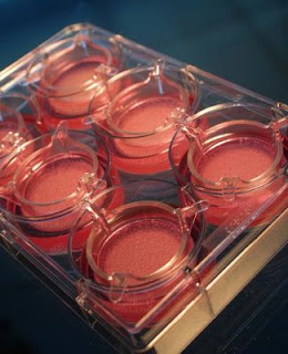
Automated Tissue Engineering on Demand
Automated Tissue Engineering on Demand
Monday, 18 May 2009
Skin from a factory – this has long been the dream of pharmacologists, chemists and doctors. Research has an urgent need for large quantities of 'skin models', which can be used to determine if products such as creams and soaps, cleaning agents, medicines and adhesive bandages are compatible with skin, or if they instead will lead to irritation or allergic reactions for the consumer. Such test results are seen as more meaningful than those from animal experiments are, and can even make such experiments largely superfluous.
But artificial skin is rare.
"The production is complex and involves a great deal of manual work. At this time, even the market's established international companies cannot produce more than 2,000 tiny skin pieces a month. With annual requirements of more than 6.5 million units in the EU area alone, however, the industrial demand far exceeds all currently available production capacities," reports Jörg Saxler. Together with Prof. Heike Mertsching, he is coordinating the "Automated Tissue Engineering on Demand" project within the Fraunhofer-Gesellschaft.
Tissue engineering is still in its infancy.
"Until now, the offer was limited predominantly to single-layer skin models that consist of a single cell type," explains Mertsching, who heads the Cell Systems Department at the Fraunhofer Institute for Interfacial Engineering and Biotechnology IGB.
"Thanks to developments at our institute, the project team has access to a patent-protected skin model that consists of two layers with different cell types. This gives us an almost perfect copy of human skin, and one that provides more information than any system currently available on the market."
An interdisciplinary team of Fraunhofer researchers is currently developing the first fully automatic production system for two-layer skin models.
"Our engineers and biologists are the only ones who have succeeded in fully automating the entire process chain for manufacturing two-layer skin models," explains Saxler, who is from the Fraunhofer Institute for Production Technology IPT where he is responsible for technology management and heads the "Life Science Engineering" business unit.
 In a multi-stage process, first small pieces of skin are sterilized. Then they are cut into small pieces, modified with specific enzymes, and isolated into two cell fractions, which are then propagated separately on cell culture surfaces. The next step in the process combines the two cell types into a two-layer model, with collagen added to the cells that are to form the flexible lower layer, or dermis. This gives the tissue natural elasticity. In a humid incubator kept at body temperature, it takes the cell fractions less than three weeks to grow together and form a finished skin model with a diameter of roughly one centimetre. The technique has already proven its use in practice, but until now, it has been too expensive and complicated for mass production.
Mertsching explains, "The production is associated with a great deal of manual work, and this reduces the method's efficiency."
The project team, in which engineers, scientists and technicians from four Fraunhofer institutes are cooperating, is currently working at full speed to automate the work steps. The researchers at the IGB and the Fraunhofer Institute for Cell Therapy and Immunology IZI are handling the development of the biological fundamentals and validation of the machine and its sub-modules. Experts from the Fraunhofer Institute for Manufacturing and Automation IPA and the Fraunhofer Institute for Production Technology IPT are taking care of prototype development, automation and integration of the machine into a complete functional system.
"At the beginning, our greatest challenge was to overcome existing barriers, because each discipline had its own very different approach," Saxler remembers.
"Meanwhile, the four institutes are working together very smoothly – everyone knows that progress is impossible without input from the others." After working together for one year, the project team has already initiated eight patent procedures.
At a collective Fraunhofer-Gesellschaft booth at the 2009 BIO in Atlanta, the researchers are presenting a computer model of the overall system, along with the three fundamental sub-modules. The first module prepares the tissue samples and isolates the two cell types; the second proliferates them. The finished skin models are built up and cultivated in the third, and then packed by a robot.
The researchers still have a lot of meticulous work ahead before the machine will be finished. The difference between success and failure often depends on details, such as the quality of the skin pieces, processing times of enzymes, and liquid viscosities. Furthermore, the cell cultures must be monitored throughout the entire manufacturing process in order to provide optimal process control and to allow timely detection of any contamination with fungi or bacteria. The skin factory is expected to be finished in two years.
"Our goal is a monthly production of 5,000 skin models with perfect quality, and a unit price under 34 euros. These are levels that are attractive for industry," Saxler continues.
But chemical, cosmetic, pharmaceutical, and medical technology companies who have to test the reaction of skin to their products are not the only ones interested in Automated Tissue Engineering. In transplantation medicine, surgeons require healthy tissue in order to replace destroyed skin sections when burn injuries cover large portions of the body. The two-layer models that the new machine is intended to produce are not yet suitable for this purpose, however.
"They don't have a blood supply, and are consequently rejected by the body after some time," Saxler explains.
However, IGB researchers are already working on a full-skin model that will even include blood vessels. Once the research has been completed, fully automatic production of the transplants should also be possible.
"We have designed the production system in such a way that it satisfies the high standards for good manufacturing practices (GMP) for the manufacture of products used in medicine," Mertsching explains.
"And so they are also suitable for producing artificial skin for transplants."
.........
ZenMaster
For more on stem cells and cloning, go to CellNEWS at http://cellnews-blog.blogspot.com/ and http://www.geocities.com/giantfideli/index.html
Posted by ZenMaster at Monday, May 18, 2009
Labels: collagen, Germany, human, proliferation, research, skin, tissue engineering 1 comments
Thursday, 16 April 2009
A New Method for Bone-marrow-derived Liver Stem Cells Isolation
Researchers designed a culture system to isolate, proliferate and differentiate liver stem cells directly from bone marrow cells. Thursday, 16 April 2009 Great interest has been aroused in the identification and isolation of liver stem cells from bone marrow cells. Several subsets of bone marrow cells have been found to have the potential to differentiate into hepatocytes, however, sorting based on immunological methods is difficult because of the complicated surface markers of the stem cells; furthermore, no report of successful passage has been published. A research article to be published on April 7, 2009 in the World Journal of Gastroenterology addresses this question. The research team led by Dr. Yun-Feng Cai and his colleagues from the Affiliated Foshan Hospital and the Second Affiliated Hospital of Sun Yat-sen University established a carefully designed culture system to isolate, proliferate and differentiate liver stem cells directly from bone marrow cells, and they were able to achieve six passages of the stem cells. The results suggest that BDLSCs can be purified and passaged (proliferated). The selecting culture system that contains cholestatic serum can purify BDLSCs directly from bone marrow cells, which provides an easy method to separate stem cells, by avoiding complicated immunological manipulation. The successful passage of the stem cells further verifies the proliferating ability of the cells, although the passage is limited, and further research will provide more experience. In this study, the authors used their original method to retrieve the cells, which are possibly BDLSCs. Then, they used fluorescence-activated cell sorting to determine the cells' characteristics before and after differentiation. This is an interesting and potentially important study, which suggests that bone-marrow-derived cells can be stimulated to expand and then differentiate into hepatocyte-like cells, which can possibly be used to treat liver disease. Reference: Passage of bone marrow-derived liver stem cells with a proliferating culture system Yun-Feng Cai, Ji-Sheng Chen, Shu-Ying Su, Zuo-Jun Zhen, Huan-Wei Chen World J Gastroenterol 2009, 15(13): 1630-1635 ......... ZenMaster
For more on stem cells and cloning, go to CellNEWS at http://cellnews-blog.blogspot.com/ and http://www.geocities.com/giantfideli/index.html
Posted by ZenMaster at Thursday, April 16, 2009
Labels: bone marrow, China, differentiation, liver, proliferation, rat, research, stem cells 0 comments


