Sunday 31 May 2009

Combined Stem Cell and Gene Therapy Cures Fanconi’s Anaemia

Approach cures human genetic disease in vitro Sunday, 31 May 2009 A study led by researchers at the Salk Institute for Biological Studies, has catapulted the field of regenerative medicine significantly forward, proving in principle that a human genetic disease can be cured using a combination of gene therapy and induced pluripotent stem (iPS) cell technology. The study, published in the May 31, 2009 early online edition of Nature, is a major milestone on the path from the laboratory to the clinic. "It's been ten years since human stem cells were first cultured in a Petri dish," says the study's leader Juan-Carlos Izpisúa Belmonte, Ph.D., a professor in the Gene Expression Laboratory and director of the Center of Regenerative Medicine in Barcelona (CMRB), Spain. "The hope in the field has always been that we'll be able to correct a disease genetically and then make iPS cells that differentiate into the type of tissue where the disease is manifested and bring it to clinic." Although several studies have demonstrated the efficacy of the approach in mice, its feasibility in humans had not been established. The Salk study offers the first proof that this technology can work in human cells. Belmonte's team, working with Salk colleague Inder Verma, Ph.D., a professor in the Laboratory of Genetics, and colleagues at the CMRB, and the CIEMAT in Madrid, Spain, decided to focus on Fanconi anaemia (FA), a genetic disorder responsible for a series of haematological abnormalities that impair the body's ability to fight infection, deliver oxygen, and clot blood. Caused by mutations in one of 13 Fanconi anaemia (FA) genes, the disease often leads to bone marrow failure, leukaemia, and other cancers. Even after receiving bone marrow transplants to correct the haematological problems, patients remain at high risk of developing cancer and other serious health conditions.
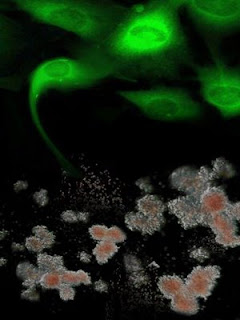

Genetically-corrected fibroblasts from Fanconi anaemia patients. Shown in green are genetically-corrected fibroblasts from Fanconi anaemia patients are reprogrammed to generate induced pluripotent stem cells, which, in turn, can be differentiated into disease-free hematopoietic progenitors, capable of producing blood cells in vitro. Credit: Courtesy of Dr. Juan-Carlos Belmonte, Salk Institute for Biological Studies.
After taking hair or skin cells from patients with Fanconi anaemia, the investigators corrected the defective gene in the patients' cells using gene therapy techniques pioneered in Verma's laboratory. They then successfully reprogrammed the repaired cells into induced pluripotent stem (iPS) cells using a combination of transcription factors, Oct4, Sox2, Klf4 and c-Myc. The resulting FA-iPS cells were indistinguishable from human embryonic stem cells and iPS cells generated from healthy donors. Since bone marrow failure as a result of the progressive decline in the numbers of functional hematopoietic stem cells is the most prominent feature of Fanconi anaemia, the researchers then tested whether patient-specific iPS cells could be used as a source for transplantable hematopoietic stem cells. They found that FA-iPS cells readily differentiated into hematopoietic progenitor cells primed to differentiate into healthy blood cells. "We haven't cured a human being, but we have cured a cell," Belmonte explains. "In theory we could transplant it into a human and cure the disease." Although hurdles still loom before that theory can become practice — in particular, preventing the reprogrammed cells from inducing tumours — in coming months Belmonte and Verma will be exploring ways to overcome that and other obstacles. In April 2009, they received a $6.6 million from the California Institute Regenerative Medicine (CIRM) to pursue research aimed at translating basic science into clinical cures. "If we can demonstrate that a combined iPS–gene therapy approach works in humans, then there is no limit to what we can do," says Verma. About the Salk Institute for Biological Studies: The Salk Institute for Biological Studies is one of the world's preeminent basic research institutions, where internationally renowned faculty probe fundamental life science questions in a unique, collaborative, and creative environment. Focused both on discovery and on mentoring future generations of researchers, Salk scientists make groundbreaking contributions to our understanding of cancer, aging, Alzheimer's, diabetes, and cardiovascular disorders by studying neuroscience, genetics, cell and plant biology, and related disciplines. Faculty achievements have been recognized with numerous honours, including Nobel Prizes and memberships in the National Academy of Sciences. Founded in 1960 by polio vaccine pioneer Jonas Salk, M.D., the Institute is an independent non-profit organization and architectural landmark. Reference: Disease-corrected haematopoietic progenitors from Fanconi anaemia induced pluripotent stem cells Ángel Raya, Ignasi Rodríguez-Pizà, Guillermo Guenechea, Rita Vassena, Susana Navarro, María José Barrero, Antonella Consiglio, Maria Castellà, Paula Río, Eduard Sleep, Federico González, Gustavo Tiscornia, Elena Garreta, Trond Aasen, Anna Veiga, Inder M. Verma, Jordi Surrallés, Juan Bueren & Juan Carlos Izpisúa Belmonte Nature advance online publication 31 May 2009, doi:10.1038/nature08129 ......... ZenMaster
For more on stem cells and cloning, go to CellNEWS at http://cellnews-blog.blogspot.com/ and http://www.geocities.com/giantfideli/index.html

Friday 29 May 2009

Adult Bone Marrow Stem Cells Injected Into Skeletal Muscle Can Repair Heart Tissue

Adult Bone Marrow Stem Cells Injected Into Skeletal Muscle Can Repair Heart Tissue Friday, 29 May 2009 University at Buffalo researchers have demonstrated for the first time that injecting adult bone marrow stem cells into skeletal muscle can repair cardiac tissue, reversing heart failure. Using an animal model, the researchers showed that this non-invasive procedure increased myocytes, or heart cells, by two-fold and reduced cardiac tissue injury by 60 percent. The therapy also improved function of the left ventricle, the primary pumping chamber of the heart, by 40 percent and reduced fibrosis, the hardening of the heart lining that impairs its ability to contract, by up to 50 percent. "This work demonstrates a novel non-invasive mesenchymal stem cell (MSC) therapeutic regimen for heart failure based on an intramuscular delivery route," said Techung Lee, Ph.D., UB associate professor of biochemistry and senior author on the paper. Mesenchymal stem cells are found in the bone marrow and can differentiate into a variety of cell types. "Injecting MSCs or factors released by MSCs improved ventricular function, promoted myocardial regeneration, lessened apoptosis (cell death) and fibrotic remodelling, recruited bone marrow progenitor cells and induced myocardial expression of multiple growth factor genes," Lee said. "These findings highlight the critical 'cross-talks' between the injected MSCs and host tissues, culminating in effective cardiac repair for the failing heart." The heart disease death rate has dropped significantly in the last three decades due to better treatments, resulting in large numbers of people living with heart failure. This advance has lead to another health hurdle: The only therapy available to reverse the decline in cardiac function is heart transplantation, and donor hearts are very scarce. Clinical trials of myocardial stem cell therapy traditionally have relied on surgery — infusing the stem cells directly into the heart or injecting them into the myocardium, the heart muscle — invasive methods that can result in harmful scar tissue, arrhythmia, calcification or small vessel blockages. "In our research with a swine model of heart failure," said Lee, "we've found that only 1-to-2 percent of MSCs infused into the myocardium grafted into the heart, and there was no evidence that they differentiated into heart muscle cells. In addition, diseased tissue is not a healthy environment for cell growth.” "For these reasons, and because patients with heart failure are not good surgical risks, it made sense to explore a non-invasive cell delivery approach," said Lee. An important feature of MSCs is their ability to produce a plethora of tissue healing effects, known as "trophic factors," which can be harnessed for stem cell therapy for heart failure. Lee noted that the multiple trophic factors produced by MSCs have been shown in the literature to be capable of reducing tissue injury, inhibiting fibrosis, promoting angiogenesis, stimulating recruitment and proliferation of tissue stem cells, and reducing inflammatory oxidative stress, a common cause of cardiovascular disease and heart failure. "Since skeletal muscle is the most abundant tissue in the body and can withstand repeated injection of large number of stem cells, we thought it would be a good method to deliver MSCs," Lee said. "We hypothesized that MSCs, via secretion of these functionally synergistic trophic factors, would be able to rescue the failing heart even when delivered away from the myocardium.” "This study proves our hypothesis," said Lee. "We've demonstrated that injecting MSCs, or trophic factors released by MSCs, into skeletal muscle improved ventricular function, promoted regeneration of heart tissue, decreased cell death and improved other factors that cause heart failure.” "This non-invasive stem cell administration regimen, if validated clinically, is expected to facilitate future stem cell therapy for heart failure." Lee said the next step is to use genetic and pharmacological engineering to make the stem cells more active, so good therapeutic effects can be achieved with fewer cells. "That is our goal. It would reduce the cost of stem cell therapy and make it more affordable for patients in the future." Reference: Heart Failure Therapy Mediated by the Trophic Activities of Bone Marrow Mesenchymal Stem Cells: A Non-invasive Therapeutic Regimen Arsalan Shabbir, David Zisa, Gen Suzuki, and Techung Lee Am J Physiol Heart Circ Physiol (April 24, 2009). doi:10.1152/ajpheart.00186.2009 ......... ZenMaster


For more on stem cells and cloning, go to CellNEWS at http://cellnews-blog.blogspot.com/ and http://www.geocities.com/giantfideli/index.html
Tuesday 26 May 2009

‘Floppy Baby’ Syndrome – Heart Protein Saves Muscle

New study shows heart muscle protein can replace its missing skeletal muscle counterpart to give mice with myopathy a long and active life Tuesday, 26 May 2009 In a world first, West Australian scientists have cured mice of a devastating muscle disease that causes a Floppy Baby Syndrome – a breakthrough that could ultimately help thousands of families across the globe. A heart muscle protein can replace its missing skeletal muscle counterpart to give mice with myopathy a long and active life. The research, published online today in the Journal of Cell Biology, reveals how a team at the Western Australian Institute for Medical Research (WAIMR) has restored muscle function in mice with one type of Floppy Baby Syndrome – a congenital myopathy disorder that causes babies to be born without the ability to properly use their muscles. The currently incurable genetic diseases render most of the affected children severely paralysed and take the lives of the majority of these children before the age of one. The contraction machinery protein, actin, exists in different forms in the adult heart and skeletal muscles. The heart form, ACTC, is also the dominant form in skeletal muscle of the foetus. However, during development, the skeletal form, ACTA1, increases in production and by birth has taken over. It is not clear why the switch occurs, or why it does not occur in the heart, but it happens in every higher vertebrate and, for that reason, has been considered vitally important. Mutations to the ACTA1 gene cause a rare but serious myopathy. Most patients die within the first year of life and some are born almost completely paralyzed. Mice lacking ACTA1 die nine days after birth. Dr Kristen Nowak, lead author on the publication, wondered if ACTC could compensate for a lack of ACTA1. The two proteins differ only slightly but, like the developmental switch in production, this difference is conserved across species. Many researchers therefore assumed such compensation would never work.


Muscle cell architecture looks normal in transgenic mice that lack ACTA1 but express human ACTC.Muscle cell architecture looks normal in transgenic mice that lack ACTA1 but express human ACTC. Credit: Nowak, K.J., et al. 2009. J. Cell Biol. doi:10.1083/jcb.200812132.


But it did. Nowak and colleagues crossed Acta1 mutant mice with transgenic mice that express human ACTC at high levels in skeletal muscle cells. The resulting mice did not die at nine days. In fact, almost all of them (93.5%) survived more than three months, and some more than two years. The mice's locomotor performance was comparable with wild type, as was their overall muscle strength (though individual muscle fibres were slightly weaker), and their endurance was actually higher — they ran faster and for longer. This begs the question, Why do we even have ACTA1? Besides pondering that, Nowak and colleagues are also working out how to boost endogenous ACTC as a possible therapy for ACTA1-lacking patients. Dr Kristen Nowak said the team was extremely encouraged that it had been able to cure a group of mice born with the condition. "The mice with Floppy Baby Syndrome were only expected to live for about nine days, but we managed to cure them so they were born with normal muscle function, allowing them to live naturally and very actively into old age," she said. "This is an important step towards one day hopefully being able to better the lives of human patients – mice who were cured of the disease lived more than two years, which is very old age for a mouse." Dr Nowak said the team was able to cure the mice with the recessive form of the genetic condition by replacing missing skeletal muscle actin – a protein integral in allowing muscles to contract – with similar actin found in the heart. "Earlier in our search to tackle these diseases, we discovered a number of children who, despite having no skeletal muscle actin in their skeletal muscle due to their genetic mutation, were not totally paralysed at birth," she said. "On closer inspection, we found it was because heart actin – another form of the protein – was abnormally "switched on" in their skeletal muscles.” "We had already begun investigating whether we could use heart actin to treat skeletal muscle actin disease, so that discovery spurred us on, and we've now proved it can be done – we can use heart actin to overcome the absence of skeletal muscle actin in mice." Heart actin is found in cardiac muscle and, during foetal development, it also works in skeletal muscles in the body, but by birth, heart actin has almost completely disappeared within skeletal muscle. Using genetic techniques, the WAIMR research team has reactivated the heart actin after birth in place of skeletal muscle actin, reversing the effects of the congenital myopathy. Head of the WAIMR research group Professor Nigel Laing said the team's next step was to apply their findings to human patients. "We are now screening more than a thousand already-approved medications looking for one that might increase heart actin in skeletal muscles, which could potentially offer a treatment for many patients," he said. "Current therapies only target the effects of these conditions, not the condition itself – we hope our approach could lead to a much greater improvement for a range of muscle diseases." This discovery is the latest for the team, which has been investigating debilitating muscle diseases for more than 20 years. The first major breakthrough for actin disease was in 1999, when the team identified that defects in the skeletal muscle actin gene, ACTA1 – responsible for producing skeletal muscle actin, cause multiple muscle diseases. Since then, the team has classified and named a new muscle disease 'Laing Myopathy' – named after Professor Nigel Laing – and helped implement worldwide screening for families at risk of genetic muscle disease. WAIMR Director Professor Peter Klinken said he was thrilled WAIMR was playing such an integral part in helping tackle devastating muscle diseases. "The persistence and determination shown by Professor Laing and his team over many, many years is nothing short of inspiring," he said. "They've asked some big questions in their quest to find a cure for this Floppy Baby Syndrome and have worked tirelessly to find the answers to those questions in the hope of helping families across the world.” "Research institutes like ours exist to help people live healthier lives and I am delighted at the important discoveries we are making in this field." About Floppy Baby Syndrome: The skeletal muscle actin mutations which cause congenital myopathies can be classified into five individual diseases which affect thousands of families worldwide. Children with recessive muscle actin diseases have no skeletal muscle actin because of mutations in the skeletal muscle actin gene which "knock out" the gene function. In Australia, dozens of families are affected by congenital myopathies, which bring high emotional costs and personal suffering, as well as financial and community burdens. This research was funded by the National Health and Medical Research Council, WAIMR and a number of patient support groups including the Association Française contre les Myopathies (French Muscular Dystrophy Association) and the US Muscular Dystrophy Association. The research project centred at the WAIMR laboratory was a collaborative effort with groups at the Medical Research Council and the University of Oxford in the United Kingdom, Cincinnati Children's Hospital Medical Center as well as the Centre for Microscopy, Characterisation and Analysis at the University of Western Australia and Perth-based Proteomics International, which have also assisted the team's work. Reference: Rescue of skeletal muscle alfa-actin–null mice by cardiac (fetal) alfa-actin Kristen J. Nowak, Gianina Ravenscroft, Connie Jackaman, Aleksandra Filipovska, Stefan M. Davies, Esther M. Lim, Sarah E. Squire, Allyson C. Potter, Elizabeth Baker, Sophie Clément, Caroline A. Sewry, Victoria Fabian, Kelly Crawford, James L. Lessard, Lisa M. Griffiths, John M. Papadimitriou, Yun Shen, Grant Morahan, Anthony J. Bakker, Kay E. Davies, and Nigel G. Laing J. Cell Biol., May 25, 2009: jcb.200812132v1 ......... ZenMaster


For more on stem cells and cloning, go to CellNEWS at http://cellnews-blog.blogspot.com/ and http://www.geocities.com/giantfideli/index.html
Friday 22 May 2009

Gene Therapy Could Expand Stem Cells' Promise

Researchers discuss combined approach to maximize benefits and minimize risks of stem cell therapy Friday, 22 May 2009 Once placed into a patient's body, stem cells intended to treat or cure a disease could end up wreaking havoc simply because they are no longer under the control of the clinician. But gene therapy has the potential to solve this problem, according to a perspective article from physician-scientists at NewYork-Presbyterian Hospital/Weill Cornell Medical Center published in a recent issue of the journal Cell Stem Cell. The paper details strategies for genetically modifying stem cells prior to transplantation in order to ensure their safety. "Stem cell therapy offers enormous potential to treat and even cure serious diseases. But wayward stem cells can turn into a runaway train without a conductor. This is an issue that can be dealt with and we have the technology to do that in the form of gene therapy," says senior author Dr. Ronald G. Crystal, chief of the Division of Pulmonary and Critical Care Medicine at NewYork-Presbyterian Hospital/Weill Cornell Medical Center, and the Bruce Webster Professor of Internal Medicine and Professor of Genetic Medicine at Weill Cornell Medical College. Stem cells have the capacity to differentiate into any of the different tissues making up the human body, thus holding the promise of treating or curing diseases such as multiple sclerosis or spinal-cord injury by replacing diseased cells with healthy cells. However, unlike other therapies such as chemotherapy, antibiotics or aspirin, stem cells have no expiration date, and that poses a real problem. "Almost all therapeutics we use have a half life. They only last a certain amount of time," Dr. Crystal says. "Stem cells are the opposite. Once the future stem cell therapist does the therapy, stem cells have the innate potential to produce more cells." The challenge takes on even more urgency with recent developments, including a federal administration now more open to exploring the potential of stem cells, the recent FDA approval of a human trial involving embryonic stem cells, as well as the reported case of a young boy who developed a brain tumour four years after receiving a stem-cell treatment for a rare genetic disorder. As evidenced by this boy's experience, one of the biggest potential problems with stem cell therapy is the development of tumours. But there are other problems as well. Stem cells directed to become beating heart cells might mistakenly end up in the brain. Or insulin-producing beta cells which can't stop means the body can no longer regulate insulin levels. "You've totally lost control," Dr. Crystal says. "What do you do?" The best chance of circumventing these issues is genetic modification of the stem cells prior to actually transplanting them, Dr. Crystal says. Theoretically, this is similar to how gene therapy is used to treat cancer, but with important improvements. "Instead of gene therapy being done in the patient, as is the case in cancer, it's being done in the cells in a laboratory before doctors use them for therapy so that they still have control of these cells," Dr. Crystal explains. Therapists would rig certain genes to respond to a "remote control" signal. For instance, giving a certain drug could prompt a "suicide" gene to kill a budding tumour. But gene therapy also needs to be carefully done and, ideally, two independent gene-manipulation systems would be used to ensure that stem cells remain firmly in control of clinicians. ......... ZenMaster For more on stem cells and cloning, go to CellNEWS at http://cellnews-blog.blogspot.com/ and http://www.geocities.com/giantfideli/index.html

Clinical Studies for Parkinson’s and ALS

New stem cell research unlocks unknown therapies Friday, 22 May 2009 New treatments for the devastating Parkinson’s disease and ALS are in clinical studies in Sweden, thanks to breaking new stem cell research. This news was presented by Dr. Jonas Frisén, Professor of stem cell research at Karolinska Institutet, at the world's largest biotech convention, BIO 2009 in Atlanta. Jonas Frisén."Stem cell research and regenerative medicine are in an extremely exciting phase right now. We are gaining knowledge very fast and many companies are being formed and are starting clinical trials in different areas," says Dr Jonas Frisén. As an example, a first-in-human study was just initiated for Parkinson's disease patients with the drug product, sNN0031, from the Swedish company NeuroNova. The drug, which is administered into the fluid-filled cavities of the brain, has shown long lasting recovery and formation of new cells in animal models of Parkinson's disease. Last year, a treatment for ALS entered the clinical trial phase. Disorders in the brain and nervous system result in more hospitalizations than any other disease group, and treatments entail large costs to society. The research field of neuroscience is one of Sweden's finest. This had resulted in achievements within numerous areas of basic science with considerable scope to direct clinical applications. These include research advances concerning the origin and repair of nerve cell damage following stroke and spinal cord injury, as well as research into major degenerative diseases such as Parkinson's and Alzheimer's. Dr Frisén is one of Sweden's leading stem cell researchers, since many years with a focus on nerve stem cells. Among his most recent publications is an article in Science, April 3rd, 2009 where evidence is shown for renewal of heart muscle cells in humans, a result that can be used to develop therapeutic strategies for cardiac pathologies. About NeuroNova: NeuroNova AB is a Swedish biopharmaceutical company working with neurogenesis and neuroprotection for treatment of several currently incurable neurodegenerative diseases. Dr Jonas Frisén is the scientific founder of NeuroNova. ......... ZenMaster


For more on stem cells and cloning, go to CellNEWS at http://cellnews-blog.blogspot.com/ and http://www.geocities.com/giantfideli/index.html

Wednesday 20 May 2009

Bone Marrow Therapy Helps Heart Patients

Bone marrow cell therapy may be beneficial for patients with ischemic heart disease Wednesday, 20 May 2009 The injection of bone marrow cells into the heart of patients with chronic myocardial ischemia (reduced blood flow to some areas of the heart) was associated with modest improvements in blood flow and function of the left ventricle, according to a study in the May 20 issue of JAMA. Bone marrow cell therapy is currently being investigated as a new therapeutic option for patients with ischemic heart disease. Two small-sized studies assessed the effect of this therapy in patients with chronic myocardial ischemia, but with varying results, according to background information in the article. Jan van Ramshorst, M.D., of Leiden University Medical Center, the Netherlands, and colleagues assessed the effect of intramyocardial (within the heart wall) bone marrow cell injection on myocardial perfusion (the flow of blood to the heart muscle) and left ventricular (LV) function in patients with chronic ischemia who were not eligible for conventional treatment. The trial included 50 patients (average age, 64 years; 43 men), who were randomized to receive about 8 injections of either bone marrow cells or placebo solution. At 3-month follow-up, when the two groups were compared, the improvement in summed stress score (a measure of myocardial perfusion) was significantly greater in the bone marrow-cell treated patients as compared with placebo-treated patients. Magnetic resonance imaging indicated that the absolute increase in left ventricular ejection fraction (LVEF; a measure of how well the left ventricle of the heart pumps with each contraction) was significantly larger in bone marrow cell-treated patients. A quality-of-life score increased at 3 and 6 months in bone marrow cell-treated patients, compared with a smaller increase in the placebo group. There was also greater improvement in exercise capacity in the bone marrow cell group. "In summary, the results of this randomized, double-blind, placebo-controlled trial demonstrate that intramyocardial bone marrow cell injection in patients with chronic ischemia is associated with significant improvements in anginal symptoms, myocardial perfusion, and LV function," the authors write. Reference: Intramyocardial Bone Marrow Cell Injection for Chronic Myocardial Ischemia Jan van Ramshorst, Jeroen J. Bax, Saskia L. M. A. Beeres, Petra Dibbets-Schneider, Stijntje D. Roes, Marcel P. M. Stokkel, Albert de Roos, Willem E. Fibbe, Jaap J. Zwaginga, Eric Boersma, Martin J. Schalij, Douwe E. Atsma JAMA. 2009;301[19]:1997-2004 ......... ZenMaster


For more on stem cells and cloning, go to CellNEWS at http://cellnews-blog.blogspot.com/ and http://www.geocities.com/giantfideli/index.html

Monday 18 May 2009

Epigenetic Inheritance

One hundred reasons to change the way we think about genetics Monday, 18 May 2009 For years, genes have been considered the one and only way biological traits could be passed down through generations of organisms. Not anymore. Increasingly, biologists are finding that non-genetic variation acquired during the life of an organism can sometimes be passed on to offspring — a phenomenon known as epigenetic inheritance. An article forthcoming in the July issue of The Quarterly Review of Biology lists over 100 well-documented cases of epigenetic inheritance between generations of organisms, and suggests that non-DNA inheritance happens much more often than scientists previously thought. Biologists have suspected for years that some kind of epigenetic inheritance occurs at the cellular level. The different kinds of cells in our bodies provide an example. Skin cells and brain cells have different forms and functions, despite having exactly the same DNA. There must be mechanisms — other than DNA — that make sure skin cells stay skin cells when they divide. Only recently, however, have researchers begun to find molecular evidence of non-DNA inheritance between organisms as well as between cells. The main question now is: How often does it happen? "The analysis of these data shows that epigenetic inheritance is ubiquitous …," write Eva Jablonka and Gal Raz, both of Tel-Aviv University in Israel. Their article outlines inherited epigenetic variation in bacteria, protists, fungi, plants, and animals. These findings "represent the tip of a very large iceberg," the authors say. For example, Jablonka and Raz cite a study finding that when fruit flies are exposed to certain chemicals, at least 13 generations of their descendants are born with bristly outgrowths on their eyes. Another study found that exposing a pregnant rat to a chemical that alters reproductive hormones leads to generations of sick offspring. Yet another study shows higher rates of heart disease and diabetes in the children and grandchildren of people who were malnourished in adolescence. In these cases, as well as the rest of the cases Jablonka and Raz cite, the source of the variation in subsequent generations was not DNA. Rather, the new traits were carried on through epigenetic means. There are four known mechanisms for epigenetic inheritance. According to Jablonka and Raz, the best understood of these is "DNA methylation." Methyl groups, small chemical groups within cells, latch on to certain areas along the DNA strand. The methyl groups serve as a kind of switch that renders genes active or inactive. By turning genes on and off, methyl groups can have a profound impact on the form and function of cells and organisms, without changing the underlying DNA. If the normal pattern of methyl groups is altered — by a chemical agent, for example — that new pattern can be passed to future generations. The result, as in the case of the pregnant rats, can be dramatic and stick around for generations, despite the fact that underlying DNA remains unchanged. LAMARCK REVISITED New evidence for epigenetic inheritance has profound implications for the study of evolution, Jablonka and Raz say. "Incorporating epigenetic inheritance into evolutionary theory extends the scope of evolutionary thinking and leads to notions of heredity and evolution that incorporate development," they write. This is a vindication of sorts for 18th century naturalist Jean Baptiste Lamarck. Lamarck, whose writings on evolution predated Charles Darwin's, believed that evolution was driven in part by the inheritance of acquired traits. His classic example was the giraffe. Giraffe ancestors, Lamarck surmised, reached with their necks to munch leaves high in trees. The reaching caused their necks to become slightly longer — a trait that was passed on to descendants. Generation after generation inherited slightly longer necks, and the result is what we see in giraffes today. With the advent of Mendelian genetics and the later discovery of DNA, Lamarck's ideas fell out of favour entirely. Research on epigenetics, while yet to uncover anything as dramatic as Lamarck's giraffes, does suggest that acquired traits can be heritable, and that Lamarck was not so wrong after all. Reference: Transgenerational Epigenetic Inheritance: Prevalence, Mechanisms, and Implications for the Study of Heredity and Evolution Eva Jablonka and Gal Raz The Quarterly Review of Biology, June 2009, vol. 84, no. 2, DOI: 10.1086/598822 ......... ZenMaster


For more on stem cells and cloning, go to CellNEWS at http://cellnews-blog.blogspot.com/ and http://www.geocities.com/giantfideli/index.html

Automated Tissue Engineering on Demand

Automated Tissue Engineering on Demand Monday, 18 May 2009 Skin from a factory – this has long been the dream of pharmacologists, chemists and doctors. Research has an urgent need for large quantities of 'skin models', which can be used to determine if products such as creams and soaps, cleaning agents, medicines and adhesive bandages are compatible with skin, or if they instead will lead to irritation or allergic reactions for the consumer. Such test results are seen as more meaningful than those from animal experiments are, and can even make such experiments largely superfluous. But artificial skin is rare. "The production is complex and involves a great deal of manual work. At this time, even the market's established international companies cannot produce more than 2,000 tiny skin pieces a month. With annual requirements of more than 6.5 million units in the EU area alone, however, the industrial demand far exceeds all currently available production capacities," reports Jörg Saxler. Together with Prof. Heike Mertsching, he is coordinating the "Automated Tissue Engineering on Demand" project within the Fraunhofer-Gesellschaft. Tissue engineering is still in its infancy. "Until now, the offer was limited predominantly to single-layer skin models that consist of a single cell type," explains Mertsching, who heads the Cell Systems Department at the Fraunhofer Institute for Interfacial Engineering and Biotechnology IGB. "Thanks to developments at our institute, the project team has access to a patent-protected skin model that consists of two layers with different cell types. This gives us an almost perfect copy of human skin, and one that provides more information than any system currently available on the market." An interdisciplinary team of Fraunhofer researchers is currently developing the first fully automatic production system for two-layer skin models. "Our engineers and biologists are the only ones who have succeeded in fully automating the entire process chain for manufacturing two-layer skin models," explains Saxler, who is from the Fraunhofer Institute for Production Technology IPT where he is responsible for technology management and heads the "Life Science Engineering" business unit. The system is intended for the fully automatic production of skin models ready for shipping. Credit: Fraunhofer.In a multi-stage process, first small pieces of skin are sterilized. Then they are cut into small pieces, modified with specific enzymes, and isolated into two cell fractions, which are then propagated separately on cell culture surfaces. The next step in the process combines the two cell types into a two-layer model, with collagen added to the cells that are to form the flexible lower layer, or dermis. This gives the tissue natural elasticity. In a humid incubator kept at body temperature, it takes the cell fractions less than three weeks to grow together and form a finished skin model with a diameter of roughly one centimetre. The technique has already proven its use in practice, but until now, it has been too expensive and complicated for mass production. Mertsching explains, "The production is associated with a great deal of manual work, and this reduces the method's efficiency." The project team, in which engineers, scientists and technicians from four Fraunhofer institutes are cooperating, is currently working at full speed to automate the work steps. The researchers at the IGB and the Fraunhofer Institute for Cell Therapy and Immunology IZI are handling the development of the biological fundamentals and validation of the machine and its sub-modules. Experts from the Fraunhofer Institute for Manufacturing and Automation IPA and the Fraunhofer Institute for Production Technology IPT are taking care of prototype development, automation and integration of the machine into a complete functional system. "At the beginning, our greatest challenge was to overcome existing barriers, because each discipline had its own very different approach," Saxler remembers. "Meanwhile, the four institutes are working together very smoothly – everyone knows that progress is impossible without input from the others." After working together for one year, the project team has already initiated eight patent procedures. At a collective Fraunhofer-Gesellschaft booth at the 2009 BIO in Atlanta, the researchers are presenting a computer model of the overall system, along with the three fundamental sub-modules. The first module prepares the tissue samples and isolates the two cell types; the second proliferates them. The finished skin models are built up and cultivated in the third, and then packed by a robot. The researchers still have a lot of meticulous work ahead before the machine will be finished. The difference between success and failure often depends on details, such as the quality of the skin pieces, processing times of enzymes, and liquid viscosities. Furthermore, the cell cultures must be monitored throughout the entire manufacturing process in order to provide optimal process control and to allow timely detection of any contamination with fungi or bacteria. The skin factory is expected to be finished in two years. "Our goal is a monthly production of 5,000 skin models with perfect quality, and a unit price under 34 euros. These are levels that are attractive for industry," Saxler continues. But chemical, cosmetic, pharmaceutical, and medical technology companies who have to test the reaction of skin to their products are not the only ones interested in Automated Tissue Engineering. In transplantation medicine, surgeons require healthy tissue in order to replace destroyed skin sections when burn injuries cover large portions of the body. The two-layer models that the new machine is intended to produce are not yet suitable for this purpose, however. "They don't have a blood supply, and are consequently rejected by the body after some time," Saxler explains. However, IGB researchers are already working on a full-skin model that will even include blood vessels. Once the research has been completed, fully automatic production of the transplants should also be possible. "We have designed the production system in such a way that it satisfies the high standards for good manufacturing practices (GMP) for the manufacture of products used in medicine," Mertsching explains. "And so they are also suitable for producing artificial skin for transplants." ......... ZenMaster


For more on stem cells and cloning, go to CellNEWS at http://cellnews-blog.blogspot.com/ and http://www.geocities.com/giantfideli/index.html

Friday 15 May 2009

ACLU Sues over Patents on Breast Cancer Genes

ACLU – Myriad Genetics lawsuit will become landmark case Friday, 15 May 2009 The American Civil Liberties Union action in filing a lawsuit yesterday against Myriad Genetics is going to lead to one of the most important legal battles in the history of biotechnology, asserts Genetic Engineering & Biotechnology News (GEN). The ACLU charged that the patenting of two human genes linked to breast and ovarian cancer will inhibit medical research. The organization also claims that the patents are invalid and unconstitutional. "This is going to turn into one of the watershed events in the evolution of the bioindustry," says John Sterling, Editor in Chief of GEN. "The pros and cons of patenting genes have been an ongoing, and often acrimonious series of debates, since the in re Chakrabarty decision in 1980. But this particular case seems to have taken on a life of its own with over fifteen plaintiffs. For while the lawsuit specifically centres on the patentability of two cancer-related genes, the ACLU says it plans to challenge the entire concept of patenting genes. What we have here is one group, the ACLU and its allies, contending that gene patents stifle life science research and potentially harm the health of thousands of patients. On the other side are biotech companies who maintain that without gene patents research incentives are seriously diminished and innovation is smothered." Kenneth I. Berns, M.D., Ph.D., Editor in Chief of the peer-reviewed journal, Genetic Testing and Molecular Biomarkers, which is the official journal of the Genetic Alliance, says the "patenting of human genes is a bad idea and that healthcare in the U.S. would be enhanced if the ACLU suit prevails." Dr. Berns is also Director of the University of Florida Genetics Institute in Gainesville. William Warren, partner at the Sutherland law firm, thinks the ACLU, in this case, is barking up the wrong tree. "The ACLU unexpectedly based its invalidity challenge on claims to un-patentable subject matter," he says. "The ACLU might have instead considered challenging the Myriad patents for obviousness." Warren and Sutherland colleague, Lei Fang, Ph.D., M.D., have authored a legal article, which will be published in the June 1 issue of GEN entitled "Patentability of Genetic Sequences Limited." For the specific details surrounding the lawsuit, please see the article on the GEN website entitled "Myriad Genetics Comes under Legal Fire for Gene Patents", which includes pertinent comments from both research and legal professionals. ......... ZenMaster


For more on stem cells and cloning, go to CellNEWS at http://cellnews-blog.blogspot.com/ and http://www.geocities.com/giantfideli/index.html

Thursday 14 May 2009

NIH New Stem Cell Guidelines

The end of the line for existing stem cell research? Thursday, 14 May 2009 Time is short for scientists to respond to the call for comments on the National Institutes of Health (NIH) proposed guidelines for the use of human embryonic stem (ES) cell lines and their eligibility for federal funds. On May 26, the window to provide feedback will close, and the drafted rules leave the possibility that funding for almost all existing cell lines will disappear. In a Forum article to be published online on May 14 by Cell Press in the journal Cell Stem Cell, Patrick Taylor, deputy general counsel at Children's Hospital Boston, explains some of the legal implications of the NIH's new funding rules, should they be adopted as written. Since the rules are retroactive, he explains, ongoing research is threatened. "Research with almost all existing cell lines will not be fundable, leaving almost no federal funds for research using cells created ethically since 2001. This will mean a loss of much of the research benefit of the last eight years, even though that research was independently reviewed and determined to be ethical under federal standards," says Taylor. "It is vitally important that scientists are aware of this problem and that the situation is resolved as quickly as possible." Ronald McKay, director of the NIH Stem Cell Unit, agrees and points out that, as proposed, the current draft guidelines may not even allow for continued research on the 21 ES cell lines approved by President Bush in 2001. "It is important to recognize that continued access to the ES cells themselves is important for medical research," says McKay. "It is common to use the economic metaphor of the 'gold standard' when discussing the value of human ES cells. But unlike gold, stem cells will not retain value if they are locked in a bank and we cannot analyze their secrets. Continued access to these cells will ensure no delay in understanding the links between human genetics and disease," he adds. The slow pace of commenting is symptomatic of a broader tendency within the scientific community. "Despite federal encouragement and the ease of posting a comment, scientists do not seem to be participating unless the proposals directly impact their research," outlines science writer Amy Maxman in an Analysis piece to be published by Cell Press in the journal Cell on the same day. The article explains how scientists can offer their views as part of the consultation on items listed at the Federal Register, such as by providing comments on the draft human ES cell guidelines currently under consideration, to ensure that federal agencies receive a balanced perspective of public opinion. Researchers from all scientific disciplines and interested members of the general public can comment on the proposed guidelines at http://nihoerextra.nih.gov/stem_cells/add.htm until 11 p.m. EST on May 26. ......... ZenMaster


For more on stem cells and cloning, go to CellNEWS at http://cellnews-blog.blogspot.com/ and http://www.geocities.com/giantfideli/index.html

How an Enzyme Tells Stem Cells Which Way to Divide

University of Oregon biochemists report a mechanism dictating cell division is not a long cascade of events Thursday, 14 May 2009 Driving Miranda, a protein in fruit flies crucial to switch a stem cell's fate, is not as complex as biologists thought, according to University of Oregon biochemists. They've found that one enzyme (aPKC or atypical protein kinase C) stands alone and acts as a traffic cop that directs which roads daughter cells will take. Kenneth Prehoda, shown here, and doctoral student Scott Atwood found that the way a stem cell is triggered to divide properly is not as complex as once thought. Credit: Photo by Jim Barlow."Wherever aPKC is at or on a cell's cortex or membrane, Miranda isn't," says Kenneth E. Prehoda, a professor in the chemistry department and member of the UO's Institute of Molecular Biology. When a stem cell duplicates into daughter cells, the side, or cortical domain, containing aPKC continues as a stem cell, while the other domain with Miranda becomes a differentiated cell such as a neuron that forms the central nervous system. Prehoda and co-author Scott X. Atwood, who studied in Prehoda's lab and recently earned his doctorate, describe how the mechanism works in the May 12 issue of the journal Current Biology. Instead of a complex cascade of protein deactivation steps that many biologists have theorized, Prehoda said, aPKC strips phosphate off an energy-transfer nucleotide known as ATP and then attaches it to Miranda. This process forces Miranda away from aPKC and helps determine the fates of subsequent daughter cells. "This process is pretty simple," he said, when viewed from a biochemical perspective. "What happens is that Miranda gets phosphorylated by aPKC, turning it into an inactivated substrate and pushing it into another location in the cell."


Dividing Drosophila neuroblast.Dividing Drosophila neuroblast. aPKC is shown in green at the top half of a fruit fly neuroblast. Miranda, in blue, has been driven away to the opposite side. Upon division, the top half will remain a stem cell, while the bottom will become a differentiated cell. Credit: Courtesy of Kenneth Prehoda.
Much of the paper in Current Biology is devoted to discuss why the more complex scenarios are not accurate. "There have been a lot of ideas on how this works, and most seemed to be really complicated and difficult to explain. We have found it's a much simpler mechanism," Prehoda said, adding that the mechanism likely is similar in many other types of cells, not just stem cells. "It's a basic-research question. How does this polarity occur? In order to develop stem cell-specific therapeutics based on a rational methodology you have to understand the mechanism," he said. If Miranda is improperly isolated into other regions by aPKC, the stem cell divides symmetrically, with both daughter cells adopting the same fate, In turn, Prehoda said, these cells can become tumours as they continue to rapidly divide without proper polarization. ......... ZenMaster
For more on stem cells and cloning, go to CellNEWS at http://cellnews-blog.blogspot.com/ and http://www.geocities.com/giantfideli/index.html

Wednesday 13 May 2009

Embryo's Heartbeat Drives Blood Stem Cell Formation

Clues about how blood forms could yield new strategies for treating blood diseases Wednesday, 13 May 2009 Biologists have long wondered why the embryonic heart begins beating so early, before the tissues actually need to be infused with blood. Two groups of researchers from Children's Hospital Boston, Brigham and Women's Hospital, and the Harvard Stem Cell Institute (HSCI) — presenting multiple lines of evidence from zebrafish, mice and mouse embryonic stem cells — provide an intriguing answer: A beating heart and blood flow are necessary for development of the blood system, which relies on mechanical stresses to cue its formation. Their studies, published online by the journals Cell and Nature, respectively, on May 13, together offer clues that may help in treating blood diseases such as leukaemia, immune deficiency and sickle cell anaemia, suggesting new ways scientists can make the types of blood cells a patient needs. This would help patients who require marrow or cord blood transplants, who do not have a perfect donor match. One team, led by Leonard Zon, MD, of the Division of Hematology/Oncology at Children's and Director of its Stem Cell research program, used zebrafish, whose transparent embryos allow direct observation of embryonic development. Publishing in Cell, Zon and colleagues discovered that compounds that modulate blood flow had a potent impact on the expression of a master regulator of blood formation, known as Runx1, which is also a recognized marker for the blood stem cells that give rise to all the cell types in the blood system. Confirming this observation, a strain of mutant embryos that lacked a heartbeat and blood circulation exhibited severely reduced numbers of blood stem cells. Further work showed that nitric oxide, whose production is increased in the presence of blood flow, is the key biochemical regulator: Increasing nitric oxide production restored blood stem cell production in the mutant fish embryos, while inhibiting nitric oxide production led to reduced stem cell number. Zon and colleagues went on to demonstrate that nitric oxide production was coupled to the initiation of blood stem cell formation across vertebrate species. Suppression of nitric oxide production in mice, by either genetic or chemical means, similarly reduced the number of functional Runx1-expressing blood stem cells. "Nitric oxide appears to be a critical signal to start the process of blood stem cell production," says Zon, who is also affiliated with the HSCI. "This finding connects the change in blood flow with the production of new blood cells." The second team, publishing in Nature, was led by George Q. Daley, MD, PhD, director of the Stem Cell Transplantation Program at Children's Hospital Boston, and Guillermo García-Cardeña, director of the Laboratory for Systems Biology of the Center for Excellence in Vascular Biology at Brigham and Women's Hospital, along with scientists from the Indiana University School of Medicine. Intrigued by the appearance of blood progenitors in the wall of the developing aorta soon after the heart starts beating, they investigated the effects of mechanical stimulation on blood formation in cultured mouse embryonic stem cells. They showed that shear stress — the frictional force of fluid flow on the surface of cells lining the embryonic aorta — increases the expression of master regulators of blood formation, including Runx1, and of genetic markers found in blood stem cells. Shear stress also increased formation of colonies of progenitor cells that give rise to specific lineages of blood cells (red cells, lymphocytes, etc.). These findings demonstrate that biomechanical forces promote blood formation. Daley, García-Cardeña and colleagues also studied mouse embryos with a mutation that prevented initiation of the heartbeat. These embryos had a sharp reduction in progenitor blood cell colonies, along with reduced expression of genetic markers of blood stem cells. When specific cells from the mutant embryos were exposed in vitro to shear stress, markers of blood stem cells and numbers of blood cell colonies were restored. Finally, the team showed that when nitric oxide production was inhibited, in both cell cultures and live mouse embryos, the effects of shear stress on blood progenitor colony formation were reduced. "In learning how the heartbeat stimulates blood formation in embryos, we've taken a leap forward in understanding how to direct blood formation from embryonic stem cells in the Petri dish," says Daley, who is also affiliated with the HSCI. "These observations reveal an unexpected role for biomechanical forces in embryonic development," adds García-Cardeña. "Our work highlights a critical link between the formation of the cardiovascular and hematopoietic systems." The authors of the two papers speculate that drugs that mimic the effects of embryonic blood flow on blood precursor cells, or molecules involved in nitric oxide signalling, might be therapeutically beneficial for patients with blood diseases. For example, nitric oxide could be used to grow and expand blood stem cells either in the culture dish or in patients after transplantation. References: Hematopoietic Stem Cell Development Is Dependent on Blood Flow Trista E. North, Wolfram Goessling, Marian Peeters, Pulin Li, Craig Ceol, Allegra M. Lord, Gerhard J. Weber, James Harris, Claire C. Cutting, Paul Huang, Elaine Dzierzak, and Leonard I. Zon Cell 137, 736–748, May 15, 2009 Biomechanical forces promote embryonic haematopoiesis Luigi Adamo, Olaia Naveiras Pamela L. Wenzel, Shannon McKinney-Freeman, Peter J. Mack, Jorge Gracia-Sancho, Astrid Suchy-Dicey, Momoko Yoshimoto, M. William Lensch, Mervin C. Yoder, Guillermo García-Cardeña & George Q. Daley Nature AOP 13 May 2009, doi:10.1038/nature08073 Embryo's heartbeat drives generation of new blood cells Howard Hughes Medical Institute, Wednesday, 13 May 2009 ......... ZenMaster


For more on stem cells and cloning, go to CellNEWS at http://cellnews-blog.blogspot.com/ and http://www.geocities.com/giantfideli/index.html

Monday 11 May 2009

Swine Flu II

Early findings about pandemic potential reported in new study Monday, 11 May 2009 Early findings about the emerging pandemic of a new strain of influenza A (H1N1) in Mexico are published today in Science. Researchers from the MRC Centre for Outbreak Analysis and Modelling at Imperial College London, working in collaboration with the World Health Organisation and public health agencies in Mexico, have assessed the epidemic using data to the end of April. Their key findings are as follows:

  • The data so far is very consistent with what researchers would expect to find in the early stages of a pandemic.
  • The researchers' best estimate is that in Mexico, influenza A (H1N1) is fatal in around 4 in 1,000 cases, which would make this strain of influenza as lethal as the one found in the 1957 pandemic. The researchers stress that healthcare has greatly improved in most countries since 1957 and the world is now better prepared.
  • The epidemic of influenza A (H1N1) is thought to have started in Mexico on 15 February 2009. The data suggests that by the end of April, around 23,000 people were infected with the virus in Mexico and 91 of these died as a result of infection. However, the figures are uncertain – for example, some mild cases may have gone unreported. The numbers infected could be as low as 6,000 people or as high as 32,000 people.
  • The uncertainty around the numbers of people who have been infected with influenza A (H1N1) in Mexico means that the case fatality ratio (CFR) of 0.4% (4 deaths per 1000) cannot be definitely established. The CFR is in the range of 0.3% to 1.5%, but at this stage the researchers believe that 0.4% is the most likely.
  • For every person infected, it is likely that there will be between 1.2 and 1.6 secondary cases. This is high compared to normal seasonal influenza, where around 10-15 percent of the population are likely to become infected. However, it is lower than would be expected for pandemic influenza, where 20-30 percent of the population are likely to become infected.
  • In an outbreak in an isolated village called La Gloria, Mexico, children were twice as likely to become infected as adults, with 61% of those aged under 15 becoming infected, compared with 29% of those over 15. This may suggest that adults have some degree of immunity against infection, because of having been previously infected with a related strain of influenza, or it may mean that children are more susceptible to infection because they interact much more closely together, for example in school, than adults.

Professor Neil Ferguson, the corresponding author of today's research from the MRC Centre for Outbreak Analysis and Modelling at Imperial College London, said: "Our study shows that this virus is spreading just as we would expect for the early stages of a flu pandemic. So far, it has been following a very similar pattern to the flu pandemic in 1957, in terms of the proportion of people who are becoming infected and the percentage of potentially fatal cases that we are seeing.” "What we're seeing is not the same as seasonal flu and there is still cause for concern – we would expect this pandemic to at least double the burden on our healthcare systems. However, this initial modelling suggests that the H1N1 virus is not as easily transmitted or as lethal as that found in the flu pandemic in 1918," added Professor Ferguson. Reference: Pandemic Potential of a Strain of Influenza A (H1N1): Early Findings The WHO Rapid Pandemic Assessment Collaboration Science Published Online May 11, 2009, DOI: 10.1126/science.1176062 ......... ZenMaster


For more on stem cells and cloning, go to CellNEWS at http://cellnews-blog.blogspot.com/ and http://www.geocities.com/giantfideli/index.html

New Tissue Scaffold Regrow Cartilage and Bone

Work could help heal sports injuries, arthritis Monday, 11 May 2009 MIT engineers and colleagues have built a new tissue scaffold that can stimulate bone and cartilage growth when transplanted into the knees and other joints. The scaffold could offer a potential new treatment for sports injuries and other cartilage damage, such as arthritis, says Lorna Gibson, the Matoula S. Salapatas Professor of Materials Science and Engineering and co-leader of the research team with Professor William Bonfield of Cambridge University. "If someone had a damaged region in the cartilage, you could remove the cartilage and the bone below it and put our scaffold in the hole," said Gibson. The researchers describe their scaffold in a recent series of articles in the Journal of Biomedical Materials Research. The technology has been licensed to Orthomimetics, a British company launched by one of Gibson's collaborators, Andrew Lynn of Cambridge University. The company recently started clinical trials in Europe. The scaffold has two layers, one that mimics bone and one that mimics cartilage. When implanted into a joint, the scaffold can stimulate mesenchymal stem cells in the bone marrow to produce new bone and cartilage. The technology is currently limited to small defects, using scaffolds roughly 8 mm in diameter. The researchers demonstrated the scaffold's effectiveness in a 16-week study involving goats. In that study, the scaffold successfully stimulated bone and cartilage growth after being implanted in the goats' knees. The project, a collaboration enabled by the Cambridge-MIT Institute, began when the team decided to build a scaffold for bone growth. They started with an existing method to produce a skin scaffold, made of collagen (from bovine tendon) and glycosaminoglycan, a long polysaccharide chain. To mimic the structure of bone, they developed a technique to mineralize the collagen scaffold by adding sources of calcium and phosphate. Once that was done, the team decided to try to create a two-layer scaffold to regenerate both bone and cartilage (known as an osteochondral scaffold). Their method produces two layers with a gradual transition between the bone and cartilage layers. "We tried to design it so it's similar to the transition in the body. That's one of the unique things about it," said Gibson. There are currently a few different ways to treat cartilage injuries, including stimulating the bone marrow to release stem cells by drilling a hole through the cartilage into the bone; transplanting cartilage and the underlying bone from another, less highly loaded part of the joint; or removing cartilage cells from the body, stimulating them to grow in the lab and re-implanting them. The new scaffold could offer a more effective, less expensive, easier and less painful substitute for those therapies, said Gibson. ......... ZenMaster


For more on stem cells and cloning, go to CellNEWS at http://cellnews-blog.blogspot.com/ and http://www.geocities.com/giantfideli/index.html

Swine Flu I

What does it do to pigs? Monday, 11 May 2009 The effects of H1N1 swine flu have been investigated in a group of piglets. Scientists writing in BioMed Central's open access Virology Journal studied the pathology of the virus, finding that all infected animals showed flu-like symptoms between one and four days after infection and were shedding virus two days after infection. Roongroje Thanawongnuwech led a team of researchers from Chulalongkorn University, Bangkok, who infected 22-day old pigs with both the H1N1 strain of swine flu and the less dangerous H3N2 subtype. "The results demonstrated that both swine flu subtypes were able to induce flu-like symptoms and lung lesions in weanling pigs. However the severity of the disease with regard to both gross and microscopic lung lesions was greater in the H1N1-infected pigs", he said. All infected pigs developed respiratory symptoms such as nasal discharge, coughing, sneezing and conjunctivitis. Upon pathological examination, lung lesions large enough to be seen by the naked eye were observed. "These lesions were characterized by dark plum-coloured, consolidated areas on lung lobes and were most severe two days after infection, especially in the H1N1-infected pigs, where approximately a third of the lung was covered", according to Thanawongnuwech. The course of infection was limited to less than a week and none of the animals died. Reference: Pathogenesis of swine influenza virus (Thai isolates) in weanling pigs: an experimental trial Donruethai Sreta, Roongtham Kedkovid, Sophon Tuamsang, Pravina Kitikoon and Roongroje Thanawongnuwech Virology Journal 2009, 6:34 doi:10.1186/1743-422X-6-34 ......... ZenMaster


For more on stem cells and cloning, go to CellNEWS at http://cellnews-blog.blogspot.com/ and http://www.geocities.com/giantfideli/index.html

New Genes Affecting Blood-pressure Identified

Comprehensive genetic study paves way for new blood-pressure medicines Monday, 11 May 2009 Eight previously unknown genes that affect blood pressure were recently identified in a comprehensive international study comprising 34,433 Europeans. The findings of the study, in which Uppsala University researchers participated, are being presented today in the Web edition of Nature Genetics. High blood pressure is a major contributing cause of cardiovascular disorders. Previous studies have shown that it is extremely difficult to identify genes that affect blood pressure, partly because blood pressure is impacted by non-genetic factors, such as exercise and intake of salt in food, and partly because there are many genes that work together to affect blood pressure. In the present study, a concerted effort was made by 93 research teams in 14 countries to find genes that affect blood-pressure levels in healthy individuals. To identify these genes, blood-pressure levels were monitored in those participating in the study, and DNA samples were gathered. The entire human genome was mapped systematically by analyzing the DNA samples with the help of hundreds of thousands of genetic markers, so-called SNP markers. Sweden was represented in the study by three research teams: Ann-Christine Syvänen's research group and the SNP technology platform at Uppsala University, a group from the Karolinska Institute, and a group from Lund University. The study included DNA samples from 2,000 Swedes. The areas of the genome that were identified in the study contain several genes that probably play a role in regulating blood pressure. The CYP17A1 gene on chromosome 10q24, for instance, plays a key role in the biosynthesis of glucorticoid hormones, which influence the metabolism of salt. In another area on chromosome 1p36 there are genes for two peptides with sodium-expelling effects. A lesser-known gene in this area, CLCN6, codes for a chloride canal in the neurons, which was not previously associated with blood-pressure regulation, and in an area on chromosome 17q21 there is the phopholipase gene, PLCD3, which is important for signalling in smooth muscles. References: Genome-wide association study identifies eight loci associated with blood pressure Nature Genetics Published online: 10 May 2009, doi:10.1038/ng.361 Genome-wide association study of blood pressure and hypertension Nature Genetics Published online: 10 May 2009, doi:10.1038/ng.384 ......... ZenMaster


For more on stem cells and cloning, go to CellNEWS at http://cellnews-blog.blogspot.com/ and http://www.geocities.com/giantfideli/index.html

Co-transplants of BM and ESCs Prevent Tumours

Bone marrow stem cell co-transplantation prevents embryonic stem cell transplant-associated tumours Monday, 11 May 2009 Transplanted embryonic stem cells are recognized as a potential treatment for patients suffering from the effects of spinal cord injury (SCI). However, in studies using embryonic stem cells transplanted into SCI laboratory animals, a serious drawback has been the development of tumours following transplantation. Publishing in the current issue of Cell Transplantation (Vol. 18 No.1), a team of Japanese researchers describe their study that demonstrates a way to eliminate the problem of tumour growth by co-transplanting bone marrow stem cells (BMSCs) along with embryonic stem cells. "Our study results suggest that co-transplanting BMSCs induce undifferentiated embryonic stem cells to differentiate into a neuronal lineage by neurotrophic factor production, resulting in suppression of tumour formation in SCI model mice," said corresponding author Dr. Masahide Yoshikawa of the Nara Medical University. "The known multipotency of BMSCs during differentiation and their known ability to produce neurotrophic factors, such as nerve growth factor, led us to speculate that co-transplantation of ES cells and BMSCs would provide an advantage over transplantation of ES cells alone." A control group of mice that only received ES cells developed tumours at the grafted site and their behavioural improvement ceased after three weeks. No tumours developed in the co-transplantation group and behavioural improvement continued over the five-week study. To date, no effective medical therapy has been available for SCI patients. While ES cells have been thought to represent a potential resource for therapy, the hurdle of tumour formation has impeded efforts. Co-transplantation of BMSCs appears to overcome the tumour hurdle, suggesting to the researchers that their success can provide a path toward human trials. "The entire mechanism of suppressed tumour development following co-transplantation remains to be elucidated," says Dr. Yoshikawa. "We considered that the BMSCs played an important role in preventing tumours and speculate that one of the mechanisms by which BMSCs promote the differentiation of ES cells is related to secreted soluble factors, including neurotrophic factors." According to Dr. Yoshikawa, the transplanted BMSCs survived in the grafted site for at least five weeks after transplantation and maintained their ability to produce NGF. "These findings are extremely important and emphasize the need for additional study on how embryonic stem cells may be used to treat human neurological problems in the not too distant future," commented Section Editor Dr. John Sladek, professor of paediatrics and neuroscience at the University of Colorado School of Medicine. ......... ZenMaster


For more on stem cells and cloning, go to CellNEWS at http://cellnews-blog.blogspot.com/ and http://www.geocities.com/giantfideli/index.html

Monday 4 May 2009

Moving Gene Therapy Forward

Mobile DNA elements that can integrate into 'foreign' DNA Monday, 04 May 2009 Gene therapy Gene therapy is the introduction of genetic material into a patient's cells resulting in a cure or a therapeutic effect. In recent years, it has been shown that gene therapy is a promising technology to treat or even cure several fatal diseases for which there is no attractive alternative therapy. Gene therapy can be used for hereditary diseases, but also for other diseases that affect heart, brain and even for cancer. Indeed, recent results suggest that gene therapy can be beneficial for patients suffering from aggressive brain cancer that would otherwise be lethal. A safe delivery of the genes? Despite the overall progress, there is still a need to develop improved and safer approaches to deliver genes into cells. The success of gene therapy ultimately depends on these gene delivery vehicles or vectors. Most vectors have been derived from viruses that can be tailor-made to deliver therapeutic genes into the patients' cells. However, some of these viral vectors can induce side-effects, including cancer and inflammation. Marinee Chuah, Thierry VandenDriessche, Eyayu Belay and their fellow VIB researchers at K.U. Leuven in collaboration with Zsuzsanna Iszvak and Zoltan Ivics and colleagues at the Max Delbrück Center in Berlin (Germany) have now developed a new non-viral approach that overcomes some of the limitations associated with viral vectors. Lessons from evolution Using the principles of evolution and natural selection, that were initially conceived by Charles Darwin, they have now developed an efficient and safe gene delivery approach based on non-viral genetic elements, called transposons. Transposons are mobile DNA elements that can integrate into 'foreign' DNA via a 'cut-and-paste' mechanism. In a way they are natural gene delivery vehicles. The researchers constructed the transposons in such a way that they can carry the therapeutic gene into the target cell DNA. Doing so, they obviate the need to rely on viral vectors. “We show for the first time that it is now possible to efficiently deliver genes into stem cells, particularly those of the immune system, using non-viral gene delivery,” says Marinee Chuah. “Many groups have tried this for many years but without success. We are glad that we could now overcome this hurdle,” claims Thierry VandenDriessche. Zsuzsanna Izsvak and Zoltan Ivics concur: “This transposon technology may greatly simplify the way gene therapy is conducted, improve its overall safety and reduce the costs.” The VIB researchers are further testing this technology to treat specific diseases including cancer and genetic disorders, in anticipation of moving forward and treat patients suffering from these diseases. ......... ZenMaster


For more on stem cells and cloning, go to CellNEWS at http://cellnews-blog.blogspot.com/ and http://www.geocities.com/giantfideli/index.html