Thursday 30 April 2009

microRNA Regulates Pluripotency in Human Embryonic Stem Cells

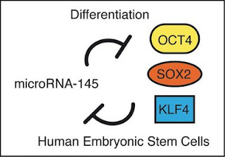
Scientists shed light on inner workings of human embryonic stem cells Thursday, 30 April 2009 Scientists at UC Santa Barbara have made a significant discovery in understanding the way human embryonic stem cells function. They explain nature's way of controlling whether these cells will renew, or will transform to become part of an ear, a liver, or any other part of the human body. The study is reported in the May 1 issue of the journal Cell. The scientists say the finding bodes well for cancer research, since tumour stem cells are the engines responsible for the growth of tumours. The discovery is also expected to help with other diseases and injuries. The study describes nature's negative feedback loop in cell biology. Professor Kenneth S. Kosik. Credit: George Foulsham/ Department of Public Affairs, UCSB."We have found an element in the cell that controls 'pluripotency,' that is the ability of the human embryonic stem cell to differentiate or become almost any cell in the body," said senior author Kenneth S. Kosik, professor in the Department of Molecular, Cellular & Developmental Biology. Kosik is also co-director and Harriman Chair in Neuroscience Research of UCSB's Neuroscience Research Institute. "The beauty and elegance of stem cells is that they have these dual properties," said Kosik. "On the one hand, they can proliferate –– they can divide and renew. On the other hand, they can also transform themselves into any tissue in the body, any type of cell in the body." The research team includes James Thomson, who provided an important proof to the research effort. Thomson, an adjunct professor at UCSB, is considered the "father of human embryonic stem cell biology." Thomson pioneered work in the isolation and culture of non-human primate and human embryonic stem cells. These cells provide researchers with unprecedented access to the cellular components of the human body, with applications in basic research, drug discovery, and transplantation medicine. With regard to human embryonic stem cells, Kosik explained that for some time he and his team have been studying a set of control genes called microRNAs. "To really understand microRNAs, the first step is to remember the central dogma of biology –– DNA is the template for RNA and RNA is translated to protein. But microRNAs stop at the RNA step and never go on to make a protein.” "The heart of the matter is that before this paper, we knew if you want to maintain a pluripotent state and allow self-renewal of embryonic stem cells, you have to sustain levels of transcription factors, including Oct4, Sox2 and Klf4," said Kosik. "We also knew that stem cells transition to a differentiated state when you decrease those factors. Now we know how that happens a little better." Transcription factors are genes that control other genes. On the other hand, microRNAs are single-stranded RNA molecules that control the activity of other genes. When microRNAs in the genome are transcribed from DNA, they target complementary messenger RNAs (mRNAs), which serve as the templates for proteins, to either encourage their degradation or prevent their translation into functional proteins. In general, one gene can be repressed by multiple microRNAs and one microRNA can repress multiple genes, the researchers explained. In a wide variety of developmental processes, microRNAs fine tune or restrict cellular identities by targeting important transcription factors or key pathways. Kosik's team found that levels of miR-145 change dramatically when human embryonic stem cells differentiate into other cell types. miR-145 was of particular interest because it had been predicted to target Oct4, Klf4 and Sox2. The new research shows that a microRNA –– a single-stranded RNA whose function is to decrease gene expression –– lowers the activity of three key ingredients, Oct4, Sox2 and Klf4, in the recipe for embryonic stem cells. The discovery may have implications for improving the efficiency of methods designed to reprogram differentiated cells into embryonic stem cell-like cells. As few as three or four genes can make cells pluripotent. Those three factors are perhaps best known as three of four ingredients originally shown to transform adult human skin cells into "induced pluripotent stem cells" (iPS cells), which behave in nearly every respect like true embryonic stem cells. That four-ingredient recipe has since been pared down to one, Oct4, in the case of neural stem cells. "We know what these genes are," Kosik said. That information was used recently for one of the most astounding breakthroughs of biology of the last couple of years –– the discovery of induced pluripotent skin cells. "You can take a cell, a skin cell, or possibly any cell of the body, and revert it back to a stem cell," Kosik said. "The way it's done, is that you take the transcription factors that are required for the pluripotent state, and you get them to express themselves in the skin cells; that's how you can restore the embryonic stem cell state. You clone a gene, you put it into what's called a vector, which means you put it into a little bit of housing that allows those genes to get into a cell, then you shoot them into a stem cell. Next, when those genes –– those very critical pluripotent cell genes –– get turned on, the skin cell starts to change; it goes back to the embryonic pluripotent stem cell state." The researchers explained that a rise in miR-145 prevents human embryonic stem cells' self-renewal and lowers the activity of genes that lend stem cells the capacity to produce other cell types. It also sends the cells on a path toward differentiation. In contrast, when miR-145 is lost, the embryonic stem cells are prevented from differentiating as the concentrations of transcription factors rise. They also show that the control between miR-145 and the "reprogramming factors" goes both ways. The promoter for miR-145 is bound and repressed by a transcription factor known as OCT4, they found. "It's a beautiful double negative feedback loop," Kosik said. "They control each other. That is the essence of this work."


Control of pluripotency of human embryonic stem cells.Human embryonic stem cells are poised between a proliferative state with the potential to become any cell in the body and a differentiated state with a more limited ability to proliferate. To maintain this delicate balance embryonic stem cells express a set of factors, including OCT4, SOX2, and KLF4, to control multiple genes that sustain the proliferative pluripotent state. A tiny RNA called miR-145 can repress these genes, and in turn, one of the transcription factors, OCT4, can repress miR-145. Thus, a double negative feedback loop sets the delicate balance.


Because there is typically less "wiggle room" in the levels of microRNA compared to mRNA, further studies are needed to quantify more precisely the copy numbers of miR-145 and its targets, to figure out exactly how this layer of control really works, Kosik said. The findings in embryonic stem cells might also have importance for cancer. "There are sets of microRNA that are widely up- or down regulated in cancers," he said, noting that several studies have specifically linked low miR-145 levels to various forms of cancer. "Tumour stem cells are the engines of tumours. If miR-145 is sustaining or maintaining a differentiated state, loss of that may have something to do with malignant transformation." Na Xu (left) and Thales Papagiannakopoulos. Credit: George Foulsham/ Department of Public Affairs, UCSB.Kosik credits the lion's share of this discovery to first author Na Xu, a postdoctoral fellow who is also supported by the California Institute for Regenerative Medicine (CIRM). "Na Xu deserves enormous credit for this work," said Kosik. "She performed nearly every experiment in the paper and was the major contributor to the ideas in the paper." Meanwhile, Thales Papagiannakopoulos, a graduate student working in the Kosik lab, was very generous in helping Na Xu with one of the experiments. He helped with one of several proofs that showed that the targets of miR-145 are the three transcription factors that are being reported, explained Kosik. Thomson provided one of several proofs for the control point of miR-145 expression, said Kosik. Reference: MicroRNA-145 Regulates OCT4, SOX2, and KLF4 and Represses Pluripotency in Human Embryonic Stem Cells Na Xu, Thales Papagiannakopoulos, Guangjin Pan, James A. Thomson and Kenneth S. Kosik Cell, 30 April 2009, doi:10.1016/j.cell.2009.02.038 ......... ZenMaster


For more on stem cells and cloning, go to CellNEWS at http://cellnews-blog.blogspot.com/ and http://www.geocities.com/giantfideli/index.html

Tuesday 28 April 2009

Making Heart Cells from Stem Cells

Gladstone scientists identify key sequence of transcription factors driving heart cell creation Tuesday, 28 April 2009 Scientists at the Gladstone Institutes of Cardiovascular Disease have identified for the first time key genetic factors that drive the process of generating new heart cells. The discovery, reported in the current issue of the journal Nature, provides important new directions on how stem cells may be used to repair damaged hearts. For decades, scientists were unable to identify a single factor that could turn non-muscle cells into beating heart cells. Using a clever approach, the research team led by Benoit Bruneau, Ph.D., found that a combination of three genes could do the trick. This is the first time any combination of factors has been found to activate cardiac differentiation in mammalian cells or tissues. "The heart has very little regenerative capacity after it has been damaged," said Dr. Bruneau. "With heart disease the leading cause of death in the Western world, this is a significant first step in understanding how we might create new cells to repair a damaged heart." Two of the three genes encode proteins called transcription factors, which are master regulators that bind to DNA and determine which genes get activated or shut off. The two transcription factors, GATA4 and TBX5, cause human heart disease when mutated and cooperate with each other to control other genes. When Dr. Bruneau and postdoctoral fellow Jun K. Takeuchi added different combinations of transcription factors to mouse cells, these two seemed important for pushing cells into heart cells — but they were not enough. "When we finally identified the key factor that could work with GATA4 and TBX5 to turn cells into beating heart cells, it was somewhat of a surprise to us," said Dr. Bruneau.


Cardiogenic factors that turn on heart genes.This represents how the cardiogenic factors turn on heart genes. The transcription factors, Tbx5 and Gata4, cannot access the DNA unless Baf60c is present. When all three are introduced, Baf60c helps open up the closed chromatin, and lets Tbx5 and Gata4 work together to turn on the heart genes. Credit: Benoit Bruneau, The Gladstone Institute of Cardiovascular Disease.
The surprising factor was a cardiac-specific protein called BAF60c, which helps determine whether transcription factors like GATA4 and TBX5 can even gain access to the DNA regions they were supposed to turn on or off. "Our previous studies had shown that chromatin remodelling complexes were important," said Dr. Bruneau. "Mice with lower levels of these complexes have severe heart defects and defective cardiac differentiation. These observations prompted us to look at Baf60c in heart differentiation." The effect was dramatic. Addition of the three factors directed differentiation of mouse mesoderm, which normally has the potential to make bone, blood, muscle, heart, and other tissues, specifically into cardiac muscle cells (cardiomyocytes) that beat rhythmically, just like normal heart cells. In fact, even cells that normally contribute to the placenta could be induced to transform into beating cardiomyocytes. "Together, these factors give us a potent mechanism to control cellular differentiation," said Dr. Bruneau. "This knowledge may help us to understand how to reprogram new cardiomyocytes for therapeutic purposes." About the Gladstone Institutes: The J. David Gladstone Institutes, an independent, non-profit biomedical research organization, affiliated with the University of California, San Francisco, is dedicated to the health and welfare of humankind through research into the causes and prevention of some of the world's most devastating diseases. Gladstone is comprised of the Gladstone Institute of Cardiovascular Disease, the Gladstone Institute of Virology and Immunology and the Gladstone Institute of Neurological Disease. Reference: Directed transdifferentiation of mouse mesoderm to heart tissue by defined factors Jun K. Takeuchi & Benoit G. Bruneau Nature advance online publication 26 April 2009, doi:10.1038/nature08039 ......... ZenMaster
For more on stem cells and cloning, go to CellNEWS at http://cellnews-blog.blogspot.com/ and http://www.geocities.com/giantfideli/index.html

Friday 24 April 2009

What makes a cow a cow?

Genome sequence sheds light on ruminant evolution Friday, 24 April 2009 Researchers report today in the journal Science that they have sequenced the bovine genome, for the first time revealing the genetic features that distinguish cattle from humans and other mammals. The six-year effort involved an international consortium of researchers and is the first full genome sequence of any ruminant species. Ruminants are distinctive in that they have a four-chambered stomach that – with the aid of a multitude of resident microbes – allows them to digest low quality forage such as grass. The bovine genome consists of at least 22,000 protein-coding genes and is more similar to that of humans than to the genomes of mice or rats, the researchers report. However, the cattle genome appears to have been significantly reorganized since its lineage diverged from those of other mammals, said University of Illinois animal sciences Professor Harris Lewin, whose lab created the high-resolution physical map of the bovine chromosomes that was used to align the sequence. Lewin, who directs the Institute for Genomic Biology, also led two teams of researchers on the sequencing project and is the author of a Perspective article in Science on the bovine genome sequence and an accompanying study by the Bovine Genome and Analysis Consortium. "Among the mammals, cattle have one of the more highly rearranged genomes," Lewin said. "They seem to have more translocations and inversions (of chromosome fragments) than other mammals, such as cats and even pigs, which are closely related to cattle.” "The human is actually a very conserved genome as compared to the ancestral genome of all placental mammals, when you look at its overall organization." The sequence of the cow's 29 pairs of chromosomes and its X chromosome (the Y chromosome was not studied) also provides new insights into bovine evolution and the unique traits that make cattle useful to humans, Lewin said.


The Hereford cow named L1 Dominette.The first cow genome to be sequenced was that of a Hereford cow named L1 Dominette, shown here with her calf. Credit: Photo courtesy of USDA Agricultural Research Service Research Geneticist Michael D. MacNeil.

For example, Illinois animal sciences research professor Denis Larkin conducted an analysis of the chromosome regions that are prone to breakage when a cell replicates its genome in preparation for the creation of sperm and egg cells. He showed that in the cattle genome these breakpoint regions are rich in repetitive sequences and segmental duplications and include species-specific variations in genes associated with lactation and immune response. A previous study from Lewin's lab published this month in Genome Research showed that the breakpoint regions of many species' chromosomes are rich in duplicated genes and that the functions of genes found in these regions differ significantly from those occurring elsewhere in the chromosomes. These repeats and segmental duplications occur by means of many different mechanisms, one of which involves sporadic and repeated insertions of short bits of genetic material, called retroposons, into the genome. "The cow genome has many types of repeats that accumulate over time," Lewin said. "And one of the things that we found is that the new ones are blasting into where the old ones are in the breakpoint regions and breaking them apart. That's the first time that that's been seen." "The repeats do a lot of things," he said. "They can change the regulation of the genes. They can make the chromosomes unstable and make them more likely to recombine with other pieces of chromosomes inappropriately." Lewin calls the breakpoint regions "hotspots of evolution in the genome." Another analysis led by Lewin, a study of metabolic genes performed by Seongwon Seo, a postdoctoral fellow in Lewin's lab and now a professor at Chungnam National University in South Korea, found that five of the 1,032 genes devoted to metabolic functions in humans are missing from the cattle genome or have radically diverged. This suggests that cattle have some unique metabolic pathways, Lewin said. These differences in metabolism, along with changes in genes devoted to reproduction, lactation and immunity are a big part of "what makes a cow a cow," Lewin said. For example, one of the changed genes, histatherin, produces a protein in cow's milk that has anti-microbial properties. The researchers also found multiple copies of a gene for an important milk protein, casein, in a breakpoint region of one of the chromosomes. “Having the genome sequence is now the window to understanding how these changes occurred, how ruminants ended up with a four-chambered stomach instead of one, how the cow’s immune system operates and how it is able to secrete large amounts of protein in its milk,” Lewin said. References: The Genome Sequence of Taurine Cattle: A Window to Ruminant Biology and Evolution The Bovine Genome Sequencing and Analysis Consortium, Christine G. Elsik, Ross L. Tellam, Kim C. Worley Science 24 April 2009, Vol. 324. no. 5926, pp. 522 – 528, DOI: 10.1126/science.1169588 Genome-Wide Survey of SNP Variation Uncovers the Genetic Structure of Cattle Breeds The Bovine HapMap Consortium Science 24 April 2009, Vol. 324. no. 5926, pp. 528 – 532, DOI: 10.1126/science.1167936 Along with these Science papers, researchers published 20 companion reports describing more detailed analyses of the domestic cattle genome sequence in journals from the open access publisher BioMed Central. All of the articles can be freely accessed at www.biomedcentral.com/series/bovine. See also: Completed cattle genome could improve beef and dairy production EurekAlert - Friday, 24 April 2009 Completed bovine genome sequence opens door to better cattle production EurekAlert - Friday, 24 April 2009 Cattle genome sequencing milestone promises health benefits EurekAlert - Friday, 24 April 2009 International science consortium publishes analysis of domestic cattle genome sequence EurekAlert - Friday, 24 April 2009 Bovine genome provides clues to possible new developments EurekAlert - Friday, 24 April 2009 Sequencing the cow's genetic code – a new agricultural era dawns EurekAlert - Friday, 24 April 2009 ......... ZenMaster


For more on stem cells and cloning, go to CellNEWS at http://cellnews-blog.blogspot.com/ and http://www.geocities.com/giantfideli/index.html

Thursday 23 April 2009

Safer Therapeutic Stem Cells Generated from Adult Cells

Scientists completely avoid problems of genetic manipulation by instead using chemical programming Thursday, 23 April 2009 The new technique solves one of the most challenging safety hurdles associated with personalized stem cell-based medicine because for the first time it enables scientists to make stem cells in the laboratory from adult cells without genetically altering them. This discovery has the potential to spark the development of many new types of therapies for humans, for diseases that range from Type 1 diabetes to Parkinson's disease. The study was published in an advance, online issue of the journal Cell Stem Cell on April 23, 2009. Associate Professor Sheng Ding."We are very excited about this breakthrough in generating embryonic-like cells from fibroblasts [cells that gives rise to connective tissue] without using any genetic material," says Scripps Research Institute Associate Professor Sheng Ding, who led the research. "Scientists have been dreaming about this for years." Normally, cells develop from stem cells into a myriad of increasingly more specialized cell types during early development and throughout a lifetime. In humans and other mammals, these developmental events are irreversible. This means that when tissues are damaged or cells are lost, there is usually no source from which to replenish them. Having a source of the most primitive stem cells available would be useful in many medical situations because these cells are "pluripotent," having the ability to become any of the body's cell types — potentially providing doctors with the ability to repair damaged tissues throughout the body. However bright this promise is, the use of stem cells in medicine has faced many hurdles. One strategy has been to work towards a therapy where doctors could take a patient's own adult cells and "reprogram" them into stem cells. This not only avoids potential ethical problems associated with the use of human embryonic stem cells, it also addresses concerns about compatibility and immune rejection that plague therapies such as organ transplantation. A few years ago, a team of researchers in Japan made a breakthrough in this general approach by converting mouse skin cells into mouse stem cells. The Japanese team accomplished this remarkable transformation by inserting a set of four genes into these skin cells. While the study was a powerful proof-of-principle, the therapeutic potential of genetically reprogrammed cells is limited because of safety issues. One obvious problem is that the four required genes and their associated foreign DNA sequences permanently reside in the cells when transplanted. Moreover, the specific genes in question are problematic because, in living tissue, they are linked to the development of cancerous tumours. Many scientists have been trying to find safer ways to generate stem cells from adult cells – developing methods that require fewer genes, or techniques that can put genes in and then take them out. However, to date all of these have still harboured significant safety concerns due to the nature of the genetic manipulations. Ding and his team previously reported the discovery of drug-like small molecules to replace some of those genes, but have also hoped to go even further and find ways to reprogram adult cells into stem cells without using any genes or genetic manipulations at all. The team of scientists accomplished this extraordinarily challenging feat by engineering and using recombinant proteins, which is proteins made from the recombination of fragments of DNA from different organisms. Many different recombinant proteins have been therapeutically and routinely used to treat human diseases. Instead of inserting the four genes into the cells they wanted to reprogram, the scientists added the purified engineered proteins and experimented with the chemically defined conditions without any genetic materials involved until they found the exact mix that allowed them to gradually reprogram the cells. The scientists found that those reprogrammed embryonic-like cells (dubbed "protein-induced pluripotent stem cells" or "piPS cells") from fibroblasts behave indistinguishably from classic embryonic stem cells in their molecular and functional features, including differentiation into various cell types, such as beating cardiac muscle cells, neurons, and pancreatic cells. Reference: Generation of Induced Pluripotent Stem Cells Using Recombinant Proteins Hongyan Zhou, Shili Wu, Jin Young Joo , Saiyong Zhu, Dong Wook Han, Tongxiang Lin, Sunia Trauger, Geoffery Bien, Susan Yao, Yong Zhu, Gary Siuzdak, Hans R. Schöler, Lingxun Duan, and Sheng Ding Cell Stem Cell, 23 April 2009, doi:10.1016/j.stem.2009.04.005 ......... ZenMaster


For more on stem cells and cloning, go to CellNEWS at http://cellnews-blog.blogspot.com/ and http://www.geocities.com/giantfideli/index.html

Wednesday 22 April 2009

What's wrong with cloning humans?

Panayiotis Zavos again claims he has cloned human embryos Wednesday, 22 April 2009 A maverick fertility doctor, Dr. Panayiotis Zavos, claims he has cloned human embryos and implanted them into women. It is not the first time he does this. See also: Fertility expert: 'I can clone a human being' The Independent - Wednesday, 22 April 2009 What's wrong with cloning humans? The Guardian - Wednesday, 22 April 2009 See previous articles on human cloning on CellNEWS: Therapeutic Cloning Gets a Boost with New Research Findings China Restrict Clinical Tests of Stem Cells Obtained by Therapeutic Cloning Chinese Researchers Make Cloned Human Blastocysts UN-GA Ban on All Human Cloning to be Reconsidered Hybrid Embryos Created in Newcastle Human Cloning Achieved Dolly Professor Abandons Human Cloning Attempts UN Analysis on Human Cloning Human Therapeutic Cloning at a Standstill Was Hwang’s Stem Cells Parthenogenetic? Seoul National University Report on Dr. Hwang Woo Suk’s Cloning Work Hwang's Stem Cell Cloning Fabricated Hwang's Team: Step Towards Therapeutic Cloning Antinori Say’s Three Cloned Babies Born Zavos Cloning Attempt Has Failed Zavos: Implant of Cloned Human Embryo Can Human Cloning be Made Safe? Antinori’s Cloning Consortium: II. Repeated Cloning Claim Dr. Panos Zavos: No implant of cloned human embryo’s yet 'A Clone Would Be Uglier, Sicker and Dimmer' Dr. Panos Zavos: Ready to Implant Human Clone Clonaid Says It Will Offer Proof of Cloned Babies Antinori’s Cloning Consortium: I. The Scientific connections The Cloning Circus Continues Dr. Panos Zavos: Where is the proof of a "baby"? Dr. Panos Zavos Question Clone Claim The Race Is On: First Cloned Baby 'EVE' Is Born ......... ZenMaster


For more on stem cells and cloning, go to CellNEWS at http://cellnews-blog.blogspot.com/ and http://www.geocities.com/giantfideli/index.html

China Breaks Ground on Regenerative Medicine Centre

China Breaks Ground on Largest Comprehensive Stem Cell Storage and Processing Facility Wednesday, 22 April 2009 Jiangsu government's China Medical City (CMC) and Shenzhen Beike Biotechnology Co. Ltd. broke ground on the 20,000 square-meter Stem Cell Regenerative Medicine Industrial Project of National Bio-Industry Base (NBPD). This facility will house China's first comprehensive regenerative medicine technology centre and its largest international stem cell bank. China Medical City South Park in Taizhou.The NBPD facility is part of multi-stage project that consists of industry partnerships aimed at providing a central research zone for China's regenerative medicine and bio-medical industry. Groups associated with this biotechnology incubation joint-project include those from Stanford University, the University of Texas Houston Medical Center, Fudan University, Huazhong Science and Technology University, Nanjing University Medical School, Jiangsu University, Shanghai Jiao Tong University and Jiangsu Provincial People's Hospital. Mr. Chen Zhu, China's Minister of Health, sent a letter of support that was read at April's ground-breaking ceremony. In his letter he stated that biotechnology is China's fastest developing technology, and stem cell research promises to improve the quality of life for people everywhere. The NBPD program was created to improve the understanding of stem cell technology and facilitate China's development as a world leader in the biotechnology industry. Located in the heart of Taizhou and occupying more than 20 square kilometres, the CMC district enjoys investment privileges as well as full support from the local, provincial and national governments. CMC is quickly becoming a world centre for biotechnology due to the geographical concentration of medical services and manufacturing, effectively increasing research efficiency and streamlining production. Jiangsu's Minister of Health Guo Qinghua explained Beike's role in the NBPD project saying: "Beike is Jiangsu's leading stem cell biotechnology company and can easily take on the task of building the NBPD. This project is set to become China's largest stem cell research centre." Shenzhen Beike Biotechnology was selected as CMC's partner to build, develop and operate the NBPD project. Beike Biotech. in TaizhouThe new stem cell processing facility includes four centres. The first is the Stem Cell Technology Transfer Center where leading scientists can collaborate and transfer their research to the clinic. The second is the Stem Cell Bank, which will be the largest stem cell bank in Asia with the capacity to store one million samples and facilities for a commercialized iPS bank. The third is the Testing Center, accredited to test the purity, safety, potency and stability of stem cell products. The fourth is the Clinical Technology Service Center, which interfaces the stem cell processing base with hospitals, distribution paths, and offers clinical support services to analyze the outcomes of the stem cell treatments. Dr. Sean Hu, chairman and CEO of Beike Biotech, praised China Medical City's foresight to develop fertile ground for stem cell technology development both from a regulatory and a funding standpoint. Dr. Hu states: "In the 1970's the US government provided the intellectual property support and venture capital companies offered the funding that allowed the US to leapfrog past Europe to become the leader in biotechnology and pharmaceuticals. China's Medical City is now doing the same and we are starting to see the results of these efforts. Beike feels honoured to be able to help China Medical City fulfil its mission of making China the world leader in clinical stem cell technology." In June 2008, Beike Biotechnology Co. Ltd. opened a 1,800 square-meter stem cell bank in Taizhou, capable of storing 100,000 samples, marking the first phase in the NBPD development process. The stem cell bank currently contracts with major hospitals to store stem cell material and provide finished stem-cell products to patients throughout China and abroad. About China Medical City in Taizhou: Jiangsu Province is considered the number one location for China's medical industry based on revenue generated over the past 5 years. The city of Taizhou in Jiangsu is not only the hometown of China's President Hu Jintao but is considered the fastest growing medical industry location in Jiangsu, with over 35% annual growth in that time. Established by the Chinese Government in 2005 and consisting of 20-25 square kilometres in the heart of Taizhou City, China Medical City is fully supported by China's local and national governments. CMC is emerging as a strong leader in China's efforts to develop a streamlined pharmaceutical and medical materials industry that concentrates all medical services and support in one location. Businesses located in CMC carry out a range of manufacturing and support services including research and development, creation and processing of medical materials, distribution, comprehensive healthcare delivery solutions and patent filing support. About Beike Biotechnology Co., Ltd.: Beike Biotechnology focuses on stem cell research and clinical application. It currently offers a full line of stem cell products derived from umbilical cord, cord blood, peripheral blood and bone marrow. The proprietary technology used in clinical production of Beike's stem cell products was developed in cooperation with the leading universities in China. See also: Stem Cell Forum in China Demonstrate Cutting Edge Research CellNEWS - Friday, 25 July 2008 Source: Beike Biotech Company Co., Ltd.. ......... ZenMaster


For more on stem cells and cloning, go to CellNEWS at http://cellnews-blog.blogspot.com/ and http://www.geocities.com/giantfideli/index.html

Monday 20 April 2009

Proposed NIH Stem Cell Guidelines Dismay Leading Stanford Researcher

Irving Weissman think therapeutic cloning should be allowed Monday, 20 April 2009 The director of stem cell research at the Stanford University School of Medicine says he is troubled by draft guidelines issued today by the National Institutes of Health that would prohibit federal funding for research on stem cell lines created through a technique sometimes referred to as “therapeutic cloning” or somatic cell nuclear transfer. Irving Weissman, MD, director of Stanford’s Institute for Stem Cell Biology and Regenerative Medicine, said the SCNT technique is one way to create disease-specific human embryonic stem cell lines on which to conduct research and test therapies. He also took issue with the assertion that the NIH consulted existing guidelines from the National Academy of Sciences and the International Society for Stem Cell Research — both of which sanction the use of SCNT-derived cell lines — in coming up with its draft recommendations. “Instead of facts, the NIH placed its own version of ethics in place of the president’s clear proclamation,” said Weissman, the Virginia & D.K. Ludwig Professor for Clinical Investigation in Cancer Research. “As head of the National Academy of Sciences' panel that unanimously endorsed research using SCNT, and as a drafter of the guidelines for the International Society for Stem Cell Research, I know that this suggested ban on federal funding of SCNT-derived human embryonic stem cell lines is against our policies and against President Obama’s March 9 comments. The NIH has not served its president well.” On March 9, President Barack Obama signed an executive order removing previous restrictions on the use of federal funds for research on any human embryonic stem cell line derived after Aug. 9, 2001. He used the ceremony to remark that it is important to ensure “that scientific data is never distorted or concealed to serve a political agenda — and that we make scientific decisions based on facts, not ideology.” In announcing the draft guidelines, acting NIH director Raynard Kington, MD, PhD, justified the restriction in part by saying that there is a lack of scientific consensus as to the necessity of funding lines derived by SCNT and that, although the technique has been used to create many embryonic stem cell lines in animals, such human embryonic stem cell lines have not yet been documented. “We believe there is strong, broad public and scientific support for the use of federal funds for research on cell lines from embryos derived through in vitro fertilization for reproductive purposes that would not otherwise be used,” said Kington, noting that similar legislation had twice passed both the House and Senate only to be vetoed by former President George W. Bush. “We do not see similar broad support for using federal funding for research on cell lines from other sources.” The somatic cell nuclear transfer technique involves removing the nucleus from an egg cell and replacing it with a nucleus from a different cell in order to create an embryonic stem cell line genetically identical to the donor nucleus. In the case of a donor who suffers from a condition like Parkinson’s disease, the SCNT process would yield an embryonic stem cell line that could be used to test specific therapies for that patient. If the draft guidelines are adopted, they would underscore the continued need for the California Institute for Regenerative Medicine, which has funded grants to several scientists working to create specific human embryonic stem cell lines for research purposes. The institute was established in 2005 by Proposition 71 to counteract the effect of President Bush’s limits on federal funding of such research. “Methods like SCNT were specifically sanctioned by Prop. 71,” said Geoff Lomax, PhD, the senior officer to the state institute’s Standards Working Group, which was instituted to develop ethical guidelines for the use of embryos in CIRM-funded research. “These potential restrictions on the range of research materials available for federal funding ensure that CIRM will continue to play a unique role in the world of stem cell research.” “For certain types of research, CIRM could remain very important,” concurred Renee Reijo-Pera, PhD, director of Stanford’s Center for Human Embryonic Stem Cell Research and Education. Reijo-Pera said she had expected the NIH guidelines to be somewhat conservative, particularly where SCNT is concerned. “I am happy that these are draft guidelines,” said Weissman, who noted that the NIH did not solicit input from either the National Academy of Sciences or the International Society for Stem Cell Research during the consensus process. “I’d like to remind the NIH of the principles enunciated by the president on March 9. Research in this area is moving very fast, and it’s not possible to say whether advances will come from work on adult-derived iPS cells or from embryonic stem cells created by nuclear transfer. Policy needs to be developed as the field develops, rather than precluding something based on ideology.” The proposed NIH guidelines will be available for public comment for 30 days, and the final guidelines will be released by the agency on or before July 7. Comments can be mailed, or submitted electronically after the guidelines are published in the Federal Register by April 24. About Stanford University School of Medicine: The Stanford University School of Medicine consistently ranks among the nation’s top 10 medical schools, integrating research, medical education, patient care and community service. The medical school is part of Stanford Medicine, which includes Stanford Hospital & Clinics and Lucile Packard Children’s Hospital. Source: Stanford University School of Medicine. ......... ZenMaster


For more on stem cells and cloning, go to CellNEWS at http://cellnews-blog.blogspot.com/ and http://www.geocities.com/giantfideli/index.html

Researchers Collaborate on Stem Cell Therapy for ALS

A team in Utah is collaborating on a stem cell therapy to fight amyotrophic lateral sclerosis, also known as Lou Gehrig’s disease. Monday, 20 April 2009 A team of researchers from the University of Utah, Salt Lake city-based Q therapeutics Inc., and the John Hopkins University School of Medicine is collaborating on a stem cell therapy to fight amyotrophic lateral sclerosis (ALS), also known as Lou Gehrig’s disease. With $5 million dollars in funding from the National Institutes of Health (NIH), Linda Kelley, Ph.D., director of the University of Utah’s Cell Therapy Facility, James Campanelli, Ph.D., of University of Utah spin-out Q Therapeutics, Inc., and Utah native Nicholas Maragakis, M.D., of The Johns Hopkins University School of Medicine, have teamed up to bring the cell-based therapy to the point of human clinical trials to treat this deadly disease. The four-year NIH grant will enable critical manufacturing and testing requirements necessary to gain U.S. Food and Drug Administration approval for human clinical trials. Kelley, principal investigator on the grant and professor of internal medicine at the University of Utah School of Medicine, said the project is a collaboration in the truest sense. “While the University will be home to the grant, the stem-cell technology that Q Therapeutics brings to the table and the clinical expertise of Dr. Maragakis are essential to the project. We are pleased to help bring this groundbreaking therapy toward human use,” Kelley said. “Our collaboration is a terrific example of how public-private partnerships can make innovative therapeutic products a reality.” Jack Brittain, University vice president for technology venture development, said: “The translational research that this funding supports – beyond basic research, but not yet in clinical trials – has been traditionally very difficult to fund. This award validates the approach being taken here at the University of Utah toward emerging technologies, such as regenerative medicine. This kind of collaboration between the University and its commercial spin-out companies is something we strive for and enthusiastically support.” ALS is a progressive neurodegenerative disease that kills certain nerve cells in the brain and spinal cord. As these cells degenerate, they lose the ability to send impulses that control muscle movement for speech, breathing, limb movement, and other functions, with death from respiratory failure typically occurring from two to five years after diagnosis. ALS affects roughly 30,000 people in this country. The cell-based ALS therapeutic originates from research at the University of Utah by Mahendra Rao, M.D., Ph.D., a co-founder of Salt Lake City-based Q Therapeutics, Inc.. “Q Therapeutics is delighted to be working with the University of Utah Cell Therapy Facility and Dr. Maragakis on this groundbreaking project,” said Campanelli, senior director of research and development for Q Therapeutics. “The Cell Therapy Facility is one-of-a-kind in the Intermountain West. We are fortunate to be able to work so closely with Dr. Kelley and her team. The close proximity of our two groups has allowed us to readily address manufacturing and processing issues that would have been a challenge to overcome had we needed to go outside Utah.” In bringing together cell therapy and neurology, the collaboration focuses on two of seven life science industry sectors identified by the State of Utah for long-term development. “Given the current economic climate, this type of grassroots effort is critical to both near-term job preservation and long-term development of Utah’s life sciences industry,” said Jason Perry, executive director of the Governor’s Office of Economic Development. “This project is perfectly aligned with the state’s targeted economic cluster for the Life Sciences and is a model for public and private collaboration.” Maragakis, a Salt Lake City native and graduate of the University of Utah School of Medicine, added: “This is an important milestone in the development of therapeutics to treat those who suffer with ALS. Given the lack of good treatment alternatives for this fatal disease, this project could lead to a first-in-class therapy that significantly alters the course of disease for many ALS patients.” Maragakis and his team of researchers at Johns Hopkins recently published results of their work in ALS in Nature Neuroscience, showing that a specific type of brain stem cell therapy can be effective in an animal model of ALS. About the University of Utah Cell Therapy Facility: Established in 1990, the University of Utah’s Cell Therapy Facility (CTF) has grown from a two-person laboratory at the University Hospital to a 18,000-square-foot cell processing and manufacturing facility that employs 40 scientists and staff. CTF provides cell processing and manufacturing services for University of Utah researchers as well as commercial entities in the cell therapy field. To date, CTF has supported two successful Investigational New Drug (IND) filings with the FDA for cell-based therapeutics. It currently supports three pre-IND cell therapeutics and has 15 contracts with commercial entities for a variety of cell processing and manufacturing services. About Q Therapeutics, Inc.: Q Therapeutics, Inc. is an emerging biopharmaceutical company, venture-backed and privately held, developing products to treat debilitating diseases of the central nervous system. The Company has exclusive rights to 17 patents arising out of work done by Mahendra Rao, M.D., Ph.D., at the University of Utah and NIH, as well as rights to pending patents from Steven Goldman, M.D., Ph.D. and the Cornell Medical Foundation. The company’s first product, Q-Cells®, is a cell-based therapeutic intended to restore or preserve normal function of neurons by providing essential support functions that occur in healthy central nervous system tissues. Q-Cells® may be applicable to a wide range of demyelinating diseases, including multiple sclerosis, transverse myelitis, cerebral palsy, and white matter stroke, as well as other neurodegenerative diseases such as ALS (Lou Gehrig’s Disease), traumatic spinal cord injury, Parkinson’s and Alzheimer’s Disease. Initial clinical targets are transverse myelitis, a rapidly paralyzing, inflammatory demyelinating spinal cord injury related to MS; and ALS, with a first IND filing targeted in 2010. Q’s pipeline includes other neural cell products for treating diseases including peripheral neuropathies, as well as use of its proprietary cells for new drug discovery. ......... ZenMaster


For more on stem cells and cloning, go to CellNEWS at http://cellnews-blog.blogspot.com/ and http://www.geocities.com/giantfideli/index.html

Human Stem Cells Promote Healing of Diabetic Ulcers

Foetal stem cells are therapeutically effective Monday, 20 April 2009 Treatment of chronic wounds is a continuing clinical problem and socio-economic burden with diabetic foot ulcers alone costing the NHS £300 million a year. Scientists in Bristol have found that human foetal stem cells can effectively be used to treat back leg ischemic ulcers in a model of type 1 diabetes. The researchers also found the culture in which the stem cells had been grown mimicked the wound-healing ability of the cells, suggesting that they could be used as a "factory" of wound-healing substances. Alternatively, the active ingredients in the culture, once identified, could be used instead; this would avoid the ethical concerns of using human foetal stem cells. In humans, diabetic patients with ischemic foot ulcers have the worst outcome of all chronic skin wounds, with higher amputation and mortality rates than patients carrying non-ischemic ulcers. Topical gels containing single growth factors have recently been used with some success in non-ischemic ulcers, but have been unsuccessful in ischemic ulcers, which are also resistant to other conventional treatment. Ischemia results when the blood supply to a tissue is greatly reduced or stopped - this can occur in diabetes since it can also cause impaired blood flow in patients. The healing activity of stem cells is recognised for their ability to separate into the various component cells of injured tissues, as well as to discharge growth factors that may encourage the formation of new blood vessels in the patient. Paolo Madeddu, Professor of Experimental Cardiovascular Medicine and colleagues at the Bristol Heart Institute, previously used stem cells in models of back leg ischemia, showing that foetal stem cells could be more therapeutically effective than adult stem cells. Foetal stem cells possess a better ability to multiply and to graft onto host tissue, and to separate into other cell types to replace those in the damaged tissue. The group led by Bristol University's Professor Madeddu have found that foetal stem cells accelerate the closure of ischemic diabetic ulcers, while stem cells from blood of adult donors are ineffective. Professor Madeddu, commenting on the research, said: "This is the first study to demonstrate the healing capacity of local therapy with CD133+ stem cells in a model of diabetic ischemic foot ulcer. The foetus-derived cells would be difficult to obtain for therapeutic applications. However, the finding that conditioned culture is also effective in stimulating wound healing may have important implications for the cure of the ischemic complications of diabetes." "Foetal CD133+ cells might be used in the future as a "factory" of therapeutic substances. Alternatively, synthetic replica of the conditioned medium could be produced to obviate ethical concerns surrounding the direct use of foetal stem cells." Karen Addington, Chief Executive of Juvenile Diabetes Research Foundation (JDRF), added: "Chronic wounds and diabetic foot ulcers are serious long-term complications of type 1 diabetes. Because of the difficulties involved in managing type 1 diabetes, people living with the condition are at an increased risk of requiring a non-traumatic limb amputation. Although more work needs to be done before we can begin to think about potential new treatments, this research represents a useful way to help identify new strategies for dealing with type 1 diabetes." The researchers discovered that a particular type of stem cell – CD133+ cells (derived from human foetal aorta) promoted blood vessel formation in order to salvage the diabetic limb. Three days following the graft consisting of collagen plus CD133+ cells, hardly any CD133+ cells were detected in the ischemic diabetic ulcer – indicating that transplanted cells had done their task in the very first days after transplantation possibly by boosting the generation of new vessels through an indirect mechanism. They found that the CD133+ cells released large amount of growth factors and cytokines endowed of pro-angiogenic and pro-survival potential. To confirm the importance of these released factors, Professor Madeddu and colleagues have grown the CD133+ cells in vitro, and then used the "conditioned" culture to reproduce the effects on wound healing and angiogenesis. These additional experiments confirmed that wound healing and angiogenesis are equally benefited by either giving stem cells or the stem cells' released product. In the attempt to explain which component of the healing cocktail were really important, they withdrew likely candidates one by one by blocking antibodies. Interestingly, they found that the vascular endothelial growth factor A (VEFG-A) and some interleukins were the crucial factors accounting for the healing effect of transplanted stem cells. Importantly, VEGF-A was recognized to be the responsible for reactivation of foetal genes, belonging to the Wingless gene family, in the wounded tissue. Withdrawal of wingless gene products also prohibited the beneficial action of conditioned medium on the wound closure and reparative angiogenesis. This discovery provides a new perspective in the use of foetal stem cells. It is known that wounds heal so well in foetuses that no scar can be visible at birth. It is therefore possible that, when foetal stem cells are transplanted onto diabetic ulcers, they reactivate a foetal program in the recipient to allow those adult ulcers to repair as efficiently as foetal wounds do. Reference: Human CD133+ Progenitor Cells Promote the Healing of Diabetic Ischemic Ulcers by Paracrine Stimulation of Angiogenesis and Activation of Wnt Signaling Lucíola S Barcelos, PhD; Cécile Duplaa, PhD; Nicolle Kränkel, PhD; Gallia Graiani, PhD; Gloria Invernici, PhD; Rajesh Katare, PhD; Mauro Siragusa, MS; Marco Meloni, PhD; Ilaria Campesi, MS; Manuela Monica, MS; Andreas Simm, PhD; Paola Campagnolo, MS; Giuseppe Mangialardi, MD; Lara Stevanato, PhD; Giulio Alessandri, PhD; Costanza Emanueli, PhD; & Paolo Madeddu, MD Circulation Research, Volume 104, 2 April 2009, doi: 10.1161/CIRCRESAHA.108.192138 ......... ZenMaster


For more on stem cells and cloning, go to CellNEWS at http://cellnews-blog.blogspot.com/ and http://www.geocities.com/giantfideli/index.html

Friday 17 April 2009

Clue to How Stem Cells Form

Emory study yields evidence of processes that erases epigenetic signals Friday, 17 April 2009 An Emory University study shows some of the first direct evidence of a process required for epigenetic reprogramming between generations – a finding that could shed more light on the mechanisms of fertilization, stem-cell formation and cloning. The journal Cell published the results of the study on the nematode C. elegans in its April 17 issue. "We believe that we have demonstrated one of the processes that erases the information in a fertilized egg, so that the offspring can begin life with a clean slate," says David Katz, lead author of the study. Katz is a post-doctoral fellow in the lab of William Kelly, associate professor of biology at Emory and a co-author of the study. "One of the most fundamental mysteries in biology is how a sperm and egg create a new organism. By looking at the process at the molecular level, we're gaining understanding of this basic question of life," Katz says. When a sperm cell fertilizes an egg cell, the specialized programming of each parent cell must be erased, in order to form a zygote that can give rise to a new organism. The process by which these two differentiated cells return to a developmental ground state in the zygote – the ultimate stem cell – is little understood. 'An amazing phenotype' The Emory researchers wanted to test the theory that removal of a particular histone protein modification involved in the packaging of DNA – dimethylation of histone H3 on lysine 4 – is involved in reprogramming the germ line. They compared successive generations of a normal strain of C. elegans – a microscopic worm commonly used for studying cell differentiation – with a mutant strain. The mutants lacked an enzyme (KDM1) that test-tube experiments have previously shown appears to play an "erasing" role – demethylating histones to remove information from the packaging of DNA.


This is KDM1 enzyme localisation in a dissected worm, C. elegans, gonad. Credit: David Katz.
In the normal strain of the worms, the histone modification the Emory researchers had targeted was not passed on to the next generation, but in the mutant strain the modification continued through 30 generations, and each generation became progressively less fertile. "That's an amazing phenotype," Katz says. "The organism gradually lost its ability to reproduce. We have shown that when this enzyme is missing, the worms can inherit the histone modification – not only from cell to cell, but from generation to generation." When the researchers re-inserted the missing enzyme into the sterile generations of mutant worms, they were able to reverse the process: the worms no longer inherited the histone modification, and they regained fertility. Showing inheritance of epigenetic event For years, it has been accepted that histone proteins help coil six-foot strands of DNA into tight balls, compact enough to fit inside the nucleus of a cell. Histone modifications have also been known to correlate with gene expression. More recently, researchers have theorized that a chemical change in the histone packaging of DNA, known as an epigenetic event, can be passed on – just as genes themselves can be inherited. "This study is one of the first demonstrations in a living organism that this theory may be true – that every generation can be affected by an epigenetic event," Kelly says. "Our work provides some of the best, direct evidence that chemical modifications in the packaging of DNA can be inherited from cell to cell," Katz added. "That indicates that these chemical modifications are not just involved in packaging – they contain information." Groundwork for stem-cell therapies A better understanding of the role of histones, and the enzymes involved in their modification, could lead to therapies for everything from cancer to infertility. "Stem-cell therapies are an incredibly promising technology for treating any problem that has to do with defective cells," Katz says. "We're hoping that our work will help this technology to develop." Katz and his colleagues are now building on the results of the study, to see if a lack of the erasing enzyme shows a similar effect in mice. About Emory University: Emory University is known for its demanding academics, outstanding undergraduate experience, highly ranked professional schools and state-of-the-art research facilities. Perennially ranked as one of the country's top 20 national universities by U.S. News & World Report, Emory encompasses nine academic divisions as well as the Carlos Museum, The Carter Center, the Yerkes National Primate Research Center and Emory Healthcare, Georgia's largest and most comprehensive health care system. Reference: A C. elegans LSD1 Demethylase Contributes to Germline Immortality by Reprogramming Epigenetic Memory David J. Katz, T. Matthew Edwards, Valerie Reinke and William G. Kelly Cell, Volume 137, Issue 2, 308-320, 17 April 2009 ......... ZenMaster
For more on stem cells and cloning, go to CellNEWS at http://cellnews-blog.blogspot.com/ and http://www.geocities.com/giantfideli/index.html

Thursday 16 April 2009

DECIPHER-ing human disease

Database provides a key to unlock the causes of illnesses Thursday, 16 April 2009 The global distribution of DECIPHER consortium members. The Decipher consortium has around 100 members from countries across the globe.Yesterday - five years after the inception of the DECIPHER database - researchers have published a report that reveals the developing role of the database in revolutionising both clinical practice and genetic research. The report explores the growing benefits of DECIPHER for researchers, clinicians and patients - highlighting how the data, provided by around 100 centres and shared openly worldwide, can benefit all three groups. DECIPHER - the Database of Chromosomal Imbalance and Phenotype in Humans using Ensembl Resources - is hosted at the Wellcome Trust Sanger Institute. It was established in 2004 to catalogue sub-microscopic structural duplications, deletions and rearrangements in the genome - called copy number variants (CNVs) - and to uncover their possible role in disease. "The first comprehensive map of human copy number variation was produced just three years ago, changing our understanding of human genetics" explains Nigel Carter, a lead member of the DECIPHER team from the Wellcome Trust Sanger Institute. "Since then, over 10,000 CNVs have been found, covering about 5 per cent of the human genome. This rate of advance has been remarkable: using new technologies, we are able to uncover the smaller, elusive variants at a 50 fold-higher resolution. But the pivotal role that DECIPHER plays is in looking at how these variants affect human health." The problem researchers face is that while many CNVs initially appear to have no visible effect on individual health, others appear to have minor effects, and some are harmful. What DECIPHER helps clinicians to do is to evaluate CNVs and determine whether or not they are linked to the patient's problems. In some cases, the findings are novel or have been observed only a handful of times before. With consent from the patient, data can be shared worldwide and clusters of people with overlapping genetic rearrangements can be identified. By looking at genetic information first in an unbiased and less subjective manner, recurrent genetic changes can be found, researchers can then seek matching symptoms. This reverses the traditional practice of identification where researchers would move from individuals with shared symptoms back to a chromosomal cause and is particularly helpful for conditions such as learning disability and congenital disorders which have a large number of different genetic causes. "We need new ways to uncover those rearrangements that cause human disease. But we must also be wary of dismissing CNVs if they appear to have no physical effect," says Charles Lee, an Associate Professor at Harvard Medical School and a Clinical Cytogeneticist at Brigham and Women's Hospital in Boston, USA. "For example, there may be variants that only affect people with a specific genetic makeup; or sometimes specific combinations of variants may result in pathology." The report provides case studies in which DECIPHER played a pivotal role. In one example a four-year-old girl with symptoms of developmental delay and poor eye contact had a novel genetic finding and remained without a clear diagnosis. However, two new cases with similar structural variants were submitted to the database one year later, to provide the elusive diagnosis. The case studies exemplify increasing value of the database as clinicians add case information. "DECIPHER is particularly useful when we look at patients with developmental delay, learning disability, dysmorphic features or congenital abnormalities, where, using genomic array technology, we can assign a diagnosis in 15 per cent of previously undiagnosed cases," explains Helen Firth, Consultant Clinical Geneticist at Addenbrookes Hospital and lead author on the study. "This improvement is dependent on a fantastic level of collaboration. More than 2000 patient cases have been contributed to the DECIPHER database since its inception: its diagnostic power strengthens as new cases are added" DECIPHER is built upon the Ensembl genome browser. It is the only open-access, web-based interactive database of its type, although data from other databases are available. The report's authors suggest that while combination of all data in one resource would be ideal, providing access to the data in one genome browser is a realistic and practical method of harnessing the combined power of the datasets. Sharing data between researchers is increasingly important. As the role of CNVs in human disease is better understood, so resources such as DECIPHER will gain momentum that will drive significant health benefits and improvements to genetic counselling. Reference: DECIPHER: Database of Chromosomal Imbalance and Phenotype in Humans Using Ensembl Resources Helen V. Firth, Shola M. Richards, A. Paul Bevan, Stephen Clayton, Manuel Corpas, Diana Rajan, Steven Van Vooren, Yves Moreau, Roger M. Pettett, Nigel P. Carter American Journal of Human Genetics, (2009), doi: 10.1016/j.ajhg.2009.03.010 ......... ZenMaster


For more on stem cells and cloning, go to CellNEWS at http://cellnews-blog.blogspot.com/ and http://www.geocities.com/giantfideli/index.html

Gene Therapy for Muscular Dystrophy Shows Promise

Safety hurdle also cleared Thursday, 16 April 2009 Researchers have cleared a safety hurdle in efforts to develop a gene therapy for a form of muscular dystrophy that disables patients by gradually weakening muscles near the hips and shoulders. Described as the first gene therapy trial in muscular dystrophy demonstrating promising findings, researchers from the University of Florida (UF), Nationwide Children's Hospital in Columbus, Ohio, and The Ohio State University report how they safely transferred a gene to produce a protein necessary for healthy muscle fibre growth into three teenagers with limb-girdle muscular dystrophy. The findings, which have relevance to genetic disorders beyond muscular dystrophy as well as conditions in which muscles atrophy, were published online today in the Annals of Neurology. "We think this is an important milestone in establishing the successful use of gene therapy in muscular dystrophy," said Jerry Mendell, MD, director of the Center for Gene Therapy in The Research Institute at Nationwide Children's Hospital and the lead author of the study. Mendell is also a professor of Paediatrics and Pathology at The Ohio State University College of Medicine. "This trial sets the stage for moving forward with treatment for this group of diseases and we are very pleased with these promising initial results. In subsequent steps we plan to deliver the gene through the circulation in hopes of reaching multiple muscles. We also want to extend the trials over longer time periods to be sure of the body's reaction." Limb-girdle muscular dystrophy actually describes more than 19 disorders that occur because patients have a faulty alpha-sarcoglycan gene. In each of the disorders, the muscle fails to produce a protein essential for muscle fibres to thrive. It can occur in children or adults, and it causes their muscles to get weaker throughout their lifetimes. The trial evaluated the safety of a modified adeno-associated virus — an apparently harmless virus known as AAV that already exists in most people — as a vector to deliver the alpha-SG gene to muscle tissue. "The safety data is accumulating because this is the same type of vector that we and other research groups have successfully used in gene therapy trials for other diseases," said Barry Byrne, MD, a UF paediatric cardiologist who is a member of the UF Genetics Institute and director of the Powell Gene Therapy Center. "In this effort, although proof of safety was the main endpoint, the added benefit was that this was an effective gene transfer. Even though we were dealing with a small area of muscle, the effect was long-lasting, and that has never been observed before." Research subjects received a dose of the gene on one side of the body and saline on the opposite side. Neither the researchers nor the patients knew which of the foot muscles received the actual treatment until the end of the experiment. The volunteers were evaluated at set intervals through 180 days, and therapy effectiveness was measured by assessing alpha-SG protein expression in the muscle, which was four to five times higher than in the muscles that received only the saline. The volunteers encountered no adverse health events, and the transferred genes continued to produce the needed protein for at least six months after treatment. In addition, scientists actually saw that muscle-fibre size increased in the treated areas, suggesting that it may be possible to combat the so-called "dystrophic process" that causes muscles to waste away during the course of the disease. Beyond muscular dystrophy, the discovery shows muscle tissue can be an effective avenue to deliver therapeutic genes for a variety of muscle disorders, including some that are resistant to treatment, such as inclusion body myositis, and in conditions where muscle is atrophied, such as in cancer and aging. "These exciting results demonstrate the feasibility of gene therapy to treat limb-girdle muscular dystrophy," said Jane Larkindale, portfolio director with Muscular Dystrophy Association Venture Philanthropy, a program that moves basic research into treatment development. "The lack of adverse events seen in this trial not only supports gene therapy for this disease, but it also supports such therapies for many other diseases." Reference: A phase I/IItrial of MYO-029 in adult subjects with muscular dystrophy Kathryn R. Wagner, James L. Fleckenstein, Anthony A. Amato, Richard J. Barohn, Katharine Bushby, Diana M. Escolar, Kevin M. Flanigan, Alan Pestronk, Rabi Tawil, Gil I. Wolfe, Michelle Eagle, Julaine M. Florence, Wendy M. King, Shree Pandya, Volker Straub, Paul Juneau, Kathleen Meyers, Cristina Csimma, Tracey Araujo, Robert Allen, Stephanie A. Parsons, John M. Wozney, Edward R. LaVallie, Jerry R. Mendell Annals of Neurology, Volume 63, Issue 5, Date: May 2008, Pages: 561-571 ......... ZenMaster


For more on stem cells and cloning, go to CellNEWS at http://cellnews-blog.blogspot.com/ and http://www.geocities.com/giantfideli/index.html