Showing posts with label Cancer. Show all posts

Monday, 5 May 2014

Ability to Isolate and Grow Breast Tissue Stem Cells Could Speed Cancer Research

Salk scientists find two key proteins that regulate the growth of mammary stem cells and could contribute to breast cancer 
Monday, 05 May 2014

By carefully controlling the levels of two proteins, researchers at the Salk Institute have discovered how to keep mammary stem cells those that can form breast tissue alive and functioning in the lab. The new ability to propagate mammary stem cells is allowing them to study both breast development and the formation of breast cancers.

Peter C. Gray, Benjamin T. Spike and Geoffrey
M. Wahl. Credit: Courtesy of the Salk Institute
for Biological Studies. 
"What we've shown is that we can take these cells out of a mouse and study them and regulate them in the laboratory by providing them with a specific factor," says Peter C. Gray, a staff scientist in Salk's Clayton Foundation Laboratories for Peptide Biology, who collaborated on the new work with Benjamin T. Spike, a senior research associate in the laboratory of Salk Professor Geoffrey M. Wahl.

The results of the study were published in the April 8th, 2014 issue of the journal Stem Cell Reports.

Mammary stem cells can give rise to new breast cells during foetal development, adolescence or lactation and may also play a role in breast cancer, so they represent a highly promising avenue for breast cancer research. But isolating the stem cells and maintaining them in the lab to study has been difficult.

"There was a lot of prior work demonstrating that mammary-specific stem cells exist, but it was virtually impossible to isolate them in numbers from an adult," says Spike.

"But we previously found we could turn to early development, when the stem cells are present in higher proportions."

When scientists add CRIPTO to a population of
breast stem cells, they retain their ability to
produce more stem cells, keeping the population
constant. But when CRIPTO's action is blocked
with the molecule ALK4, the cells differentiate
into mature cells and the population of stem cells
shrinks. Credit: Salk Institute for Biological
Studies. 
When the researchers used foetal breast tissue rather than adult tissue from mice, they were able to pinpoint which cells were stem cells but the cells would rapidly change when grown in a dish. A defining property of all stem cells is that when they divide into two new cells, they can form both stem cells and differentiated cells (cells on their way to becoming a specific type of tissue).

Spike and Gray grew the mammary stem cells in culture dishes and stained them so that new stem cells appeared a different colour from differentiated mammary cells. Then, they began testing the effects of two proteins – known as CRIPTO and GRP78 – that play significant roles in both stem cell biology and embryonic development.

"In normal conditions, we first see the cells as yellow – the combination of red and green within a single cell – then later see cells that are either red or green, showing that our cells had the capacity to make two different types of mature cells," says Spike.

"But then when we do the experiment again and start changing protein levels, the ratio of these cells becomes very different."

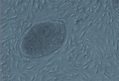
Isolated foetal mammary cells show high levels
of CRIPTO (green) and GRP78 (red), which have
been found to help control the differentiation of
mammary stem cells. Credit: Salk Institute for
Biological Studies. 
The researchers found that when they blocked CRIPTO, the cells mostly formed differentiated cells instead of new stem cells. Over time, this stem cell population shrank since they weren't repopulating themselves. When they instead boosted levels of CRIPTO, the stem cell colony grew as new stem cells were produced more often than differentiated cells.

In studies in mice, the scientists also found that CRIPTO helped the animals form new mammary tissues, which led the team to hypothesize that CRIPTO may be produced by nearby cells in the fat to spur the growth of breast tissue.

In a previous study, Gray's group had discovered that the protein GRP78 binds CRIPTO on the surface of cells and regulates CRIPTO function. This prompted the scientists to test whether GRP78 had an effect on the mammary stem cells. As they suspected, when cells lacked GRP78 on their surfaces, they didn't respond to CRIPTO.

Both CRIPTO and GRP78 have been implicated in cancers, including breast cancer and lung cancers. Scientists think high levels of either protein could encourage tumour growth using similar pathways that they use to spur breast tissue growth. With the new ability to isolate and sustain mammary stem cells, Spike and Gray hope they can uncover details on exactly what cellular programs CRIPTO and GRP78 activate. Understanding this in stem cells could further understanding on how these proteins are involved in tumour growth.

Additionally the researchers think that targeting CRIPTO and GRP78 – which are ideal drug targets since they are present outside of cells – could halt or slow cancer growth. 

"It's looking more and more like what's required to target cancer is to have many therapeutics hitting different pathways," says Gray.

"We think targeting CRIPTO and GRP78 could be a unique way of supplementing existing treatment modalities by targeting stem cell-like cells in cancer."

Source: Salk Institute
Contact: Chris Emery

Reference:
CRIPTO/GRP78 Signaling Maintains Fetal and Adult Mammary Stem Cells Ex Vivo
Benjamin T. Spike, Jonathan A. Kelber, Evan Booker, Madhuri Kalathur, Rose Rodewald, Julia Lipianskaya, Justin La, Marielle He, Tracy Wright, Richard Klemke, Geoffrey M. Wahl, Peter C. Gray
Stem Cell Report, 8 April 2014, Volume 2, Issue 4, p427–439
.........


For more on stem cells and cloning, go to CellNEWS at

Tuesday, 14 January 2014

Keeping Stem Cells Pluripotent

By blocking key signal, researchers maintain embryonic stem cells in vital, undifferentiated state
Tuesday, 14 January 2014

While the ability of human embryonic stem cells (hESCs) to become any type of mature cell, from neuron to heart to skin and bone, is indisputably crucial to human development, no less important is the mechanism needed to maintain hESCs in their pluripotent state until such change is required.

In a paper published in this week’s Online Early Edition of PNAS, researchers from the University of California, San Diego School of Medicine identify a key gene receptor and signalling pathway essential to doing just that – maintaining hESCs in an undifferentiated state.

The finding sheds new light upon the fundamental biology of hESCs – with their huge potential as a diverse therapeutic tool – but also suggests a new target for attacking cancer stem cells, which likely rely upon the same receptor and pathway to help spur their rampant, unwanted growth.

The research, led by principal investigator Karl Willert, PhD, assistant professor in the Department of Cellular and Molecular Medicine, focuses upon the role of the highly conserved WNT signalling pathway, a large family of genes long recognized as a critical regulator of stem cell self-renewal, and a particular encoded receptor known as frizzled family receptor 7 or FZD7.

“WNT signalling through FZD7 is necessary to maintain hESCs in an undifferentiated state,” said Willert.

“If we block FZD7 function, thus interfering with the WNT pathway, hESCs exit their undifferentiated and pluripotent state.”

The researchers proved this by using an antibody-like protein that binds to FZD7, hindering its function.

“Once FZD7 function is blocked with this FZD7-specific compound, hESCs are no longer able to receive the WNT signal essential to maintaining their undifferentiated state.”

FZD7 is a so-called “onco-fetal protein,” expressed only during embryonic development and by certain human tumours. Other studies have suggested that FZD7 may be a marker for cancer stem cells and play an important role in promoting tumour growth. If so, said Willert, disrupting FZD7 function in cancer cells is likely to interfere with their development and growth just as it does in hESCs.

Willert and colleagues, including co-author Dennis Carson, MD, of the Sanford Consortium for Regenerative Medicine and professor emeritus at UC San Diego, plan to further test their FZD7-blocking compound as a potential cancer treatment.

Contact: Scott LaFee

Reference:
The WNT receptor FZD7 is required for maintenance of the pluripotent state in human embryonic stem cells
Antonio Fernandez, Ian J. Huggins, Luca Perna, David Brafman, Desheng Lu, Shiyin Yao,Terry Gaasterland, Dennis A. Carson, and Karl Willert
PNAS, January 13, 2014, doi:10.1073/pnas.1323697111
.........


For more on stem cells and cloning, go to CellNEWS at
http://cellnews-blog.blogspot.com/

Thursday, 12 September 2013

Stem Cells are wired for Cooperation, Down to the DNA

Study reveals network of genes that safeguard cooperation in stem cells and the developing embryo
Thursday, 12 September 2013

We often think of human cells as tiny computers that perform assigned tasks, where disease is a result of a malfunction. But in the current issue of Science, researchers at The Mount Sinai Medical Center offer a radical view of health — seeing it more as a cooperative state among cells, while they see disease as result of cells at war that fight with each other for domination.

Their unique approach is backed by experimental evidence. The researchers show a network of genes in cells, which includes the powerful tumour suppressor p53, which enforce a cooperative state within cells — rather like the queen bee in a beehive. Disease or disorder occurs when these enforcer genes are mutated, allowing competition between cells to ensue.

"Both competition and cooperation drive evolution, and we are wired for cooperation all the way down to our genes," says the study's senior investigator, Thomas P. Zwaka, MD, PhD, Professor at the Black Family Stem Cell Institute at the Icahn School of Medicine at Mount Sinai.

The findings, if backed by future research, offer a new way to address disease, Dr. Zwaka says. Understanding the genetic basis of cooperative and competitive cellular behaviours could explain how cancer and immune system dysfunction develops, he says.

"If a cell has lost a gene that fosters communication among cells, it may dominate other cells by ignoring signals to stop proliferating. It also makes sense that the immune system might detect and attack cells that are not cooperating. Failure to cooperate may also underlie development of birth defects."

He adds that it may be possible to flip the cooperation switch back on therapeutically, or to manipulate stem-like cells to misbehave in a way that produces replacement cells for regenerative medicine.

"Cell misbehave, they are unpredictable. They do not operate like little machines," he says.

"What our study suggests is that cooperation is so central to our evolution that we have genetic mechanisms to protect us against cheating and dominating behaviour."

A network of genes with an ancient function
The research team, which also includes study first author Marion Dejosez, PhD, Assistant Professor at the Icahn School at Mount Sinai, took a long view toward the behaviour of cells. They wondered how it was that cells, which lived on earth as single units for hundreds of millions of years, could effectively bundle themselves together to perform specific tasks.

"Cells started somehow to form alliances, and to cooperate, and obviously this multicellularity had certain advantages."

But they also questioned what happened to the "cheating" behaviour that can be seen in single cells, such as amoeba, that live in colonies — competitive behaviour that allows the cell to gain a reproductive advantage without contributing its fair share to the community.

They conducted a genetic screen in stem cells to look for mutants that allow cells to "misbehave — to become a little antisocial and do things they wouldn't normally do," Dr. Zwaka says.

The screen picked up about 100 genes, which seem to cluster together into a network.

The team focused on three of those genes — p53, long known as the guardian of the genome, Topoisomerase 1 (Top1), which control genomic stability, and olfactory receptors involved in the sensation of smell.

"We could understand that p53 might foster cooperation, because loss of p53 function is a step in the development of many cancers. But finding that top1 and olfactory receptors may have the same function was a surprise," he says.

"We think these genes have the ancient function of safeguarding multicellular organisms by helping cells to coordinate their activities."

The scientists then tested the effects of knocking down these genes in developing mouse embryos. To their surprise, p53 and Top1 knockdown embryos developed normally — perhaps because other intact social enforcement genes took over.

"This showed us that mutant cells only misbehave when they are around normal cells. They become competitive, perhaps promoting an evolutionary advance," Dr. Zwaka says.

"When all the cells are the same, either all mutated or all normal, they cooperate with each other.”

"This study suggests that cell cooperation, altruistic behaviour, cheating, and other so-called social behaviours are wired into cells via the genome at the early primitive stage," he says.

"Perhaps there is no coincidence that amoeba, insects, animals, the human culture and society, generally follow innate rules of cooperation. Darwin's explanation of evolution as a struggle for existence needs to be tempered with an acknowledgment of the importance of cooperation in the evolution of complexity."

Contact: Press Office
.........


For more on stem cells and cloning, go to CellNEWS at
http://cellnews-blog.blogspot.com/

Wednesday, 11 September 2013

Reprogramming in situ: Spanish Team is First to Produce Embryonic Stem Cells in Living Adult Organisms

The characteristics of the obtained stem cells correspond to a primitive totipotent state that has never before been obtained
Wednesday, 11 September 2013

A team from the Spanish National Cancer Research Centre (CNIO) has become the first to make adult cells from a living organism retreat in their evolutionary development to recover the characteristics of embryonic stem cells.

Pictured are Manuel Serrano and Maria Abad
in his laboratory at the CNIO. Credit: Spanish
National Cancer Research Center (CNIO).
Researchers have also discovered that these embryonic stem cells, obtained directly from the inside of the organism, have a broader capacity for differentiation than those obtained via in vitro culture. Specifically, they have the characteristics of totipotent cells: a primitive state never before obtained in a laboratory.

The study, carried out by CNIO, was led by Manuel Serrano, the director of the Molecular Oncology Programme and head of the Tumoural Suppression Laboratory. The study was supported by Manuel Manzanares's team from the Spanish National Cardiovascular Research Centre (CNIC).

Embryonic stem cells are the main focus for the future of regenerative medicine. They are the only ones capable of generating any cell type from the hundreds of cell types that make up an adult organism, so they are the first step towards curing illnesses such as Alzheimer, Parkinson's disease or diabetes. Nevertheless, this type of cell has a very short lifespan, limited to the first days of embryonic development, and they do not exist in any part of an adult organism.

One of the greatest achievements in recent biomedical research was in 2006 when Shinya Yamanaka managed to create embryonic stem cells (pluripotent stem cells, induced in vitro or in vitro iPSCs) in a laboratory from adult cells, via a cocktail of just four genes. Yamanaka's discovery, for which he was awarded the Nobel Prize in Medicine in 2012, opened a new horizon in regenerative medicine.

CNIO researchers have taken another step forward, by achieving the same as Yamanaka, but this time within the same organism, in mice, without the need to pass through in vitro culture dishes. Generating these cells within an organism brings this technology even closer to regenerative medicine.

The first challenge for CNIO researchers was to reproduce the Yamanaka experiment in a living being. They chose a mouse as a model organism. Using genetic manipulation techniques, researchers created mice in which Yamanaka's four genes could be activated at will. When these genes were activated, they observed that the adult cells were able to retreat in their evolutionary development to become embryonic stem cells in multiple tissues and organs.

María Abad, the lead author of the article and a researcher in Serrano's group, said:
"This change of direction in development has never been observed in nature. We have demonstrated that we can also obtain embryonic stem cells in adult organisms and not only in the laboratory".

Manuel Serrano added that:
"We can now start to think about methods for inducing regeneration locally and in a transitory manner for a particular damaged tissue".

Stem cells obtained in mice also show totipotent characteristics never generated in a laboratory, equivalent to those present in human embryos at the 72-hour stage of development, when they are composed of just 16 cells.

In comparison with the cells obtained with the technique developed by Yamanaka, the stem cells obtained by CNIO therefore represent an even earlier embryonic state, with greater capacity for differentiation.

The authors were even able to induce the formation of pseudo-embryonic structures (teratomas) in the thoracic and abdominal cavities of the mice. These pseudo-embryos displayed the three layers typical of embryos (ectoderm, mesoderm and endoderm), and extra-embryonic structures such as the Vitelline membrane and even signs of blood cell formation.

"This data tell us that our stem cells are much more versatile than Yamanaka's in vitro iPSCs, whose potency generates the different layers of the embryo but never tissues that sustain the development of a new embryo, like the placenta", said the CNIO researcher.

The authors emphasise that the possible therapeutic applications of their work are still distant, but they admit that, without doubt, it might mean a change of direction for stem cell research, for regenerative medicine or for tissue engineering.

"Our stem cells also survive outside of mice, in a culture, so we can also manipulate them in a laboratory", said Abad.

"The next step is studying if these new stem cells are capable of efficiently generating different tissues such as that of the pancreas, liver or kidney".

Contact: Juan J. Gomez

Reference:
Reprogramming in vivo produces teratomas and iPSCs with totipotency features
María Abad, Lluc Mosteiro, Cristina Pantoja, Marta Cañamero, Teresa Rayón, Inmaculada Ors, Osvaldo Graña, Diego Megías, Orlando Domínguez, Dolores Martínez, Miguel Manzanares, Sagrario Ortega, Manuel Serrano
Nature (2013), DOI: 10.1038/nature12586
.........


For more on stem cells and cloning, go to CellNEWS at

Monday, 12 August 2013

Tumour Suppressor is needed for Stem Cells to Mature into Neurons

Tumour Suppressor is needed for Stem Cells to Mature into Neurons
Monday, 12 August 2013

CHD5 has previously been proposed as a tumour suppressor, acting as a brake that prevents healthy cells from developing into cancer cells. But the part played by the protein in healthy tissue, and whether this role is important for its ability to counter tumour growth, has remained largely uncharted. Working with colleagues at Trinity College in Dublin and BRIC in Copenhagen, researchers at Karolinska Institute have revealed its function in normal nervous system development and as a tumour suppressor.

The recently published study shows that when stem cells approach the final phase of their specialisation as neurons, CHD5 begins to be expressed at high levels. CHD5 can reshape the chromatin, in which DNA is packed around proteins, and in so doing either facilitate or obstruct the expression of genes. Ulrika Nyman, postdoc researcher in Dr Johan Holmberg's research group and one of the main authors of the current study, explains that on switching off CHD5 in the stem cells of mice embryos during the period in which the brain develops and the majority of neurons are formed, they found was that without CHD5, a stem cell is unable to silence the expression of a number of stem cell genes and genes that are actually to be expressed in muscle, blood or intestinal cells. They also observed an inability in the stem cell to switch on the expression of genes necessary for it to mature into a neuron, leaving it trapped in a stage between stem cell and neuron.

The gene that codes for CHD5 is found on part of chromosome 1 (1p36), which is often lost in tumour cells in a number of cancers, particularly neuroblastoma, a disease that strikes almost only children and which is thought to arise during the development of the peripheral nervous system. Neuroblastoma lacking this section of chromosome and thus also CHD5 are often more aggressive and more rapidly fatal. Treatment with retinoic acid can make immature nerve cells and some neuroblastoma cells mature into specialised nerve cells, but when the researchers prevented neuroblastoma cells from up-regulating CHD5, the tumours no longer responded to retinoic acid treatment.

"In the absence of CHD5, neural tumour cells cannot mature into harmless neurons, but continue to divide, making the tumour more malignant and much harder to treat," says Dr Holmberg at the Department of Cell and Molecular Biology.

"We now hope to be able to restore the ability to up-regulate CHD5 in aggressive tumour cells and make them mature into harmless nerve cells."

Contact: Johan Holmberg

Reference:
CHD5 Is Required for Neurogenesis and Has a Dual Role in Facilitating Gene Expression and Polycomb Gene Repression
Chris M. Egan, Ulrika Nyman, Julie Skotte, Gundula Streubel, Siobhán Turner, David J. O’Connell, Vilma Rraklli, Michael J. Dolan, Naomi Chadderton, Klaus Hansen, Gwyneth Jane Farrar, Kristian Helin, Johan Holmberg, Adrian P. Bracken
Developmental Cell, Volume 26, Issue 3, 223-236, 12 August 2013 10.1016/j.devcel.2013.07.008
.........


For more on stem cells and cloning, go to CellNEWS at

Tuesday, 25 June 2013

Enhancing RNA Interference

Enhancing RNA Interference
Tuesday, 25 June 2013

Nanoparticles that deliver short strands of RNA offer a way to treat cancer and other diseases by shutting off malfunctioning genes. Although this approach has shown some promise, scientists are still not sure exactly what happens to the nanoparticles once they get inside their target cells.

A new study from MIT sheds light on the nanoparticles' fate and suggests new ways to maximize delivery of the RNA strands they are carrying, known as short interfering RNA (siRNA).

Lipid nanoparticles (carrying siRNA) are 
shown as they are transported inside cells 
using endocytic vesicles. Credit: Daria 
Alakhova and Gaurav Sahay. 
"We've been able to develop nanoparticles that can deliver payloads into cells, but we didn't really understand how they do it," says Daniel Anderson, the Samuel Goldblith Associate Professor of Chemical Engineering at MIT.

"Once you know how it works, there's potential that you can tinker with the system and make it work better."

Anderson, a member of MIT's Koch Institute for Integrative Cancer Research and MIT's Institute for Medical Engineering and Science, is the leader of a research team that set out to examine how the nanoparticles and their drug payloads are processed at a cellular and subcellular level. Their findings appear in the June 23 issue of Nature Biotechnology. Robert Langer, the David H. Koch Institute Professor at MIT, is also an author of the paper.

One RNA-delivery approach that has shown particular promise is packaging the strands with a lipid like material; similar particles are now in clinical development for liver cancer and other diseases.

Through a process called RNA interference, siRNA targets messenger RNA (mRNA), which carries genetic instructions from a cell's DNA to the rest of the cell. When siRNA binds to mRNA, the message carried by that mRNA is destroyed. Exploiting that process could allow scientists to turn off genes that allow cancer cells to grow unchecked.

Scientists already knew that siRNA-carrying nanoparticles enter cells through a process, called endocytosis, by which cells engulf large molecules. The MIT team found that once the nanoparticles enter cells they become trapped in bubbles known as endocytic vesicles. This prevents most of the siRNA from reaching its target mRNA, which is located in the cell's cytosol (the main body of the cell).

This happens even with the most effective siRNA delivery materials, suggesting that there is a lot of room to improve the delivery rate, Anderson says.

"We believe that these particles can be made more efficient. They're already very efficient, to the point where micrograms of drug per kilogram of animal can work, but these types of studies give us clues as to how to improve performance," Anderson says.

Molecular traffic jam
The researchers found that once cells absorb the lipid-RNA nanoparticles, they are broken down within about an hour and excreted from the cells.

They also identified a protein called Niemann Pick type C1 (NPC1) as one of the major factors in the nanoparticle-recycling process. Without this protein, the particles could not be excreted from the cells, giving the siRNA more time to reach its targets.

"In the absence of the NPC1, there's a traffic jam, and siRNA gets more time to escape from that traffic jam because there is a backlog," says Gaurav Sahay, an MIT postdoc and lead author of the Nature Biotechnology paper.

In studies of cells grown in the lab without NPC1, the researchers found that the level of gene silencing achieved with RNA interference was 10 to 15 times greater than that in normal cells.

Lack of NPC1 also causes a rare lysosomal storage disorder that is usually fatal in childhood. The findings suggest that patients with this disorder might benefit greatly from potential RNA interference therapy delivered by this type of nanoparticle, the researchers say. They are now planning to study the effects of knocking out the NPC1 gene on siRNA delivery in animals, with an eye toward testing possible siRNA treatments for the disorder.

The researchers are also looking for other factors involved in nanoparticle recycling that could make good targets for possibly slowing down or blocking the recycling process, which they believe could help make RNA interference drugs much more potent. Possible ways to do that could include giving a drug that interferes with nanoparticle recycling, or creating nanoparticle materials that can more effectively evade the recycling process.

Contact: Sarah McDonnell
Written by: Anne Trafton, MIT News Office

Reference:
Efficiency of siRNA delivery by lipid nanoparticles is limited by endocytic recycling
Gaurav Sahay, William Querbes, Christopher Alabi, Ahmed Eltoukhy, Sovan Sarkar, Christopher Zurenko, Emmanouil Karagiannis, Kevin Love, Delai Chen, Roberto Zoncu, Yosef Buganim, Avi Schroeder, Robert Langer & Daniel G Anderson
Nature Biotechnology (2013), doi:10.1038/nbt.2614
.........


For more on stem cells and cloning, go to CellNEWS at
http://cellnews-blog.blogspot.com/

Friday, 23 March 2012

Embryonic Stem Cells Shift Metabolism in Cancer-like Way upon Implanting in Uterus

Switch may release fuel and materials for rapid growth and formation of layers that later become organs 
Friday, 23 March 2012

This is stem cell biologist Dr. Hannele
Ruohola-Baker of the University of
Washington in Seattle. Credit: Univ.
of Wash..
Shortly after a mouse embryo starts to form, some of its stem cells undergo a dramatic metabolic shift to enter the next stage of development, Seattle researchers report today. These stem cells start using and producing energy like cancer cells.

This discovery is published today in EMBO Journal, the European Molecular Biology Organization journal.

"These findings not only have implications for stem cell research and the study of how embryos grow and take shape, but also for cancer therapy," said the senior author of the study, Dr. Hannele Ruohola-Baker, University of Washington professor of biochemistry. The study was collaborative among several research labs in Seattle.

The metabolic transition they discovered occurs very early as the mouse embryo, barely more than a speck of dividing cells, implants in the mother's uterus. The change is driven by low oxygen conditions, Ruohola-Baker explained.

The researchers also saw a specific type of biochemical slowdown in the stem cells' mitochondria – the cells' powerhouses. The phenomenon previously was associated with aging and disease. This was the first example of the same downshift controlling normal early embryonic development.
This is a microscopic image from the mouse
embryonic stem cell metabolism study in
Seattle. Credit: Hannele Ruohola-Baker lab.
"This downshift coincides with the time when the germ line, the keeper of the genome for the next generation, is set aside," Ruohola-Baker said.

"Hence reduction of mitochondrial reactive oxygen species may be nature's way to protect the future."

Embryonic stem cells are called pluripotent because they have the ability to renew themselves and have the potential to become any cell in the body. Self-sustaining and versatile are qualities necessary for the growth, repair and maintenance of the body – and for regenerative medicine therapies.

Although they share these sought-after qualities, "Pluripotent stem cells come in several flavours," Ruohola-Baker explained. They differ in subtle ways that expand or shrink their capacities as the raw living material from which animals are shaped.

There's a big reason why the researchers wanted to understand the distinction between the stem cells that make up the inner cell mass of the free-floating mouse embryo, and those in the epiblast, or implantation stage. Mouse embryonic cells at the epiblast stage more closely resemble human embryonic stem cells - and cancer cells.

Human stem cells and mouse epiblast stem cells have lower mitochondrial respiration activity than do earlier stage mouse stem cells. This reduction occurs despite the fact that the later stage stem cells have more mature mitochondria. The researchers confirmed that certain genes that control mitochondria are turned down during the transition from inner cells mass to epiblast cells.

Instead, the transitioning cells obtain their energy exclusively from breaking down a sugar, glucose. In contrast, the earlier stage mouse embryonic stem cells have more energy options, dynamically switching from mitochondrial respiration to glucose breakdown on demand.

As the embryo enlarges from a few dividing cells to a dense mass that buries into uterus for further development, oxygen comes at a premium.

The researchers discovered that the low-oxygen conditions activate a transcription factor called hypoxia-inducible factor 1alpha. This factor is sufficient to drive mouse embryonic stem cells to rely exclusively on glucose metabolism for their energy. The next challenge is to reveal whether the metabolic switch is deterministic for the fate of these stem cells, in normal as well as in cancer development.

This forced metabolic switch may determine the functional fate of some of the tiny mass of cells making up the primordial embryo. They transition first into epiblast stem cells and, afterward produce the entire developing embryo.

In cancer cells, the shift to a sugar-busting metabolism is known as the Warburg effect, the researchers explain. The Warburg effect sets in motion the biochemical activities that provide the fuel and materials required for rapid tumour cell growth and division.

The Warburg effect in embryonic cells, the researcher proposed, "may serve a similar function in preparation for the dramatic burst of embryonic growth and for the formation of the layers of the early embryo that later will become organs and other body structures."

Contact: Leila Gray

Reference:
HIF1α induced switch from bivalent to exclusively glycolytic metabolism during ESC-to-EpiSC/hESC transition
Wenyu Zhou, Michael Choi, Daciana Margineantu, Lilyana Margaretha, Jennifer Hesson, Christopher Cavanaugh, C Anthony Blau, Marshall S Horwitz, David Hockenbery, Carol Ware and Hannele Ruohola-Baker
The EMBO Journal advance online publication 23 March 2012; doi:10.1038/emboj.2012.71
.........

ZenMaster


For more on stem cells and cloning, go to CellNEWS at

http://cellnews-blog.blogspot.com/

Thursday, 5 May 2011

Normal Stem Cells Made to Look and Act Like Cancer Stem Cells

Normal Stem Cells Made to Look and Act Like Cancer Stem Cells
Thursday, 05 May 2011

Researchers at the University of North Carolina School of Medicine at Chapel Hill, after isolating normal stem cells that form the developing placenta, have given them the same properties of stem cells associated with an aggressive type of breast cancer.

From left to right are study co-first
authors Nicole Vincent Jordan and
Amy N. Abell, Ph.D. Credit: Photo
by Les Lang/UNC School of
Medicine.
The scientific first opens the door for developing novel targeted therapies aimed at triple negative breast cancer. Known also as TNBC, this highly recurrent tumour spreads aggressively beyond its original site in the breast and carries a poor prognosis for patients who have it.

The study will be published online Friday, May 6, by the journal Cell Stem Cell.

"We changed only one amino acid in normal tissue stem cells, trophoblast stem cells. While they maintained their self-renewal, these mutant stem cells had properties very similar to what people predict in cancer stem cells: they were highly mobile and highly invasive," said Gary Johnson, PhD, professor and chair of pharmacology at UNC and senior study author.

"No one has ever isolated a stem cell like that." Johnson is also a member of the UNC Lineberger Comprehensive Cancer Center.

In normal development, epithelial stem cells called trophoblasts are involved in the formation of placental tissue. To do so, they must undergo a conversion to tissue-like cells. These then travel to the site in the uterus where they revert to a non-invasive tissue cell.

"But the mutant trophoblast stem cells made in our lab, which would normally invade the uterus and then stop, just keep going," Johnson said.

The study led by the first authors, research assistant professor Amy N. Abell, PhD and graduate student Nicole Vincent Jordan, both working in Johnson's lab, showed that similar to triple-negative breast cancer stem cells, normal tissue stem cells also go through the same program of molecular changes during organ development called epithelial mesenchymal transition, or EMT. This suggests that breast cancer cells utilize this tissue stem cell molecular program for tumour metastasis, or cancer spread.

The discovery was made using a unique mouse model of tissue stem cell EMT developed in the Johnson laboratory. The study identified two proteins that regulate the expression of specific genes in tissue stem cells during organ development that control normal EMT. Inactivation of the proteins MAP3K4 and CBP in trophoblast stem cells causes them to become hyper invasive.

In collaboration with Aleix Prat, PhD and Charles Perou, PhD in the UNC Lineberger Comprehensive Cancer Center, the research team made another discovery: an overlap between the gene expression signature of the mutant tissue stem cells properties during EMT and the triple-negative human breast cancer gene signature that's predictive of invasiveness. The same genes were down regulated.

"This significant genetic intersection between tissue stem cells and TNBC has identified previously unrecognized genes that likely contribute to breast cancer metastasis," said Johnson.

"This newly identified gene signature is currently being investigated in different models of breast cancer with the goal of developing new therapeutic interventions for the treatment of TNBC."

Source: University of North Carolina School of Medicine
Contact: Les Lang
.........


ZenMaster

For more on stem cells and cloning, go to CellNEWS at
http://cellnews-blog.blogspot.com/

Thursday, 10 March 2011

Reprogrammed Stem Cells Hit Genomic Roadblock

An international study shows that reprogramming cells leads to genomic aberrations
Thursday, 10 March 2011

It's a discordant note in the symphony of good news that usually accompanies stem cell research announcements. Stem cells hold enormous promise in regenerative medicine, thanks to their ability to regenerate diseased or damaged tissues. They have made it possible to markedly improve the effectiveness of many medical treatments – muscle regeneration in cases of dystrophy, skin grafts for treating burn victims, and the treatment of leukaemia via bone marrow transplants.

The problem is obtaining them. Those that are the true source of life, in the first days of embryonic development, are of course the most highly sought after; still undifferentiated, they are "pluripotent," meaning they can evolve into liver, muscle, eye – any kind of cell. But the issue of how to obtain them clearly raises insurmountable ethical questions.

"In this regard, the recent discovery of the "reprogramming" phenomenon, by which somatic cells can be induced to convert to a pluripotent state simply by forcing the expression of a few genes, opens a phenomenal number of possibilities in regenerative medicine," says Didier Trono, Dean of the EPFL School of Life Sciences.

"Imagine, for example, collecting a few cells from the hair follicle of a haemophiliac patient, reprogramming them to the pluripotency of their embryonic precursor, correcting the mutation responsible for the coagulation disorder that plagues the patient, and then re-administering them, genetically "cured," after having orchestrated a differentiation into fully functional progeny."

Increased risks for cancer?
But a study that has just been published in the journal Cell Death and Differentiation, to be followed by two articles in the journal Nature, is dampening those hopes. Conducted by the Department of Biochemistry at the University of Geneva and the European Institute of Oncology in Milan, with the participation of Trono's laboratory, it concludes that these reprogrammed cells exhibit a "genomic instability" that appears to be caused by the process used to return the cells to their embryonic state. Even more serious, the genetic mutations observed resemble mutations that are found in cancer cells. The scientists draw the conclusion that reprogrammed stem cells need to be extensively investigated before they can even be considered for use in regenerative medicine.

The experiments were done using mouse mammary and fibroblast cells. The researchers used three different processes for reprogramming the cells to a "stem," or embryonic, state. The first method was developed expressly for this study, and the others have already been well documented.

Yet all the processes led to the same, implacable conclusion: the genetic anomalies multiplied, in a manner that seems to indicate that they are inherent to the reprogramming process itself, which typically makes use of oncogenes.

"Interestingly, oncogenes have the potential to induce genomic instability," the authors explain.

These results underline the necessity of conducting further studies. First, to see if the genetic anomalies are serious enough to compromise the function and stability of cells regenerated using the reprogrammed cells; and second, to "refine the methods used for generating induced pluripotent cells, in order to avoid this problem. These results will thus motivate scientists to come up with a solution," concludes Trono.

Source: Ecole Polytechnique Fédérale de Lausanne
Contact: Emmanuel Barraud
.........


ZenMaster


For more on stem cells and cloning, go to CellNEWS at
http://cellnews-blog.blogspot.com/

Friday, 15 October 2010

Gene Identified that Prevents Stem Cells from Turning Cancerous

Gene Identified that Prevents Stem Cells from Turning Cancerous
Friday, 15 October 2010

Stem cells, the prodigious precursors of all the tissues in our body, can make almost anything, given the right circumstances. Including, unfortunately, cancer. Now research from Rockefeller University shows that having too many stem cells, or stem cells that live for too long, can increase the odds of developing cancer. By identifying a mechanism that regulates programmed cell death in precursor cells for blood, or hematopoietic stem cells, the work is the first to connect the death of such cells to a later susceptibility to tumours in mice. It also provides evidence of the potentially carcinogenic downside to stem cell treatments, and suggests that nature has sought to balance stem cells' regenerative power against their potentially lethal potency.

Research associate Maria Garcia-Fernandez, Hermann Steller, head of the Strang Laboratory of Apoptosis and Cancer Biology, and their colleagues explored the activity of a gene called Sept4, which encodes a protein, ARTS, that increases programmed cell death, or apoptosis, by antagonizing other proteins that prevent cell death. ARTS was originally discovered by Sarit Larisch, a visiting professor at Rockefeller, and is found to be lacking in human leukaemia and other cancers, suggesting it suppresses tumours. To study the role of ARTS, the experimenters bred a line of mice genetically engineered to lack the Sept4 gene.

For several years, Garcia-Fernandez studied cells that lacked ARTS, looking for signs of trouble relating to cell death. In mature B and T cells, she could not find any, however, so she began to look at cells earlier and earlier in development, until finally she was comparing hematopoietic progenitor and stem cells. Here she found crucial differences, to be published Friday in Genes and Development.

Newborn ARTS-deprived mice had about twice as many hematopoietic stem cells as their normal, ARTS-endowed peers, and those stem cells were extraordinary in their ability to survive experimentally induced mutations.

"The increase in the number of hematopoietic progenitor and stem cells in Sept4-deficient mice brings with it the possibility of accelerating the accumulation of mutations in stem cells," says Garcia-Fernandez.

"They have more stem cells with enhanced resistance to apoptosis. In the end, that leads to more cells accumulating mutations that cannot be eliminated."

Indeed, the ARTS-deprived mice developed spontaneous tumours at about twice the rate of their controls.

"We make a connection between apoptosis, stem cells and cancer that has not been made in this way before: this pathway is critically important in stem cell death and in reducing tumour risk," Steller says.

"The work supports the idea that the stem cell is the seed of the tumour and that the transition from a normal stem cell to a cancer stem cell involves increased resistance to apoptosis."

ARTS interferes with molecules called inhibitor of apoptosis proteins (IAPs), which prevent cells from killing themselves. By inhibiting these inhibitors, under the right circumstances ARTS helps to take the brakes off the process of apoptosis, permitting the cell to die on schedule. Pharmaceutical companies are working to develop small molecule IAP antagonists, but this research is the first to show that inactivating a natural IAP antagonist actually causes tumours to grow, Steller says. It also suggests that the premature silencing of the Sept4/ARTS pathway at the stem cell level may herald cancer to come.

"This work not only defines the role of the ARTS gene in the underlying mechanism of mammalian tumour cell resistance to programmed cell death, but also links this gene to another hallmark of cancer, stem and progenitor cell proliferation," said Marion Zatz, who oversees cell death grants, including Steller's, at the NIH's National Institute of General Medical Sciences.

"The identification of the ARTS gene and its role in cancer cell death provides a potential target for new therapeutic approaches."

Source: Rockefeller University
Contact: Brett Norman

Reference:
Sept4/ARTS is required for stem cell apoptosis and tumor suppression
Maria Garcia-Fernandez, Holger Kissel, Samara Brown, Travis Gorenc, Andrew J. Schile, Shahin Rafii, Sarit Larisch, and Hermann Steller
Genes and Development, Oct 15, 2010; 24 (20)
.........


ZenMaster


For more on stem cells and cloning, go to CellNEWS at
http://cellnews-blog.blogspot.com/

Friday, 3 September 2010

Cancer-causing Gene Crucial in Stem Cell Development

Cancer-causing Gene Crucial in Stem Cell Development
Friday, 03 September 2010

Stem cells might be thought of as trunks in the tree of life. All multi-cellular organisms have them, and they can turn into a dazzling variety other cells — kidney, brain, heart or skin, for example. One class, pluripotent stem cells, has the capacity to turn into virtually any cell type in the body, making them a focal point in the development of cell therapies, the conquering of age-old diseases or even re-growing defective body parts.

Now, a research team at the University of Georgia has shown for the first time that a gene called Myc may be far more important in the development and persistence of stem cells than was known before. Myc is traditionally thought of as a cancer-causing gene, or oncogene, but recent studies from the UGA team have established critical roles for it in stem cell biology. The discovery has important implications for the basic understanding of developmental processes and how stem cells can be used for therapeutic purposes.

"This new research has uncovered a really unexpected role for Myc," said Stephen Dalton, GRA Eminent Scholar of Molecular Cell Biology and Georgia Cancer Coalition Distinguished Scientist at UGA.

"Our work here represents the first mechanistic characterization of how Myc controls the pluripotent stem cell state."

The research was published today in the journal Cell Stem Cell. Other authors of the paper include Keriayn Smith and Amar Singh of the Dalton lab at UGA. Smith left recently to begin a postdoc at the University of North Carolina. Dalton also is a member of the department of biochemistry and molecular biology in the Franklin College of Arts and Sciences and is affiliated with the UGA Cancer Center and the Biomedical and Health Sciences Institute.

In previous work, Dalton and his colleagues showed that Myc is critical for stem cell maintenance and that it affects widespread changes in gene expression. This latter function is crucial when stem cells differentiate into more specific cell types. In the new research, Dalton's team showed that Myc sustains the important pluripotency process by repressing a "master regulator" gene called GATA6.

"Pluripotency is the inherent property of a cell to create all cell types, from an embryo to an adult organism," said Dalton.

"It's an extremely important biological process, and knowing how it is controlled is crucial not only from a basic developmental perspective but also so that we can harness the potential of stem cells for the development of therapies, including those for diabetes, cardiovascular disease and a range of neurological disorders. Through a detailed understanding of early development, we hope to apply this information so that pluripotent stem cells can be differentiated into therapeutically useful cell types."

"These cells can then be used in a clinical setting to cure degenerative diseases and treat acute injury."

The finding that Myc inhibits GATA6 came as a big surprise to the Dalton team and points out that researchers have only seen the tip of the "molecular iceberg" in terms of what Myc does in stem cells. It now seems likely that understanding Myc's role in further detail will reshape current ideas about the basic biology of stem cells.

Dalton's new work addressed the uncertainty about how Myc maintains the pluripotency of stem cells by examining what happens when two forms of Myc — c-Myc and N-Myc — are inactivated in pluripotent stem cells. What he found was that either c- or N-Myc is sufficient to maintain pluripotency, but that the absence of both triggers the differentiation of pluripotent stem cells. Myc is therefore acting as a "brake" to restrain differentiation. When the "differentiation brake" is removed, cells lose their stem cell properties, and, potentially, they can become any one of over a hundred different cell types.

Pluripotent stem cells can now be made from skin fibroblasts and even from blood samples. (Fibroblasts are cells common in connective tissues of animals and play an important role in the healing of wounds, among many functions.) The conversion of mature fibroblast or blood cells back to pluripotent stem cells is called "reprogramming." Myc also has a critical role in this process. The ability to make stem cells from a patient's blood or skin is going to revolutionize medicine as it opens the way for patient-specific stem cells that would circumvent problems associated with immune rejection, said Dalton.

"During the reprogramming of cells, Myc represses genes associated with the differentiated state and primes them for the expression of stem cell genes," he said.

"We now speculate that during the early reprogramming stage, Myc serves to change the cell cycle so that stem cells can divide for long periods of time without aging. This is also what Myc does in cancer cells."

Dalton said that there is an intriguing relationship between normal stem cells and cancer cells. Since Myc is crucial for maintenance of stem cells and for the development of cancer, pluripotent stem cells represent a good model for tumour biologists. Cancer is thought to be initiated by rogue stem cells found in different tissues, further highlighting the link between stem cell biology, cancer and Myc.

"This is clearly going to be a major area of research for many years to come," Dalton said.

Source: University of Georgia
Contact: Stephen Dalton

Reference:
Myc Represses Primitive Endoderm Differentiation in Pluripotent Stem Cells
Keriayn N. Smith, Amar M. Singh, Stephen Dalton
Cell Stem Cell, Volume 7, Issue 3, 343-354, 3 September 2010, 10.1016/j.stem.2010.06.023
.........


ZenMaster


For more on stem cells and cloning, go to CellNEWS at
http://cellnews-blog.blogspot.com/