Friday 23 March 2012

Embryonic Stem Cells Shift Metabolism in Cancer-like Way upon Implanting in Uterus

Switch may release fuel and materials for rapid growth and formation of layers that later become organs 
Friday, 23 March 2012

This is stem cell biologist Dr. Hannele
Ruohola-Baker of the University of
Washington in Seattle. Credit: Univ.
of Wash..
Shortly after a mouse embryo starts to form, some of its stem cells undergo a dramatic metabolic shift to enter the next stage of development, Seattle researchers report today. These stem cells start using and producing energy like cancer cells.

This discovery is published today in EMBO Journal, the European Molecular Biology Organization journal.

"These findings not only have implications for stem cell research and the study of how embryos grow and take shape, but also for cancer therapy," said the senior author of the study, Dr. Hannele Ruohola-Baker, University of Washington professor of biochemistry. The study was collaborative among several research labs in Seattle.

The metabolic transition they discovered occurs very early as the mouse embryo, barely more than a speck of dividing cells, implants in the mother's uterus. The change is driven by low oxygen conditions, Ruohola-Baker explained.

The researchers also saw a specific type of biochemical slowdown in the stem cells' mitochondria – the cells' powerhouses. The phenomenon previously was associated with aging and disease. This was the first example of the same downshift controlling normal early embryonic development.
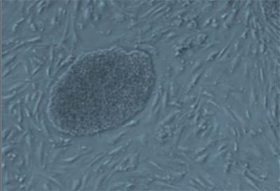
This is a microscopic image from the mouse
embryonic stem cell metabolism study in
Seattle. Credit: Hannele Ruohola-Baker lab.
"This downshift coincides with the time when the germ line, the keeper of the genome for the next generation, is set aside," Ruohola-Baker said.

"Hence reduction of mitochondrial reactive oxygen species may be nature's way to protect the future."

Embryonic stem cells are called pluripotent because they have the ability to renew themselves and have the potential to become any cell in the body. Self-sustaining and versatile are qualities necessary for the growth, repair and maintenance of the body – and for regenerative medicine therapies.

Although they share these sought-after qualities, "Pluripotent stem cells come in several flavours," Ruohola-Baker explained. They differ in subtle ways that expand or shrink their capacities as the raw living material from which animals are shaped.

There's a big reason why the researchers wanted to understand the distinction between the stem cells that make up the inner cell mass of the free-floating mouse embryo, and those in the epiblast, or implantation stage. Mouse embryonic cells at the epiblast stage more closely resemble human embryonic stem cells - and cancer cells.

Human stem cells and mouse epiblast stem cells have lower mitochondrial respiration activity than do earlier stage mouse stem cells. This reduction occurs despite the fact that the later stage stem cells have more mature mitochondria. The researchers confirmed that certain genes that control mitochondria are turned down during the transition from inner cells mass to epiblast cells.

Instead, the transitioning cells obtain their energy exclusively from breaking down a sugar, glucose. In contrast, the earlier stage mouse embryonic stem cells have more energy options, dynamically switching from mitochondrial respiration to glucose breakdown on demand.

As the embryo enlarges from a few dividing cells to a dense mass that buries into uterus for further development, oxygen comes at a premium.

The researchers discovered that the low-oxygen conditions activate a transcription factor called hypoxia-inducible factor 1alpha. This factor is sufficient to drive mouse embryonic stem cells to rely exclusively on glucose metabolism for their energy. The next challenge is to reveal whether the metabolic switch is deterministic for the fate of these stem cells, in normal as well as in cancer development.

This forced metabolic switch may determine the functional fate of some of the tiny mass of cells making up the primordial embryo. They transition first into epiblast stem cells and, afterward produce the entire developing embryo.

In cancer cells, the shift to a sugar-busting metabolism is known as the Warburg effect, the researchers explain. The Warburg effect sets in motion the biochemical activities that provide the fuel and materials required for rapid tumour cell growth and division.

The Warburg effect in embryonic cells, the researcher proposed, "may serve a similar function in preparation for the dramatic burst of embryonic growth and for the formation of the layers of the early embryo that later will become organs and other body structures."

Contact: Leila Gray

Reference:
HIF1α induced switch from bivalent to exclusively glycolytic metabolism during ESC-to-EpiSC/hESC transition
Wenyu Zhou, Michael Choi, Daciana Margineantu, Lilyana Margaretha, Jennifer Hesson, Christopher Cavanaugh, C Anthony Blau, Marshall S Horwitz, David Hockenbery, Carol Ware and Hannele Ruohola-Baker
The EMBO Journal advance online publication 23 March 2012; doi:10.1038/emboj.2012.71
.........

ZenMaster


For more on stem cells and cloning, go to CellNEWS at

http://cellnews-blog.blogspot.com/

Somatic Stem Cells Obtained from Skin Cells for First Time Ever

Skipping pluripotency 'detour,' Max Planck researcher Schoeler again takes lead in stem cell research 
Thursday, 22 March 2012

Breaking new ground, scientists at the Max Planck Institute for Molecular Biomedicine in Münster, Germany, have succeeded in obtaining somatic stem cells from fully differentiated somatic cells. Stem cell researcher Hans Schöler and his team took skin cells from mice and, using a unique combination of growth factors while ensuring appropriate culturing conditions, have managed to induce the cells' differentiation into neuronal somatic stem cells.
induced neural stem cells
This is an immunofluorescence microscopy
image of the induced neural stem cells
(iNSCs) using antibodies against two neural
stem cell markers SSEA1 (red colour) and
Olig2 (green colour). Credit: MPI for
Molecular Biomedicine.
"Our research shows that reprogramming somatic cells does not require passing through a pluripotent stage," explains Schöler. "Thanks to this new approach, tissue regeneration is becoming a more streamlined - and safer - process."

Up until now, pluripotent stem cells were considered the 'be-all and end-all' of stem cell science. Historically, researchers have obtained these 'jack-of-all-trades' cells from fully differentiated somatic cells. Given the proper environmental cues, pluripotent stem cells are capable of differentiating into every type of cell in the body, but their pluripotency also holds certain disadvantages, which preclude their widespread application in medicine.

According to Schöler, "pluripotent stem cells exhibit such a high degree of plasticity that under the wrong circumstances they may form tumours instead of regenerating a tissue or an organ."

Schöler's somatic stem cells offer a way out of this dilemma: they are 'only' multipotent, which means that they cannot give rise to all cell types but merely to a select subset of them - in this case, a type of cell found in neural tissue - a property, which affords them an edge in terms of their therapeutic potential.

To allow them to interconvert somatic cells into somatic stem cells, the Max Planck researchers cleverly combined a number of different growth factors, proteins that guide cellular growth.

"One factor in particular, called Brn4, which had never been used before in this type of research, turned out to be a genuine 'captain' who very quickly and efficiently took command of his ship - the skin cell - guiding it in the right direction so that it could be converted into a neuronal somatic stem cell," explains Schöler. This interconversion turns out to be even more effective if the cells, stimulated by growth factors and exposed to just the right environmental conditions, divide more frequently.

"Gradually, the cells lose their molecular memory that they were once skin cells," explains Schöler. It seems that even after only a few cycles of cell division the newly produced neuronal somatic stem cells are practically indistinguishable from stem cells normally found in the tissue.

Schöler's findings suggest that these cells hold great long-term medical potential:

"The fact that these cells are multipotent dramatically reduces the risk of neoplasm formation, which means that in the not-too-distant future they could be used to regenerate tissues damaged or destroyed by disease or old age; until we get to that point, substantial research efforts will have to be made."

So far, insights are based on experiments using murine skin cells; the next steps now are to perform the same experiments using actual human cells. In addition, it is imperative that the stem cells' long-term behaviour is thoroughly characterized to determine whether they retain their stability over long periods of time.

"Our discoveries are a testament to the unparalleled degree of rigor of research conducted here at the Münster Institute," says Schöler.

"We should realize that this is our chance to be instrumental in helping shape the future of medicine."

At this point, the project is still in its initial, basic science stage although "through systematic, continued development in close collaboration with the pharmaceutical industry, the transition from the basic to the applied sciences could be hugely successful, for this as well as for other, related, future projects," emphasizes Schöler.

This, then, is the reason why a suitable infrastructure framework must be created now rather than later.

"The blueprints for this framework are all prepped and ready to go - all we need now are for the right political measures to be ratified to pave the way towards medical applicability."

Contact: Dr. Hans Schoeler

Reference:
Direct Reprogramming of Fibroblasts into Neural Stem Cells by Defined Factors
Han D.W., Tapia N., Hermann A., Hemmer K., Höing S., Araúzo-Bravo M.J., Zaehres H., Frank S., Moritz S., Greber B., Yang J.H., Lee H.T., Schwamborn J.C., Storch A., Schöler H.R.
Cell Stem Cell, 22 March 2012, 10.1016/j.stem.2012.02.021
.........

ZenMaster

For more on stem cells and cloning, go to CellNEWS at http://cellnews-blog.blogspot.com/

Thursday 22 March 2012

A Shortcut for Stem Cell Programming

Researchers at the University of Bonn artificially derive brain stem cells directly from the connective tissue of mice 
Thursday, 22 March 2012

These stem cells can reproduce and be converted into various types of brain cells. To date, only reprogramming in brain cells that were already fully developed or which had only a limited ability to divide was possible. The new reprogramming method presented by the Bonn scientists and submitted for publication in July 2011 now enables derivation of brain stem cells that are still immature and able to undergo practically unlimited division to be extracted from conventional body cells. The results have now been published in the current edition of the journal Cell Stem Cell.


Induced neural stem cells produced artificially.
The picture shows the superposition of a
microscopic image (grayscale) with two
fluorescence dye staining. Red indicates a
neural nuclear protein (Olig2), and stained
green is a distinct neural stem cells protein
(nestin). Credit: University of Bonn.
The Japanese stem cell researcher Professor Shinya Yamanaka and his team produced stem cells from the connective tissue cells of mice for the first time in 2006; these cells can differentiate into all types of body cells. These induced pluripotent stem cells (iPS cells) develop via reprogramming into a type of embryonic stage. This result made the scientific community sit up and take notice. If as many stem cells as desired can be produced from conventional body cells, this holds great potential for medical developments and drug research.

"Now a team of scientists from the University of Bonn has proven a variant for this method in a mouse model," report Dr. Frank Edenhofer and his team at the Institute of Reconstructive Neurobiology (Director: Dr. Oliver Brüstle) of the University of Bonn. Also involved were the epileptologists and the Institute of Human Genetics of the University of Bonn, led by Dr. Markus Nöthen, who is also a member of the German Center for Neurodegenerative Diseases.

Edenhofer and his co-workers Marc Thier, Philipp Wörsdörfer and Yenal B. Lakes used connective tissue cells from mice as a starting material. Just as Yamanaka did, they initiated the conversion with a combination of four genes.

"We however deliberately targeted the production of neural stem cells or brain stem cells, not pluripotent iPS multipurpose cells," says Edenhofer. These cells are known as somatic or adult stem cells, which can develop into the cells typical of the nervous system, neurons, oligodendrocytes and astrocytes.

The gene "Oct4" is the central control factor
The gene "Oct4" is a crucial control factor.

"First, it prepares the connective tissue cell for reprogramming, later, however, Oct4 appears to prevent destabilized cells from becoming brain stem cells" reports the Bonn stem cell researcher. While this factor is switched on during reprogramming of iPS cells over a longer period of time, the Bonn researchers activate the factor with special techniques for only a few days.

"If this molecular switch is toggled over a limited period of time, the brain stem cells, which we refer to as induced neural stem cells (iNS cells), can be reached directly," said Edenhofer.

"Oct4 activates the process, destabilizes the cells and clears them for the direct reprogramming. However, we still need to analyze the exact mechanism of the cellular conversion."

The scientists at the University of Bonn have thus found a new way to reprogram cells, which is considerably faster and also safer in comparison to the iPS cells and embryonic stem cells.

"Since we cut down on the reprogramming of the cells via the embryonic stage, our method is about two to three times faster than the method used to produce iPS cells," stresses Edenhofer. Thus the work involved and the costs are also much lower. In addition, the novel Bonn method is associated with a dramatically lower risk of tumors. As compared to other approaches, the Bonn scientists' method stands out due to the production of neural cells that can be multiplied to a nearly unlimited degree.

Low risk of tumor and unlimited self-renewal
A low risk of tumor formation is important because in the distant future, neural cells will replace defective cells of the nervous system. A vision of the various international scientific teams is to eventually create adult stem cells for example from skin or hair root cells, differentiate these further for therapeutic purposes, and then implant them in damaged areas.

"But that is still a long way off," says Edenhofer.

However, the scientists have a rather urgent need today for a simple way to obtain brain stem cells from the patient to use them to study various neurodegenerative diseases and test drugs in a Petri dish.

"Our work could form the basis for providing practically unlimited quantities of the patient's own cells." T

he current study was initially conducted on mice.

"We are now extremely eager to see whether these results can also be applied to humans," says the Bonn scientist.

Contact: Dr. Frank Edenhofer

Reference:
Direct Conversion of Fibroblasts into Stably Expandable Neural Stem Cells
Marc Thier, Philipp Wörsdörfer, Yenal B. Lakes, Raphaela Gorris, Stefan Herms, Thoralf Opitz, Dominic Seiferling, Tamara Quandel, Per Hoffmann, Markus M. Nöthen, Oliver Brüstle, and Frank Edenhofer
Cell Stem Cell, DOI: 10.1016/j.stem.2012.03.003
.........


For more on stem cells and cloning, go to CellNEWS at
http://cellnews-blog.blogspot.com/

Monday 19 March 2012

Researchers Print Live Cells with a Standard Inkjet Printer

Researchers Print Live Cells with a Standard Inkjet Printer
Monday, 19 March 2012

A fibroblast printed with the modified
inkjet printer. The interior of the cell
shows that the fluorescently tagged
actin monomers have been incorporated.
Credit: © The Journal of Visualized
Experiments. To watch the full video
article, please click here.

Researchers from Clemson University have found a way to create temporary holes in the membranes of live cells using a standard inkjet printer. The method will be published in JoVE, the Journal of Visualized Experiments, on March 16.

"We first had the idea for this method when we wanted to be able to visualize changes in the cytoskeleton arrangement due to applied forces on cells," said paper-author Dr. Delphine Dean.

She said other researchers have been using this method to print cells onto slides, but that they have only recently discovered that printing the cells causes the disruption in their membranes for a few hours. Creating temporary pores allow researchers to put molecules inside of cells that wouldn't otherwise fit, and study how the cells react.

"The authors have used an extremely innovative approach for bio-printing cells. Moreover, this approach can be used for applications other than cell printing," said JoVE Science Editor, Dr. Nandita Singh.

"Matrix proteins can be printed onto substrates with this technique for cell patterning. This JoVE publication will make this approach simple and approachable and enable other labs to replicate the procedure."

The printer is modified by removing the paper feed mechanism and adding a "stage" from which to feed the slides. The ink is replaced with a cell solution, and the cells are printed directly on to the slides.

Using this method, the researchers are able to process thousands of cells in a matter of minutes. Dr. Dean's team used the holes to introduce fluorescent molecules that illuminate the skeleton of the cell.

"We are actually interested in the cell mechanics of compressed cells. This method allows us to push on the cells and watch the response easily," said Dr. Dean.

"We are interested in cardiovascular cells, and how they respond to mechanical force."

Dr. Dean chose to submit her method to JoVE, the only peer reviewed, PubMed-indexed science journal to publish all of its content in both text and video format, because, according to her, "until you've seen it done, it's hard to understand the process."

Contact: Katherine Scott

Reference:
Creating Transient Cell Membrane Pores Using a Standard Inkjet Printer
Owczarczak, A. B., Shuford, S. O., Wood, S. T., Deitch, S., Dean, D.
J. Vis. Exp. (61), e3681, DOI: 10.3791/3681 (2012)
.........

ZenMaster

For more on stem cells and cloning, go to CellNEWS at
http://cellnews-blog.blogspot.com/

Saturday 17 March 2012

Stem Cells Hint at Potential Treatment for Huntington's Disease

Stem Cells Hint at Potential Treatment for Huntington's Disease
Saturday, 17 March 2012

A GABA neuron made from human
stem cells in the lab of University of
Wisconsin-Madison neuroscientist
Su-Chun Zhang. GABA neurons are
the brain cells whose degradation causes
Huntington’s disease, a condition
characterized by severely degraded
motor function, among other things.
Zhang and his colleagues have shown
that the severe motor deficits observed
in a mouse model of Huntington’s can
be corrected by implanting the lab made
cells. Credit: Su-Chun Zhang.
Huntington's disease, the debilitating congenital neurological disorder that progressively robs patients of muscle coordination and cognitive ability, is a condition without effective treatment, a slow death sentence.

But if researchers can build on new research reported this week (March 15, 2012) in the journal Cell Stem Cell, a special type of brain cell forged from stem cells could help restore the muscle coordination deficits that cause the uncontrollable spasms characteristic of the disease.

"This is really something unexpected," says Su-Chun Zhang, a University of Wisconsin-Madison neuroscientist and the senior author of the new study, which showed that locomotion could be restored in mice with a Huntington's-like condition.

Zhang is an expert at making different types of brain cells from human embryonic or induced pluripotent stem cells. In the new study, his group focused on what are known as GABA neurons, cells whose degradation is responsible for disruption of a key neural circuit and loss of motor function in Huntington's patients. GABA neurons, Zhang explains, produce a key neurotransmitter, a chemical that helps underpin the communication network in the brain that coordinates movement.

In the laboratory, Zhang and his colleagues at the UW-Madison Waisman Center have learned how to make large amounts of GABA neurons from human embryonic stem cells, which they sought to test in a mouse model of Huntington's disease. The goal of the study, Zhang notes, was simply to see if the cells would safely integrate into the mouse brain. To their astonishment, the cells not only integrated but also project to the right target and effectively reestablished the broken communication network, restoring motor function.

The results of the study were surprising, Zhang explains, because GABA neurons reside in one part of the brain, the basal ganglia, which plays a key role in voluntary motor coordination. But the GABA neurons exert their influence at a distance on cells in the midbrain through the circuit fueled by the GABA neuron chemical neurotransmitter.

"This circuitry is essential for motor coordination," Zhang says, "and it is what is broken in Huntington patients. The GABA neurons exert their influence at a distance through this circuit. Their cell targets are far away."

That the transplanted cells could effectively reestablish the circuit was completely unexpected.

"Many in the field feel that successful cell transplants would be impossible because it would require rebuilding the circuitry. But what we've shown is that the GABA neurons can remake the circuitry and produce the right neurotransmitter."

The implications of the new study are important not only because they suggest it may one day be possible to use cell therapy to treat Huntington's, but also because it suggests the adult brain may be more malleable than previously believed.

The adult brain, notes Zhang, is considered by neuroscientists to be stable, and not easily susceptible to therapies that seek to correct things like the broken circuits at the root of conditions like Huntington's. For a therapy to work, it has to be engineered so that only cells of interest are affected.

"The brain is wired in such a precise way that if a neuron projects the wrong way, it could be chaotic."

Zhang stresses that while the new research is promising, working up from the mouse model to human patients will take much time and effort. But for a disease that now has no effective treatment, the work could become the next best hope for those with Huntington's.

Contact: Su-Chun Zhang
.........


For more on stem cells and cloning, go to CellNEWS at
http://cellnews-blog.blogspot.com/