![]() |
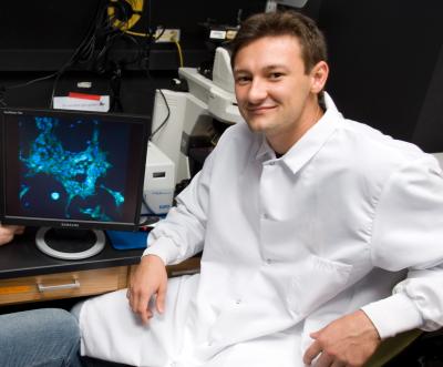
Andres Bratt-Leal, a former graduate
student
in the laboratory of Todd McDevitt,
analyzes
stem cells using a microscope. Credit: Georgia
Tech Photo: Rob Felt.
|
Wednesday, 10 July 2013
Micro-particles Create Localized Control of Stem Cell Differentiation
Posted by ZenMaster at Wednesday, July 10, 2013
Labels: BMP-4, differentiation, embryonic, mouse, research, stem cells, tissue engineering 0 comments
Sunday, 21 November 2010
Fibrodysplasia Ossificans Progressiva: A Rare Disease Reveals New Path for Creating Stem Cells
As debilitating as disease can be, sometimes it acts as a teacher
Sunday, 21 November 2010
Researchers at Harvard Medical School and the Harvard School of Dental Medicine have found that by mimicking a rare genetic disorder in a dish, they can rewind the internal clock of a mature cell and drive it back into an adult stem-cell stage. This new "stem cell" can then branch out into a variety of differentiated cell types, both in culture and in animal models.
"This certainly has implications for personalized medicine, especially in the area of tissue engineering," says Bjorn Olsen, the Hersey Professor of Cell Biology at Harvard Medical School and Dean of Research at the Harvard School of Dental Medicine.
These findings appear November 21, online in Nature Medicine.
Fibrodysplasia Ossificans Progressiva (FOP), which affect fewer than 1,000 people worldwide, is a horrific genetic disease in which acute inflammation causes soft tissue to morph into cartilage and bone. Over the course of a few decades, patients gradually become thoroughly ossified, as though parts of their body have turned to stone. There is no cure or treatment.
Damian Medici, an instructor of medicine at Harvard Medical School and Beth Israel Deaconess Medical Center, found that, unlike normal skeletal tissue, the pathological cartilage and bone cells from these patients contained biomarkers specific for endothelial cells — cells that line the interior of blood vessels. This led him to question whether or not the cartilage and bone growing in soft tissues of FOP patients had an endothelial origin.
Medici and his colleagues transferred the mutated gene that causes FOP into normal endothelial cells. Unexpectedly, the endothelial cells converted into a cell type nearly identical to what are called mesenchymal stem cells, or adult stem cells that can differentiate into bone, cartilage, muscle, fat, and even nerve cells. (Embryonic stem cells have the potential to become any type of cell, whereas adult stem cells are limited.)
What's more, through further experiments the researchers found that instead of using the mutated gene to induce the transformation, they could incubate endothelial cells with either one of two specific proteins (growth factors TGF-beta2 and BMP4) whose cellular interactions mimicked the effects of the mutated gene, providing a more efficient way to reprogram the cells.
Afterwards, Medici was able to take these reprogrammed cells and, in both culture dishes and animal models, coax them into developing into a group of related tissue types.
"It's important to clarify that these new cells are not exactly the same as mesenchymal stem cells from bone marrow," says Medici.
"There are some important differences. However, they appear to have all the potential and plasticity of mesenchymal stem cells."
"The power of this system is that we are simply repeating and honing a process that occurs in nature," says Olsen.
"In that sense, it's less artificial than other current methods for reprogramming cells."
According to study collaborator Frederick Kaplan, Isaac & Rose Nassau Professor of Orthopaedic Molecular Medicine at the University of Pennsylvania School of Medicine and a world expert on FOP:
"While we want to use this knowledge to stop the renegade bone formation of FOP, these new findings provide the first glimpse of how to recruit and harness the process to build extra bone for those who desperately need it."
Medici and Olsen echo this, stating that the most direct application for these findings is the field of tissue engineering and personalized medicine. It is conceivable that transplant patients may one day have some of their own endothelial cells extracted, reprogrammed, and then grown into the desired tissue type for implantation. Host rejection would not be an issue.
Source: Harvard Medical School
Contact: David Cameron
Reference:
Conversion of vascular endothelial cells into multipotent stem-like cells
Damian Medici, Eileen M Shore, Vitali Y Lounev, Frederick S Kaplan, Raghu Kalluri & Bjorn R Olsen
Nature Medicine, early online publication, Nov 21, 2010, doi:10.1038/nm.2252
.........
ZenMaster
For more on stem cells and cloning, go to CellNEWS at
http://cellnews-blog.blogspot.com/
Posted by ZenMaster at Sunday, November 21, 2010
Labels: BMP-4, human, reprogram, research, stem cells, US 0 comments
Thursday, 6 November 2008
Key Trigger of Embryonic Stem Cell Differentiation
Key Trigger of Embryonic Stem Cell Differentiation Thursday, 06 November 2008 Clusters of mouse embryonic stem cells called embryoid bodies more closely approximate true embryos in organization and structure than previously thought, according to researchers at the Stanford University School of Medicine. Harnessing the signals that influence the cells' fate may help researchers more accurately direct the differentiation of embryonic stem cells for use in therapy. The researchers found that embryoid bodies have hallmarks of gastrulation - a remarkable developmental step that launches a hollow ball of cells toward becoming an organism with three distinct types of precursor cells. The scientists showed that this process is initiated by a single signalling pathway in embryoid bodies and in real embryos. Enhancing or blocking this signal affects what the cells become, the scientists found. "A lot of embryonic stem cell research is aimed at devising ways to help the cells differentiate along a particular path," said Roeland Nusse, PhD, professor of developmental biology. "But it's very difficult to know how to do this. We're learning that they do more things in culture than we previously thought; at the same time, we're developing more tools to control what they become." Nusse is the senior author of the research, which will be published in the Nov. 6 issue of the journal Cell Stem Cell. He is also a Howard Hughes Medical Institute investigator and a member of Stanford's Cancer Center. The study was funded in part by a grant from the California Institute of Regenerative Medicine intended to clarify the role of a common group of cell signalling molecules called the Wnt family in the differentiation of embryonic stem cells. Nusse and the first authors of the paper, postdoctoral scholar Derk ten Berge, PhD, and undergraduate student Wouter Koole, used easily tracked reporter genes that are expressed only when cells are responding to Wnt signals to figure out when and where Wnt is active in mouse embryos and embryoid bodies. Embryoid bodies are clumps of embryonic stem cells that can to begin to differentiate into different tissues but they are not true embryos. Using this system, the researchers learned that Wnt-responsive cells first appear in 6.5-day-old embryos in an area called the primitive streak that forms on what will become the posterior side of the embryo. It is the first step toward gastrulation, in which an outer layer of cells dimples inward at what will be either the mouth or anus to form the three distinct precursor cell types shared by most animals: the ectoderm, or outer layer, which forms neurons, skin cells and pigment; the endoderm, or inner layer, which forms many of the organs; and mesoderm, or middle layer, which forms muscle and red blood cells. More importantly, Nusse and his colleagues determined that Wnt-responsive cells in the embryoid bodies also spontaneously form a primitive streak, though they never truly gastrulate. Supplementing the naturally occurring Wnt signal with "extra" Wnt protein accelerated the formation of the primitive streak, and adding proteins that blocked Wnt activity inhibited it. "We knew that embryoid bodies did exhibit some self-organization," said Nusse. "They form a hollow cavity with inner and outer cell layers. But the primitive streak is the first indication we have that they can develop the kind of asymmetry that is seen in embryos." Furthermore, the extra Wnt caused the Wnt-responsive cells to differentiate primarily into mesendodermal precursors (which can become either mesoderm or endoderm and is associated with the posterior of the embryo) and inhibited the formation of neurectoderm (ectoderm destined to become cells of the nervous system that are mostly associated with the embryo's anterior). Blocking Wnt activity tipped the balance in the other direction, causing the cells to shun mesendoderm and become mainly neurectoderm. The first step to controlling cell fate is to understand which protein in the normal cocktail of growth factors used to maintain the embryoid bodies is responsible for triggering the cells' Wnt-responsive pathways. The researchers identified one specific factor, called BMP, that gets the ball rolling. Inhibiting this factor stops the spontaneous formation of the primitive streak in the embryoid bodies and gives the researcher more precise control over the cells' differentiation. "Differentiation is a step-wise process," said Nusse. "To get to a particular endpoint, you need to know all the steps along the way. Our research indicates that embryoid bodies are a better-than-expected model of what happens in the embryo, and suggests how we may be able to manipulate those steps to our advantage to get pure populations of certain types of cells for research or therapy." Reference: Wnt Signaling Mediates Self-Organization and Axis Formation in Embryoid Bodies Derk ten Berge, Wouter Koole, Christophe Fuerer, Matt Fish, Elif Eroglu and Roel Nusse Cell Stem Cell, Volume 3, Issue 5, 508-518, 6 November 2008, doi:10.1016/j.stem.2008.09.013 ......... ZenMaster
For more on stem cells and cloning, go to CellNEWS at http://cellnews-blog.blogspot.com/ and http://www.geocities.com/giantfideli/index.html
Posted by ZenMaster at Thursday, November 06, 2008
Labels: BMP-4, California, differentiation, embryonic, stem cells, Wnt 0 comments
Wednesday, 9 April 2008
Human ESC Research Reveals Earliest Step in Human Development
Human ESC Research Reveals Earliest Step in Human Development Wednesday, 09 April 2008 Researchers at Johns Hopkins have uncovered the molecular underpinnings of one of the earliest steps in human development using human embryonic stem cells. Their identification of a critical signal mediated by the protein BMP-4 that drives the differentiation of stem cells into what will become the placenta, will be published in the April issue of Cell Stem Cell. The finding, they say, also highlights one aspect of human cell biology that has not been replicated in other animal model systems. It is virtually impossible to use anything other than human embryonic stem cells to gather information of this kind. One reason for the excitement, the investigators say, is that the system can provide a research model to study very early human development, including the formation of placenta which develops from the same early embryo. “The findings was serendipitous and at the same time a very important addition to our understanding of early human development,” says Linzhao Cheng, Ph.D., an associate professor of gynaecology and obstetrics and co-director of the stem cell program of the Johns Hopkins Institute for Cell Engineering. “This is one area of stem cell biology where human and mouse differs significantly and we never would have discovered this if we had limited our studies to using only mouse embryonic stem cells. Adult human stem cells just didn’t work for this.” The research team uncovered their finding during efforts to study a rare human blood disorder caused by mutations in a gene called PIG-A. According to Cheng, a good model to study the disease does not exist as engineered mice without the gene either die before birth, or do not reproduce symptoms found in patients. So using a conventional genetic engineering tool, the researchers tried for years – literally – to knock out PIG-A in adult stem cells, without success. They then turned to knocking out PIG-A in human embryonic stem cells. “Only with the human embryonic stem cells could we grow out the rare cells engineered to lack PIG-A,” says Cheng. The result was the growth of two human embryonic stem cell lines that lack PIG-A, and therefore do not contain any proteins known as glycosylphosphatidylinositol (GPI) anchor proteins on the cell’s surface. GPI anchor proteins attach many different types of proteins involved in cell communication to a cell’s outside surface. Without certain GPI proteins, cells may not function properly. Then the researchers took one more step to verify that their engineered embryonic stem cells behaved like normal stem cells. “We just wanted to make sure that our knockout cells could still differentiate and specialise,” says Cheng. One of the earliest steps of embryonic stem cell differentiation in normal embryonic development is the development of the trophoblast, a layer of seed cells that later develops into the placenta. Trophoblast differentiation, according to Cheng, occurs when embryonic stem cells are exposed to BMP-4 protein, either naturally or in the lab. To their surprise, however, when they treated their knockout cells with BMP-4, the cells did not become trophoblasts. Only when they added the PIG-A gene back into their cells did BMP-4 do its work and cause the cells to become trophoblasts, allowing the researchers to conclude that trophoblast differentiation depends on certain cell surface proteins to receive the BMP-4 signal. Reference: Trophoblast Differentiation Defect in Human Embryonic Stem Cells Lacking PIG-A and GPI-Anchored Cell-Surface Proteins Guibin Chen, Zhaohui Ye, Xiaobing Yu, Jizhong Zou, Prashant Mali, Robert A. Brodsky, and Linzhao Cheng Cell Stem Cell, Vol 2, 345-355, 10 April 2008 ......... ZenMaster
For more on stem cells and cloning, go to CellNEWS at http://www.geocities.com/giantfideli/index.html
Posted by ZenMaster at Wednesday, April 09, 2008
Labels: BMP-4, differentiation, embryo, embryonic, hESCs, research, stem cells 0 comments