Showing posts with label extracellular matrix. Show all posts

Thursday, 18 December 2014

Stem Cells Faulty in Duchenne Muscular Dystrophy

Stem Cells Faulty in Duchenne Muscular Dystrophy
Thursday, 18 December 2014

Like human patients, mice with a form of Duchenne muscular dystrophy undergo progressive muscle degeneration and accumulate connective tissue as they age. Now, researchers at the Stanford University School of Medicine have found that the fault may lie at least partly in the stem cells that surround the muscle fibres.

They've found that during the course of the disease, the stem cells become less able to make new muscle and instead begin to express genes involved in the formation of connective tissue. Excess connective tissue – a condition called fibrosis – can accumulate in many organs, including the lungs, liver and heart, in many different disorders. In the skeletal muscles of people with muscular dystrophy, the fibrotic tissue impairs the function of the muscle fibres and leads to increasing weakness and stiffness, which are hallmarks of the disease.

The researchers discovered that this abnormal change in stem cells could be inhibited in laboratory mice by giving the animals a drug that is already approved for use in humans. The drug works by blocking a signalling pathway involved in the development of fibrosis. Although much more research is needed, the scientists are hopeful that a similar approach may one day work in children with muscular dystrophy.

"These cells are losing their ability to produce muscle, and are beginning to look more like fibroblasts, which secrete connective tissue," said Thomas Rando, MD, PhD, professor of neurology and neurological sciences.

"It's possible that if we could prevent this transition in the muscle stem cells, we could slow or ameliorate the fibrosis seen in muscular dystrophy in humans."

A paper describing the researchers' findings will be published Dec. 17 in Science Translational Medicine. Rando, the paper's senior author, is director of the Glenn Laboratories for the Biology of Aging and founding director of the Muscular Dystrophy Association Clinic at Stanford. Former postdoctoral scholar Stefano Biressi, PhD, is the lead author. Biressi is now at the Centre for Integrative Biology at the University of Trento in Italy.

A devastating disease
Duchenne muscular dystrophy is a devastating disease that affects about 1 in every 3,600 boys born in the United States. Patients usually experience severe, progressive muscle weakness that confines them to a wheelchair in early adolescence and eventually leads to paralysis. It's caused by mutations in the dystrophin gene, which encodes the dystrophin protein. The dystrophin protein serves to connect muscle fibres to the surrounding external matrix. This connection stabilizes the fibres, enhancing their strength and preventing injury. Sufferers are nearly always boys because the dystrophin gene is located on the X chromosome. (Girls would need to inherit two faulty copies, which is unlikely because male carriers often die in early adulthood.)

Under normal conditions, muscle stem cells respond to muscle damage by dividing into cells, one of which becomes new muscle, while the other remains a stem cell. However, in the mice missing the dystrophin gene, the muscle stem cells slowly assume a different fate. They begin to resemble fibroblasts instead of muscle-making machines.

To conduct the research, Biressi and Rando used a strain of laboratory mice in which the muscle stem cells were engineered to glow with a fluorescent light when treated with a drug called tamoxifen. They then bred the mice with another strain in which the dystrophin gene is mutated, and followed the fate of the cells over time.

The researchers found that the expression of myogenic genes, which are associated with the regeneration of muscle in response to injury, was nearly completely lacking in many of the muscle stem cells in the mice after just 11 months, while the expression of fibrotic genes had increased compared with that of control animals. The cells from the dystrophic animals were also oddly located: Rather than being nestled next to the muscle fibres, they had begun to move away into the spaces between tissues.

The role of a signalling pathway
Rando and Biressi knew that a similar, but much less pronounced, accumulation of connective tissue in muscle fibres occurs during normal aging. That process is governed by signalling proteins, which include the Wnt and TGF-beta protein families. Wnt plays a critical role in embryonic development and cancer; TGF-beta controls cell division and specialization. They wondered whether blocking the Wnt/TGF-beta pathway in the dystrophic mice would inhibit fibrosis in the animals' muscles.

The researchers turned to a drug called losartan, which is used to treat high blood pressure. Losartan inhibits the expression of the genes for TGF-beta types 1 and 2. The researchers thought it would probably interrupt the signalling pathway that leads the muscle stem cells astray.

Treating the mice with losartan, they found, did in fact prevent the muscle stem cells from expressing fibrosis-associated genes and partially maintained their ability to form new muscle.

"This scar tissue, or fibrosis, leaves the muscle less elastic and impairs muscle function," Rando said.

"So we'd like to understand why it happens, and how to prevent it. It's also important to limit fibrosis to increase the likelihood of success with other possible therapies, such as cell therapy or gene therapy."

Next steps
Because TGF-beta type 1 plays many roles throughout the body, the researchers are now working to find ways to specifically inhibit TGF-beta type 2, which is involved in the transition of the muscle stem cells. They're also interested in learning how to translate the research to other diseases.

"Fibrosis seems to occur in a vicious cycle," Rando said.

"As the muscle stem cells become less able to regenerate new muscle, the tissue is less able to repair itself after damage. This leads to fibrosis, which then further impairs muscle formation. Understanding the biological basis of fibrosis could have a profound effect on many other diseases."

Contact: Krista Conger
.........


For more on stem cells and cloning, go to CellNEWS at

Sunday, 10 August 2014

Matrix Stiffness is an Essential Tool in Stem Cell Differentiation

Matrix Stiffness is an Essential Tool in Stem Cell Differentiation
Sunday, 10 August 2014

Cells grown on hydrogels of the same stiffness 
all display fat cell markers and deform the
underlying matrix material the same way.
Credit: Adam Engler, UC San Diego Jacobs
School of Engineering. 
Bioengineers at the University of California, San Diego have proven that when it comes to guiding stem cells into a specific cell type, the stiffness of the extracellular matrix used to culture them really does matter. When placed in a dish of a very stiff material, or hydrogel, most stem cells become bone-like cells. By comparison, soft materials tend to steer stem cells into soft tissues such as neurons and fat cells. The research team, led by bioengineering professor Adam Engler, also found that a protein binding the stem cell to the hydrogel is not a factor in the differentiation of the stem cell as previously suggested. The protein layer is merely an adhesive, the team reported Aug. 10 in the advance online edition of the journal Nature Materials.

Their findings affirm Engler's prior work on the relationship between matrix stiffness and stem cell differentiations.

Cells grown on three hydrogels of the same
stiffness all display fat cell markers and deform
the underlying matrix material in the same way.
Credit: Adam Engler, UC San Diego Jacobs
School of Engineering. 
"What's remarkable is that you can see that the cells have made the first decisions to become bone cells, with just this one cue. That's why this is important for tissue engineering," said Engler, a professor at the UC San Diego Jacobs School of Engineering.

Engler's team, which includes bioengineering graduate student researchers Ludovic Vincent and Jessica Wen, found that the stem cell differentiation is a response to the mechanical deformation of the hydrogel from the force exerted by the cell. In a series of experiments, the team found that this happens whether the protein tethering the cell to the matrix is tight, loose or non-existent. To illustrate the concept, Vincent described the pores in the matrix as holes in a sponge covered with ropes of protein fibres. Imagine that a rope is draped over a number of these holes, tethered loosely with only a few anchors or tightly with many anchors. Across multiple samples using a stiff matrix, while varying the degree of tethering, the researchers found no difference in the rate at which stem cells showed signs of turning into bone-like cells. The team also found that the size of the pores in the matrix also had no effect on the differentiation of the stem cells as long as the stiffness of the hydrogel remained the same.

Cells grown on three hydrogels of the same
stiffness all display fat cell markers and deform
the underlying matrix material in the same way.
Credit: Adam Engler/UC San Diego Jacobs
School of Engineering. 
"We made the stiffness the same and changed how the protein is presented to the cells (by varying the size of the pores and tethering) and ask whether or not the cells change their behaviour," Vincent said.

"Do they respond only to the stiffness? Neither the tethering nor the pore size changed the cells."

"We're only giving them one cue out of dozens that are important in stem cell differentiation," said Engler.

"That doesn't mean the other cues are irrelevant; they may still push the cells into a specific cell type. We have just ruled out porosity and tethering, and further emphasized stiffness in this process."

Contact: Catherine Hockmuth

Reference:
Interplay of matrix stiffness and protein tethering in stem cell differentiation
Jessica H. Wen, Ludovic G. Vincent, Alexander Fuhrmann, Yu Suk Choi, Kolin Hribar, Hermes Taylor-Weiner, Shaochen Chen and Adam J. Engler
Nature Materials, Aug. 10 in the Advance online edition, Aug. 10 2014.
.........


For more on stem cells and cloning, go to CellNEWS at
http://cellnews-blog.blogspot.com/

Wednesday, 19 February 2014

An Essential Step Toward Printing Living Tissues

New method enables scientists to print tissue constructs with blood vessels
Wednesday, 19 February 2014

A new 3-D printing method developed by Wyss
Core Faculty member Jennifer Lewis and her
team uses multiple print heads and customized
"inks" to create complex living tissue constructs,
complete with tiny blood vessels. To see how it
works, visit http://wyss.harvard.edu/. Credit: 
Wyss Institute and Harvard School of Engineering
and Applied Sciences. 
A new bio-printing method developed at the Wyss Institute for Biologically Inspired Engineering at Harvard University and the Harvard School of Engineering and Applied Sciences (SEAS) creates intricately patterned 3D-tissue constructs with multiple types of cells and tiny blood vessels. The work represents a major step toward a longstanding goal of tissue engineers: creating human tissue constructs realistic enough to test drug safety and effectiveness.

The method also represents an early but important step toward building fully functional replacements for injured or diseased tissue that can be designed from CAT scan data using computer-aided design (CAD), printed in 3D at the push of a button, and used by surgeons to repair or replace damaged tissue.

"This is the foundational step toward creating 3D living tissue," said Jennifer Lewis, Ph.D., senior author of the study, who is a Core Faculty Member of the Wyss Institute for Biologically Inspired Engineering at Harvard University, and the Hansjörg Wyss Professor of Biologically Inspired Engineering at Harvard SEAS. Along with lead author David Kolesky, a graduate student in SEAS and the Wyss Institute, her team reported the results February 18 in the journal Advanced Materials.

Tissue engineers have tried for years to produce lab-grown vascularized human tissues robust enough to serve as replacements for damaged human tissue. Others have printed human tissue before, but they have been limited to thin slices of tissue about a third as thick as a dime. When scientists try to print thicker layers of tissue, cells on the interior starve for oxygen and nutrients, and have no good way of removing carbon dioxide and other waste. So they suffocate and die.

Nature gets around this problem by permeating tissue with a network of tiny, thin-walled blood vessels that nourish the tissue and remove waste, so Kolesky and Lewis set out to mimic this key function.

3D printing excels at creating intricately detailed 3D structures, typically from inert materials like plastic or metal. In the past, Lewis and her team have pioneered a broad range of novel inks that solidify into materials with useful electrical and mechanical properties. These inks enable 3D printing to go beyond form to embed functionality.

To print 3D tissue constructs with a predefined pattern, the researchers needed functional inks with useful biological properties, so they developed several "bio-inks" — tissue-friendly inks containing key ingredients of living tissues. One ink contained extracellular matrix, the biological material that knits cells into tissues. A second ink contained both extracellular matrix and living cells.

To create blood vessels, they developed a third ink with an unusual property: it melts as it cools, rather than as it warms. This allowed the scientists to first print an interconnected network of filaments, then melt them by chilling the material and suction the liquid out to create a network of hollow tubes, or vessels.

The Harvard team then road-tested the method to assess its power and versatility. They printed 3D tissue constructs with a variety of architectures, culminating in an intricately patterned construct containing blood vessels and three different types of cells – a structure approaching the complexity of solid tissues.

Moreover, when they injected human endothelial cells into the vascular network, those cells regrew the blood-vessel lining. Keeping cells alive and growing in the tissue construct represents an important step toward printing human tissues.

"Ideally, we want biology to do as much of the job of as possible," Lewis said.

Lewis and her team are now focused on creating functional 3D tissues that are realistic enough to screen drugs for safety and effectiveness.

"That's where the immediate potential for impact is," Lewis said.

Scientists could also use the printed tissue constructs to shed light on activities of living tissue that require complex architecture, such as wound healing, blood vessel growth, or tumour development.

"Tissue engineers have been waiting for a method like this," said Don Ingber, M.D., Ph.D., Wyss Institute Founding Director.

"The ability to form functional vascular networks in 3D tissues before they are implanted not only enables thicker tissues to be formed, it also raises the possibility of surgically connecting these networks to the natural vasculature to promote immediate perfusion of the implanted tissue, which should greatly increase their engraftment and survival".

Contact: Dan Ferber

Reference:
3D Bioprinting of Vascularized, Heterogeneous Cell-Laden Tissue Constructs
David B. Kolesky, Ryan L. Truby, A. Sydney Gladman, Travis A. Busbee, Kimberly A. Homan and Jennifer A. Lewis
Advanced Materials, 18 FEB 2014, DOI: 10.1002/adma.201305506
.........


For more on stem cells and cloning, go to CellNEWS at
http://cellnews-blog.blogspot.com/

Thursday, 6 February 2014

Pivotal Role of the Stem Cell Environment during Brain Development

Max Planck researchers explain why iodine deficiency during pregnancy may have disastrous consequences
Thursday, 06 February 2014

Higher mammals, such as humans, have markedly larger brains than other mammals. Scientists from the Max Planck Institute of Molecular Cell Biology and Genetics in Dresden recently discovered a new mechanism governing brain stem cell proliferation. It serves to boost the production of neurons during development, thus causing the enlargement of the cerebral cortex – the part of the brain that enables us humans to speak, think and dream. The surprising discovery made by the Dresden-based researchers: two components in the stem cell environment – the extracellular matrix and thyroid hormones – work together with a protein molecule found on the stem cell surface, a so-called integrin. This likely explains why iodine deficiency in pregnant women has disastrous consequences for the unborn child, affecting its brain development adversely – without iodine, no thyroid hormones are produced.

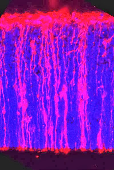
This image shows stem cells in the cortex of a
mouse embryo (cell nuclei: blue). Credit: MPI
für Molecular Cell Biology and Genetics/

D. Stenzel. 
"Our study highlights this relationship and provides a potential explanation for the condition neurologists refer to as cretinism", says Wieland Huttner, Director at the Max Planck Institute in Dresden. This neurological disorder severely impairs the mental abilities of a person.

In the course of evolution, certain mammals, notably humans, have developed larger brains than others, and therefore more advanced cognitive abilities. Mice, for example, have brains that are around a thousand times smaller than the human one. In their study, which was conducted in cooperation with the Fritz Lipmann Institute in Jena, the researchers in Dresden wanted to identify factors that determine brain development, and understand how larger brains have evolved.

A cosy bed for brain stem cells
Brain neurons are generated from stem cells called basal progenitors that are able to proliferate in humans, but not in mice. In humans, basal progenitors are surrounded by a special environment, a so-called extracellular matrix (ECM), which is produced by the progenitors themselves. Like a cosy bed, it accommodates the proliferating cells. Mice lack such ECM, which means that they generate fewer neurons and have a smaller brain.

The scientists therefore conducted tests to see whether in mice, basal progenitors start to proliferate if a comparable cell environment is simulated.

"We simulated an extracellular matrix for the brain stem cells using a stimulating antibody. This antibody activates an integrin on the cell surface of basal progenitors and thus stimulates their proliferation", explains Denise Stenzel, who headed the experiments.

Because a requirement of thyroid hormones for proper brain development was previously known, the researchers blocked the production of these hormones in pregnant rats to see if their absence would inhibit basal progenitor proliferation in the embryos. Indeed, fewer progenitors and, consequently, neurons were produced, likely explaining the abnormal brain development in the absence of thyroid hormones. When the action of these hormones on the integrin was blocked, the ECM-simulating antibody alone was no longer able to induce basal progenitor proliferation.

A combination of ECM and thyroid hormones thus appears necessary for basal progenitors to proliferate and produce enough neurons for brain development. Human brain stem cells produce the suitable environment naturally.

"That is probably how, in the course of evolution, we humans developed larger brains", says Wieland Huttner, summing up the study.

The research produced another important finding too.

"We were able to explain the role of iodine in embryonic brain development at the cellular level", says Denise Stenzel. Iodine is essential for the production of thyroid hormones, and an iodine deficiency in pregnant women is known to have adverse effects on the brain development of the unborn child.

Contact: Dr. Wieland B. Huttner

Reference:
Integrin αvβ3 and thyroid hormones promote expansion of progenitors in embryonic neocortex 
Stenzel, Denise; Wilsch-Bräuninger, Michaela; Wong, Fong Kuan; Heuer, Heike; Huttner, Wieland B. 
 Development 2014 141:795-806; doi: 10.1242/dev.101907
.........


For more on stem cells and cloning, go to CellNEWS at

Tuesday, 7 January 2014

Stem Cells on the Road to Specialization

Stem Cells on the Road to Specialization
Tuesday, 07 January 2014

Extracellular matrix. 
Scientists at the University of Copenhagen have gained new insight into how both early embryonic cells and embryonic stem cells are directed into becoming specialised cell types, like pancreatic and liver cells. The results have just been published in the scientific journal eLife.

This latest research from the Danish Stem Cell Center (Danstem) at the University of Copenhagen, helps identify how stem cells create so called pathways and roads supporting their own specialization. This understanding is an important step towards stem cell-based cell therapies for conditions like diabetes and liver diseases.

"The new insight that we have gained into the impact of the physical environment on cell development is highly valuable," says Professor Joshua Brickman from DanStem,

"It enables us to create the optimal physical environment in the laboratory for stem cells and progenitor cells to develop into specific, mature cells."

On the road
Developing cells constantly move and while moving around, they organise and build a physical environment very much like a small city with pathways and roads. The new research published in the scientific journal eLife shows two important things. Firstly the embryonic cells receive signals from other cells that actually instruct them in how to organise and build the road leading the cells towards early stages of pancreas and liver cells.

Professor Brickman and his team also found that they could isolate these roads from the developing stem cells and literally freeze them. The saved roads were then used in a separate experiment which showed that in the absence of an important cell signal, the road alone can be used to improve the cells' development and differentiation towards mature cells.

"Apart from gaining new important insight into cell development, our work also suggests that some of the current approaches to human embryonic stem cells specialization towards both pancreatic and liver cells may not have been effective, because the important role of these roads, the so called extra-cellular matrix, was ignored," says Joshua Brickman.

Contact: Joshua Brickmann

Reference:
PI3K/Akt1 signalling specifies foregut precursors by generating regionalized extra-cellular matrix
S Nahuel Villegas, Michaela Rothová, Martin E Barrios-Llerena, Maria Pulina, Anna-Katerina Hadjantonakis, Thierry Le Bihan, Sophie Astrof, Joshua M Brickman
eLife 2013;2:e00806, DOI: http://dx.doi.org/10.7554/eLife.00806
.........


For more on stem cells and cloning, go to CellNEWS at