Friday, 18 September 2009

Memories of the Way They Used to Be

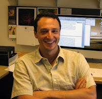
Human iPS Cells Retain Some Gene Expression of Donor Cells Friday, 18 September 2009 A mosaic of human iPS cells generated by A team of researchers from the University of California, San Diego School of Medicine and the Salk Institute for Biological Studies in La Jolla have developed a safe strategy for reprogramming cells to a pluripotent state without use of viral vectors or genomic insertions. Their studies reveal that these induced pluripotent stem cells (iPSCs) are very similar to human embryonic stem cells, yet maintain a “transcriptional signature.” In essence, these cells retain some memory of the donor cells they once were. The study, led by UCSD Stem Cell Program researcher Alysson R. Muotri, PhD, assistant professor in the Departments of Pediatrics at UCSD and Rady Children’s Hospital and UCSD’s Department of Cellular and Molecular Medicine, will be published online in PLoS ONE on September 17. Alysson R. Muotri, PhD. Credit: UC. San Diego Medical Center.“Working with neural stem cells, we discovered that a single factor can be used to re-program a human cell into a pluripotent state, one with the ability to differentiate into any type of cell in the body” said Muotri. Traditionally, a combination of four factors was used to create iPSCs, in a technology using viral vectors – viruses with the potential to affect the transcriptional profile of cells, sometimes inducing cell death or tumours. In addition, while both mouse and human iPSCs have been shown to be similar to embryonic stem cells in terms of cell behaviour, gene expression and their potential to differentiate into different types of cells, researchers had not achieved a comprehensive analysis to compare iPSCs and embryonic stem cells. “One reason is that previous methodologies used to derive iPSCs weren’t ‘footprint free,’” Muotri explained. “Viruses could integrate into the genome of the cell, possibly affecting or disrupting genes.” "In order to take full advantage of reprogramming, it is essential to develop methods to induce pluripotency in the absence of permanent changes in the genome," added Fred H. Gage, PhD, a professor in the Laboratory for Genetics at the Salk Institute and the Vi and John Adler Chair for Research on Age-Related Neurodegenerative Diseases. By creating iPSCs from human neural stem cells without the use of viruses, the scientists learned something new. While the genetic transcriptional profile of the new iPSCs was closer to that of embryonic stem cells than to human neural stem cells, the iPSCs still carried a transcriptional “signature” of the original neural cell. “While most of the original genetic memory was erased when the cells were reprogrammed, some were retained,” said Muotri. He added that, in the past, it wasn’t known if this was caused by the use of viral vectors. “By using a footprint-free methodology, we have shown a safe way to generate human iPSCs for clinical purposes and basic research. We’ve also raised an interesting question about what, if any, effect the ‘memory retention’ of these cells might have.” The research was supported by start-up funds from the UCSD Stem Cell Research Program, and by grants from the California Institute of Regenerative Medicine and The Lookout Fund Foundation. Reference: Transcriptional Signature and Memory Retention of Human-Induced Pluripotent Stem Cells Maria C. N. Marchetto, Gene W. Yeo, Osamu Kainohana, Martin Marsala, Fred H. Gage, Alysson R. Muotri PLoS ONE 4(9): e7076. doi:10.1371/journal.pone.0007076 ......... ZenMaster

For more on stem cells and cloning, go to CellNEWS at http://cellnews-blog.blogspot.com/ and http://www.geocities.com/giantfideli/index.html

Thursday, 17 September 2009

Rare Genetic Disease Successfully Reversed using Stem Cell Transplantation

Scripps Research scientists correct gene defect in mice that causes lethal symptoms in children Thursday, 17 September 2009 A recent study by Scripps Research Institute scientists offers good news for families of children afflicted with the rare genetic disorder, cystinosis. In research that holds out hope for one day developing a potential therapy to treat the fatal disorder, the study shows that the genetic defect in mice can be corrected with stem cell transplantation. "After meeting the children who suffer from this disease, like an 18-year-old who has already had three kidney transplants, and the families who are desperately searching for help, our team is committed to moving toward a cure for cystinosis, a lysosomal storage disorder," says principal investigator Stephanie Cherqui, assistant professor in the Department of Molecular and Experimental Medicine. "This study is an important step toward that goal." In the study, which is published in the September 17, 2009 print edition of the journal Blood, the Scripps Research team used bone marrow stem cell transplantation to address symptoms of cystinosis in a mouse model. The procedure virtually halted the cystine accumulation responsible for the disease and the cascade of cell death that follows. Cystine is a by-product of the break down of cellular components the body no longer needs in the cell's "housekeeping" organelles, called lysosomes. Normally, cystine is shunted out of cells, but in cystinosis a gene defect of the lysosomal cystine transporter causes it to build up, forming crystals that are especially damaging to the kidneys and eyes. A Rare But Devastating Disease While cystinosis is rare — affecting an estimated 500 people in the United States and 2,000 worldwide — it is devastating. Three types of cystinosis have been described based on the age at diagnosis and the amount of cystine in cells: infantile onset, adolescent onset, and adult onset. Children as young as six months can begin to suffer renal dysfunction, which grows progressively worse with time. Other symptoms include diabetes, muscular disease, neurological dysfunction, and retinopathy. Infantile onset is the most common, as well as the most severe, form of the disease. The only available drug to treat cystinosis, cysteamine, while slowing the progression of kidney degradation, does not prevent it, and end-stage kidney failure is inevitable. "Cysteamine must be given every six hours, so children have to be woken up each night to take this drug, which has unpleasant side effects, and many others to treat various symptoms," Cherqui says. "So although there is treatment, it is difficult treatment that does not cure the disease." "Surprised and Encouraged" In the new study, the researchers found that transplanted bone marrow stem cells carrying the normal lysosomal cystine transporter gene abundantly engrafted into every tissue of the experimental mice. This led to an average drop in cystine levels of about 80 percent in every organ. In addition to preventing kidney dysfunction, there was less deposition of cystine crystals in the cornea, less bone demineralization, and an improvement in motor function. "The results really surprised and encouraged us," says Cherqui, who as a doctoral student in France in 1998 helped discover the gene involved in cystinosis. "Because the defect is present in every cell of the body, we did not expect a bone marrow stem cell transplant to be so widespread and effective." Cherqui, who generated the mouse model in 2000 that is currently used to study cystinosis, says that adult bone marrow stem cell therapy is particularly well suited as a potential treatment for cystinosis because these cells target all types of tissues. In addition, stem cells reside in the bone marrow for the duration of a patient's life, becoming active as needed, a particular benefit for a progressive disease like cystinosis. The work of Cherqui and her colleagues may have wider applications for other genetic diseases, providing proof of principle that adult stem cell transplants may be successful in humans for genetic diseases with systemic defects, especially those of a progressive nature. Cherqui expects to spend the next several years analyzing the safety of genetically modified autologous (obtained from the same individual) bone marrow transplants in the cystinosis mouse and other models before moving on to human clinical trials. This work was funded by the Cystinosis Research Foundation. Reference: Successful treatment of the murine model of cystinosis using bone marrow cell transplantation Kimberly Syres, Frank Harrison, Matthew Tadlock, James V. Jester, Jennifer Simpson, Subhojit Roy, Daniel R. Salomon, and Stephanie Cherqui Blood 2009 114: 2542-2552, DOI 10.1182/blood-2009-03-213934 ......... ZenMaster


For more on stem cells and cloning, go to CellNEWS at http://cellnews-blog.blogspot.com/ and http://www.geocities.com/giantfideli/index.html

How microRNAs Drive Tumour Progression

How microRNAs Drive Tumour Progression Thursday, 17 September 2009 UCSF researchers have identified collections of tiny molecules known as microRNAs that affect distinct processes critical for the progression of cancer. The findings, they say, expand researchers' understanding of the important regulatory function of microRNAs in tumour biology and point to new directions for future study and potential treatments. The researchers refer to these microRNA collections as signatures, and their study results are reported in the September 15 issue of "Genes & Development.'' The study was led by the laboratory of Douglas Hanahan, PhD, an American Cancer Society Research Professor in the Department of Biochemistry and Biophysics at UCSF. Approximately five percent of all known human genes encode, or produce, microRNAs, yet scientists are only now — nearly a decade after their discovery — beginning to unlock the mystery of their functions. MicroRNAs are snippets of single-stranded RNAs that prevent a gene's code from being translated from messenger RNA into proteins, which are essential for cell growth and development. Produced in the nucleus and released into the cytoplasm, they home in on messenger RNAs that possess a stretch that is complementary to their genetic sequence. When they locate them, they latch on, preventing the messenger RNA from being processed by the protein-making machines known as ribosomes. As such, microRNAs are able to ratchet down a cell's production of a given protein. Over the last several years, several groups have identified hundreds of microRNAs that are deregulated between normal tissue and tumours, however researchers only understand what a handful of these powerful regulators are doing to drive tumour formation. "Virtually all cancers acquire approximately six distinct capabilities en route to tumour formation," said lead author Peter Olson, PhD, a postdoctoral fellow in the Diabetes Center and Helen Diller Family Comprehensive Cancer Center at UCSF. "When a cancer researcher observes a gene or microRNA go awry, it can be challenging to understand how that microRNA impacts tumourigenesis." To home in on the question, the authors turned to a mouse model of pancreatic neuroendocrine tumours in which lesions go through discrete stages before culminating in invasive and metastatic carcinomas. In the three-year microRNA study, they found that cells in the mouse model developed and functioned normally but started to replicate uncontrollably at five weeks. Several weeks later, some pancreatic islets had become angiogenic (forming new blood vessels) — a step in the journey from a dormant state to a malignant state — though had not yet formed a tumour. By 10 weeks, a subset of angiogenic lesions had progressed to the tumour stage, and by week 16, a small percentage of mice had developed liver metastasis. "This represents the spectrum of stages that we think are important for all tumours, including human disease," said Olson. By measuring the expression level of all known microRNA in pre-tumour stages, tumours and metastases, the authors were able to associate deregulated microRNAs with processes such as hyper-proliferation, angiogenesis and metastasis. Focusing on the metastatic signature, researchers found — in one of the most striking observations of the project — that tumours bore a startlingly divergent microRNA expression pattern compared to primary tumours. Moreover, a subset of primary tumours showed more similarity to metastases than to other primary tumours. "If you can identify tumours that have an increased propensity to metastasize, then it would have a very important clinical application," said Olson. "A lively debate in metastatic research has centred around whether primary tumour cells must suffer an additional mutation that endows that cell with a metastatic capability, or whether certain mutational combinations that are responsible for primary tumour formation also significantly increase the propensity of that cell to metastasize. These data provide evidence for the latter.'' Reference: MicroRNA dynamics in the stages of tumorigenesis correlate with hallmark capabilities of cancer Peter Olson, Jun Lu, Hao Zhang, Anny Shai, Matthew G. Chun, Yucheng Wang, Steven K. Libutti, Eric K. Nakakura, Todd R. Golub, and Douglas Hanahan Genes Dev. September 15, 2009 23: 2152-2165; doi:10.1101/gad.1820109 ......... ZenMaster


For more on stem cells and cloning, go to CellNEWS at http://cellnews-blog.blogspot.com/ and http://www.geocities.com/giantfideli/index.html

Wednesday, 16 September 2009

Cure for Colour Blindness in Monkeys

Gene therapy used to treat adult vision disorders involving cone cells Wednesday, 16 September 2009 Researchers from the University of Washington and the University of Florida used gene therapy to cure two squirrel monkeys of colour blindness — the most common genetic disorder in people. Writing online Wednesday in the journal Nature, scientists cast a rosy light on the potential for gene therapy to treat adult vision disorders involving cone cells — the most important cells for vision in people. "We've added red sensitivity to cone cells in animals that are born with a condition that is exactly like human colour blindness," said William W. Hauswirth, Ph.D., a professor of ophthalmic molecular genetics at the UF College of Medicine and a member of the UF Genetics Institute and the Powell Gene Therapy Center. "Although colour blindness is only moderately life-altering, we've shown we can cure a cone disease in a primate, and that it can be done very safely. That's extremely encouraging for the development of therapies for human cone diseases that really are blinding." The finding is also likely to intrigue millions of people around the world who are colour-blind, including about 3.5 million people in the United States, more than 13 million in India and more than 16 million in China. The problem mostly affects men, leaving about 8 percent of Caucasian men in the United States incapable of discerning red and green hues that are important for everyday things like recognizing traffic lights. "People who are colour-blind feel that they are missing out," said Jay Neitz, Ph.D., a professor of ophthalmology at the University of Washington. "If we could find a way to do this with complete safety in human eyes, as we did with monkeys, I think there would be a lot of people who would want it. Beyond that, we hope this technology will be useful in correcting lots of different vision disorders." Here is one of the squirrel monkeys, Dalton, who was treated for red-green colour blindness enjoying a feast of coloured fruits and vegetables. The image on the left was digitally altered to simulate what the scene would look like to a person (or monkey) with red-green colour blindness. Credit: Neitz Lab, Washington University.The discovery comes about 10 years after Neitz and his wife Maureen Neitz, Ph.D., a professor of ophthalmology at the University of Washington and senior author of the study, began training two squirrel monkeys named Dalton and Sam. In addition to teaching the animals, the Neitz research group worked with the makers of a standard vision-testing technique called the Cambridge Colour Test to perfect a way the monkeys could "tell" them which colours they were seeing. The tests are similar to ones given to elementary children the world over, in which students are asked to identify a specific pattern of coloured dots among a field of dots that vary in size, colour and intensity. The researchers devised a computer touch screen the monkeys could use to trace the colour patterns. When the animals chose correctly, they received a reward of grape juice. Likewise, decades were spent by Hauswirth and colleagues at the University of Florida, to develop the gene-transfer technique that uses a harmless adeno-associated virus to deliver corrective genes to produce a desired protein. In this case, researchers wanted to produce a substance called long-wavelength opsin in the retinas of the monkeys. This particular form of opsin is a colourless protein that works in the retina to make pigments that are sensitive to red and green. "We used human DNA’s, so we won't have to switch to human genes as we move toward clinical treatments," said Hauswirth, who is also involved in a clinical trial with human patients to test gene therapy for the treatment of Leber congenital amaurosis, a form of blindness that strikes children. About five weeks after the treatment, the monkeys began to acquire colour vision, almost as if it occurred overnight. "Nothing happened for the first 20 weeks," Neitz said. "But we knew right away when it began to work. It was if they woke up and saw these new colours. The treated animals unquestionably responded to colours that had been invisible to them." It took more than a year and a half to test the monkeys' ability to discern 16 hues, with some of the hues varying as much as 11-fold in intensity. Dalton is named for John Dalton, an English chemist who realized he was colour-blind and published the first paper about the condition in 1798. "We've had Dalton and Sam for 10 years. They are like our children," Neitz said. "This species are friendly, docile monkeys that we just love. We think it is useful to continue to follow them — it's been two years now that they've been seeing in colour, and continuing to check their vision and allowing them to play with the computer is part of their enrichment." With the discovery, the researchers are the first to address a vision disorder in primates in which all photoreceptors is intact and healthy, providing a hint of gene therapy's full potential to restore vision. About 1 in 30,000 Americans have a hereditary form of blindness called achromatopsia, which causes nearly complete colour blindness and extremely poor central vision. "Those patients would be targets for almost exactly the same treatment," Hauswirth said. Even in common types of blindness such as age-related macular degeneration and diabetic retinopathy, vision could potentially be rescued by targeting cone cells, he said. "The major thrust of the study is you can ameliorate if not cure colour blindness with gene therapy," said Gerald H. Jacobs, Ph.D., a research professor of psychology at the University of California, Santa Barbara, who was not involved in the research. "There are still questions about safety, but in these monkeys at least, there were no untoward effects. Those who are motivated to ameliorate their colour defect might take some hope from the findings.” "This is also another example of how utterly plastic the visual system is to change," Jacobs said. "The nervous system can extract information from alterations to photo-pigments and make use of it almost instantaneously." Reference: Gene therapy for red–green colour blindness in adult primates Katherine Mancuso, William W. Hauswirth, Qiuhong Li, Thomas B. Connor, James A. Kuchenbecker, Matthew C. Mauck, Jay Neitz & Maureen Neitz Nature advance online publication 16 September 2009, doi:10.1038/nature08401 ......... ZenMaster


For more on stem cells and cloning, go to CellNEWS at http://cellnews-blog.blogspot.com/ and http://www.geocities.com/giantfideli/index.html

Tuesday, 15 September 2009

Benefits and Limits of Personal Genetics

Dartmouth researchers get personal with genetics Tuesday, 15 September 2009 Two recent studies by Dartmouth College researchers use individual genetic data to reveal the powers and limits of our current understanding of how the genome influences human health and what genes can reveal about the ancestry of the people of New Hampshire. Published in the Sept. 11 issue of the American Journal of Human Genetics, Dartmouth Professor Jason Moore and Vanderbilt Professor Scott Williams analyzed how personal genetic testing companies are using still-nascent genome data to judge the health of their customers. People can now buy inexpensive kits, submit a DNA sample (often a swab from the inside of a cheek or a little bit of saliva), and receive data about their susceptibility to a number of gene-influenced ailments, such as prostate cancer, Alzheimer's, or type II diabetes. Moore and Williams argue that our knowledge of the human genome and its relationship to human health, while growing by leaps and bounds, is still in its infancy. "The relationship between health and genetics is very complex," says Moore, professor of genetics and of community and family medicine at Dartmouth Medical School (DMS). "It's often a combination of multiple genes and multiple environmental factors that work together to increase or decrease your risk of disease. I don't think the knowledge base is sufficient to put genetics in the hands of the public quite yet." Moore is also the Frank Lane Research Scholar in Computational Genetics and Director of Bioinformatics at DMS The authors admit that genetic research is progressing, and they cite the example of the discovery of the BRCA1 and BRCA2 genes and their role in breast cancer. However, the authors caution that, while there is no question these genes are involved in breast cancer, the underlying mechanisms behind the genetic risk are still being worked out. "There is a perception that these tests can provide answers," says Moore. "I used my own genetic material for this study, and my results didn't really tell me anything I didn't know, based on family history." Moore and Williams call for refocusing and stepping up the research on gene-to-gene and gene-to-environment interactions. They explain that for many years, researchers have focused on single genes and clinical endpoints. The time has come, they say, to embrace rather than ignore the complexity of human traits as they are expressed by the whole genome working in concert. "Although genetic testing for common human diseases is not yet useful, using genetic testing results to reveal an individual's ancestry is increasingly reliable," says Moore. He and PhD candidate Chantel Sloan recently mined some genetic data for a study that examined the population structure of New Hampshire residents. Published in the September issue of PLoS ONE (a journal of the Public Library of Science), they study by Sloan and Moore and their colleagues analyzed more than 1,000 genetic markers from 864 people in New Hampshire. They discovered six subgroups of people with distinct genetic backgrounds including a group of Finnish and Russian/Polish/Lithuanian ancestry. "I knew that people would be primarily European," says Sloan. "What I didn't expect was the strong connection between genetic structure and people of Eastern European ancestry, which I learned is consistent with New Hampshire census and immigration data from 1870 to 1930." Sloan used data initially compiled for a cancer study, so the genetic markers were cancer susceptibility genes rather than known ancestral genes, and the population being analyzed was not racially or geographically distinct. The results challenge the assumption that large numbers of special genetic markers are needed to discover genetically distinct groups of people. "This is an example of how personal genetic data can be used to help inform people of their ancestry," says Moore. "Informing people of their future health is still out of reach, though." References: Epistasis and Its Implications for Personal Genetics Jason H. Moore, Scott M. Williams AJHG, Volume 85, Issue 3, 309-320, 11 September 2009, doi:10.1016/j.ajhg.2009.08.006 Genetic Population Structure Analysis in New Hampshire Reveals Eastern European Ancestry Chantel D. Sloan, Angeline D. Andrew, Eric J. Duell, Scott M. Williams, Margaret R. Karagas, Jason H. Moore PLoS ONE 4(9): e6928. doi:10.1371/journal.pone.0006928 ......... ZenMaster


For more on stem cells and cloning, go to CellNEWS at http://cellnews-blog.blogspot.com/ and http://www.geocities.com/giantfideli/index.html

Sunday, 13 September 2009

How Stem Cells Make Skin

EMBL scientists come a step closer to understanding skin, breast and other cancers. Sunday, 13 September 2009 Stem cells have a unique ability: when they divide, they can either give rise to more stem cells, or to a variety of specialised cell types. In both mice and humans, a layer of cells at the base of the skin contains stem cells that can develop into the specialised cells in the layers above. Scientists at the European Molecular Biology Laboratory (EMBL) in Monterotondo, in collaboration with colleagues at the Centro de Investigaciones Energéticas, Medioambientales y Tecnologicas (CIEMAT) in Madrid, have discovered two proteins that control when and how these stem cells switch to being skin cells. The findings, published online today in Nature Cell Biology, shed light on the basic mechanisms involved not only in formation of skin, but also on skin cancer and other epithelial cancers. At some point in their lives, the stem cells at the base of the skin stop proliferating and start differentiating into the cells that form the skin itself. To do so, they must turn off the 'stem cell programme' in their genes and turn on the 'skin cell programme'. Researchers suspected that a family of proteins called C/EBPs might be involved in this process, as they were known to regulate it in other types of stem cell, but had so far failed to identify which C/EBP protein controlled the switch in skin. Claus Nerlov and his group at EMBL Monterotondo discovered it was not one protein, but two: C/EBPα and C/EBPβ.


Skin cell differentiation. In normal skin (left), the stem cells at the base, shown in green, differentiate into skin cells, shown in red. In mice whose skin has neither C/EBP-α nor C/EBP-β (middle), this differentiation is blocked: green-labelled stem cells appear in upper layers of skin, and there are no differentiated skin cells (no red staining). This also happens at the initial stages of basal cell carcinomas. In skin where C/EBP-β is present but has lost its capacity to interact with E2F, a molecule that regulates the cell cycle (right), skin cells start differentiating abnormally, before they have properly exited the stem cell "program" (yellow/orange). This is similar to what is observed in the initial stages of squamous cell carcinomas, a more aggressive and invasive skin tumour. Credit: Claus Nerlov/EMBL.
The EMBL researchers used genetic engineering techniques to delete the genes that encode C/EBPα and β specifically in the skin of mouse embryos, and found that without these proteins the skin of the mice did not form properly. "Mice with neither C/EBPα nor β had taut and shiny skin that couldn't keep the water inside their bodies, they lacked many of the proteins that make skin mechanically strong and water tight, and they died of de-hydration shortly after birth," Nerlov explains. However, a single working copy of either the gene for C/EBPα or the gene for C/EBPβ was enough to ensure that skin developed properly. This means that the two proteins normally do the same job in the skin's stem cells - an unexpected redundancy, which may have arisen because there are so many stem cells in skin that a tight control on proliferation is needed to avoid problems like cancer. Or it may simply be a by-product of the fact that these two proteins have different functions in other situations, such as wound healing or repair of sunlight-induced skin damage. One of the hallmarks of epithelial cancers - which include skin, breast, and oral cancers - is that they have genes turned on which would normally only be expressed in embryonic stem cells, and which may help cancer cells divide indefinitely. Such genes become re-expressed in the skin in the absence of C/EBPs. Therefore, by understanding how C/EBPα and β turn off such 'stem cell' programmes, researchers hope to come a step closer to finding ways to fight such cancers. When Nerlov and colleagues looked at how C/EBPα and -β work in the skin, they found that these proteins also regulate a number of other molecules that control skin development. Several important pathways known to control skin and hair formation were improperly activated in the mice lacking C/EBPα and -β. "This is a very important discovery", says Nerlov. "It opens up a lot of new areas, because we can see how these proteins control virtually every other molecule known to regulate skin cell differentiation. It seems to be a key piece in the puzzle of how our skin is formed and maintained throughout life." Reference: C/EBPα and β couple interfollicular keratinocyte proliferation arrest to commitment and terminal differentiation Rodolphe G. Lopez, Susana Garcia-Silva, Susan J. Moore, Oksana Bereshchenko, Ana B. Martinez-Cruz, Olga Ermakova, Elke Kurz, Jesus M. Paramio & Claus Nerlov Nature Cell Biology Published online: 13 September 2009, doi:10.1038/ncb1960 ......... ZenMaster
For more on stem cells and cloning, go to CellNEWS at http://cellnews-blog.blogspot.com/ and http://www.geocities.com/giantfideli/index.html

Saturday, 12 September 2009

MicroRNAs Are Themselves Regulated

RNAs taking centre stage Saturday, 12 September 2009 In an article published in Nature, scientists at the Friedrich Miescher Institute for Biomedical Research (part of the Novartis Research Foundation) have shown that short strands of ribonucleic acid (RNA) which regulate protein production are themselves also regulated. This additional layer of regulation opens up new perspectives for therapeutic approaches. RNAs, serving as a mere intermediary between DNA and proteins, were long regarded as a poor relation by researchers, attracting little interest. However, following the discovery of small RNAs known as microRNAs, they have increasingly been moving into the limelight. MicroRNAs bind to messenger RNA (mRNA), thereby regulating the translation of genes into proteins. Recently, various studies have shown that the production of microRNAs is tightly controlled, but their subsequent fate was not clear. It was assumed that mature microRNAs remained stable in the cell for days, and that their possible functions were therefore restricted: a microRNA persisting for a relatively long period cannot be involved in any processes in the cell requiring rapid adaptation. Regulated regulators The study carried out by Helge Grosshans, a Research Group Leader at the Friedrich Miescher Institute, has now finally shifted attention away from DNA, spotlighting the key role played by microRNAs in the theatre of cellular processes. As Grosshans and his team report in the current issue of the renowned journal Nature, they discovered a mechanism for active degradation of microRNAs and showed that this mechanism is itself regulated. Explaining his findings, Grosshans says: "What was formerly conceived of as a direct, straightforward pathway is gradually turning out to be a dense network of regulatory mechanisms: genes are not simply translated into proteins via mRNA. MicroRNAs control the translation of mRNAs into proteins, and proteins in turn regulate the microRNAs at various levels." In addition, the FMI researchers showed in the nematode Caenorhabditis elegans that, via regulation of degradation, it is possible to influence microRNA activity. This means that microRNAs may, after all, be involved in the regulation of rapidly occurring processes.

The meteoric rise of microRNAs MicroRNAs are short, single-stranded RNA molecules which interact with mRNAs in a sequence-dependent manner. They thus inhibit translation of mRNAs into proteins. MicroRNAs were first described in 1993 in the nematode Caenorhabditis elegans. They were subsequently also shown to play an important role in regulating development processes and in pathogenesis in higher organisms. The findings of recent years and now also Helge Grosshans's study have shifted attention away from DNA toward RNAs, which are taking centre stage. The term "microRNA" was only introduced in 2001.
Targeted degradation of disease-causing RNAs But the findings are also relevant in another respect. As microRNAs have been implicated in the development of diseases, efforts to date have focused on replacing disease-causing microRNAs with other microRNAs, or inactivating them with the aid of complementary RNA strands. Unfortunately, it is extremely difficult to deliver RNAs to target cells for therapeutic purposes. Accordingly, the prospects of success for these novel treatment approaches have been uncertain. In his study, however, Grosshans identified a protein that specifically degrades microRNAs. If it now proves possible to specifically activate or inhibit this protein and its partners that could provide an approach, which is closer to classical and well-established forms of therapy. "We now assume that a large number of human genes are regulated by microRNAs, so the regulatory mechanism we've discovered has a great potential to significantly influence numerous processes in human cells," Grosshans comments. About the FMI The Friedrich Miescher Institute for Biomedical Research (FMI), based in Basel, Switzerland, is a world-class centre for basic research in life sciences. It was founded in 1970 as a joint effort of two Basel-based pharmaceutical companies and is now part of the Novartis Research Foundation. The FMI is devoted to the pursuit of fundamental biomedical research. Areas of expertise are neurobiology, growth control, which includes signalling pathways, and the epigenetics of stem cell development and cell differentiation. The institute counts 320 collaborators. The FMI also offers training in biomedical research to PhD students and postdoctoral fellows from around the world. In addition, the FMI is affiliated with the University of Basel. The Director of the FMI since 2004 is Prof. Susan Gasser. Reference: Active turnover modulates mature microRNA activity in C. elegans Chatterjee S & H. Grosshans Nature, 24 August 2009, doi:10.1038/nature08349 ......... ZenMaster
For more on stem cells and cloning, go to CellNEWS at http://cellnews-blog.blogspot.com/ and http://www.geocities.com/giantfideli/index.html

Friday, 11 September 2009

See CIRMTV's Channel on YouTube

California's stem cell research funding agency is using YouTube to tell its story. Friday, 11 September 2009 The California Institute for Regenerative Medicine's CIRMTV tells the story of stem cell research through short videos about the work of researchers and doctors and — most compellingly — patients. The 22 videos on the site range from Stanford University stem cell research director Irv Weissman talking about the differences between adult and embryonic stem cells to, most recently, an update about the progress and promise in Parkinson's disease treatments. See CIRMTV's Channel: YouTube - CIRMTV's Channel ......... ZenMaster


For more on stem cells and cloning, go to CellNEWS at http://cellnews-blog.blogspot.com/ and http://www.geocities.com/giantfideli/index.html

Tuesday, 8 September 2009

'Liposuction Leftovers' Easily Converted to iPS Cells

'Liposuction Leftovers' Easily Converted to iPS Cells Tuesday, 08 September 2009 Globs of human fat removed during liposuction conceal versatile cells that are more quickly and easily coaxed to become induced pluripotent stem cells, or iPS cells, than are the skin cells most often used by researchers, according to a new study from Stanford's School of Medicine. "We've identified a great natural resource," said Stanford surgery professor and co-author of the research, Michael Longaker, MD, who has called the readily available liposuction leftovers "liquid gold." Reprogramming adult cells to function like embryonic stem cells is one way researchers hope to create patient-specific cell lines to regenerate tissue or to study specific diseases in the laboratory. "Thirty to 40 percent of adults in this country are obese," agreed cardiologist Joseph Wu, MD, PhD, the paper's senior author. "Not only can we start with a lot of cells, we can reprogram them much more efficiently. Fibroblasts, or skin cells, must be grown in the lab for three weeks or more before they can be reprogrammed. But these stem cells from fat are ready to go right away." The fact that the cells can also be converted without the need for mouse-derived "feeder cells" may make them an ideal starting material for human therapies. Feeder cells are often used when growing human skin cells outside the body, but physicians worry that cross-species contamination could make them unsuitable for human use. The findings will be published online Sept. 7 in the Proceedings of the National Academy of Sciences. Longaker is the deputy director of Stanford’s Stem Cell Biology and Regenerative Medicine Institute and director of children’s surgical research at Lucile Packard Children’s Hospital. Wu is an assistant professor of cardiology and radiology, and a member of Stanford’s Cardiovascular Institute. Even those of us who are not obese would probably be happy to part with a couple of pounds (or more) of flab. Nestled within this unwanted latticework of fat cells and collagen are multipotent cells called adipose, or fat, stem cells. Unlike highly specialized skin-cell fibroblasts, these cells in the fat have a relatively wide portfolio of differentiation options — becoming fat, bone or muscle as needed. It's this pre-existing flexibility, the researchers believe, that gives these cell an edge over the skin cells. "These cells are not as far along on the differentiation pathway, so they're easier to back up to an earlier state," said first author and postdoctoral scholar Ning Sun, PhD, who conducted the research in both Longaker's and Wu's laboratories. "They are more embryonic-like than fibroblasts, which take more effort to reprogram." These reprogrammed iPS cells are usually created by expressing four genes, called Yamanaka factors, normally unexpressed (or expressed at very low levels) in adult cells. Sun found that the fat stem cells actually express higher starting levels of two of the four reprogramming genes than do adult skin cells — suggesting that these cells are already primed for change. When he added all four genes, about 0.01 percent of the skin-cell fibroblasts eventually became iPS cells but about 0.2 percent of the fat stem cells did so — a 20-fold improvement in efficiency. The new iPS cells passed the standard tests for pluripotency: They formed tumours called teratomas when injected into immunocompromised mice, and they could differentiate into cells from the three main tissue types in the body, including neurons, muscle and gut epithelium. The researchers are now investigating whether the gene expression profiles of the fat stem cells could be used to identify a subpopulation that could be reprogrammed even more efficiently. "The idea of reprogramming a cell from your body to become anything your body needs is very exciting," said Longaker, who emphasized that the work involved not just a collaboration between his lab and Wu's, but also between the two Stanford institutes. "The field now needs to move forward in ways that the Food and Drug Administration would approve — with cells that can be efficiently reprogrammed without the risk of cross-species contamination — and Stanford is an ideal place for that to happen." "Imagine if we could isolate fat cells from a patient with some type of congenital cardiac disease," said Wu. "We could then differentiate them into cardiac cells, study how they respond to different drugs or stimuli and see how they compare to normal cells. This would be a great advance." ......... ZenMaster


For more on stem cells and cloning, go to CellNEWS at http://cellnews-blog.blogspot.com/ and http://www.geocities.com/giantfideli/index.html

Friday, 4 September 2009

Transplanted Human Stem Cells Prolong Survival in Mouse Model of Batten Disease

Transplanted Human Stem Cells Prolong Survival in Mouse Model of Batten Disease Friday, 04 September 2009 A new study finds substantial improvement in a mouse model of a rare, hereditary neurodegenerative disease after transplantation of normal human neural stem cells. The research findings, published by Cell Press in the September 4th issue of the journal Cell Stem Cell, show that the transplanted cells provided a critical enzyme that was missing in the brains of the experimental mice and represent an important step toward what may be a successful therapeutic approach for a currently untreatable and devastating disease. Infantile neuronal ceroid lipofuscinosis (INCL), commonly known as Batten disease, is a fatal neurodegenerative disease in children. It is caused by a mutation in the gene that makes a crucial enzyme called palmitoyl protein thioesterase-1 (PPT1). A deficiency of PPT1 in the brain causes the abnormal accumulation of a cellular lipid storage material called lipofuscin, which leads to neuron death, a decline in cognitive and motor skills, visual impairment, seizures and premature death. Unfortunately, intravenous enzyme replacement therapy is not a viable treatment approach as it is nearly impossible to get the PPT1 enzyme into the brain. Although there is currently no effective treatment for INCL, it has been hypothesized that transplanted donor cells might be able to secrete the needed enzyme directly into the host brain. A mouse model of INCL that mimics many aspects of the human disease has been developed and provides an excellent experimental model for testing whether a human neural stem cell transplant may be a beneficial disease treatment. Dr. Nobuko Uchida from StemCells, Inc., in Palo Alto, California led a study that tested this hypothesis with banked human neural stem cells that had been purified, expanded, and preserved. "We took a novel approach and transplanted normal, non-tumourigenic, and non-genetically modified human neural stem cells to deliver the deficient enzyme in the mouse model of INCL," explains Dr. Uchida. "We transplanted self-renewing human neural stem cells because, theoretically, these transplants can provide life-long production of the missing enzyme." Dr. Uchida and colleagues found that the purified human neural stem cells engrafted to the brain of INCL mice, migrated extensively, and produced enough PPT1 in the host mice to elicit significant improvement. Specifically, the INCL mice exhibited reduced lipofuscin, widespread neuroprotection, and a delayed loss of motor coordination. "Early intervention with neural stem cell transplants into the brains of INCL patients may supply a continuous and long-lasting source of the missing PPT1 and provide some therapeutic benefit through protection of endogenous neurons," concludes Dr. Uchida. "These data support our rationale for continued development in humans and the potential for a medical breakthrough in this deadly disease." Notably, StemCells, Inc., recently reported positive results from the first Phase 1 clinical trials assessing the safety of these human neural stem cells as a potential treatment for Batten disease. Reference: Neuroprotection of Host Cells by Human Central Nervous System Stem Cells in a Mouse Model of Infantile Neuronal Ceroid Lipofuscinosis Stanley J. Tamaki, Yakop Jacobs, Monika Dohse, Alexandra Capela, Jonathan D. Cooper, Michael Reitsma, Dongping He, Robert Tushinski, Pavel V. Belichenko, Ahmad Salehi, William Mobley, Fred H. Gage, Stephen Huhn, Ann S. Tsukamoto, Irving L. Weissman and Nobuko Uchida Cell Stem Cell, Volume 5, Issue 3, 310-319, 4 September 2009, doi:10.1016/j.stem.2009.05.022 ......... ZenMaster


For more on stem cells and cloning, go to CellNEWS at http://cellnews-blog.blogspot.com/ and http://www.geocities.com/giantfideli/index.html

Wednesday, 2 September 2009

What Makes Us Uniquely Human?

Discovery of novel genes could unlock the mystery Wednesday, 02 September 2009 Humans and chimpanzees are genetically very similar, yet it is not difficult to identify the many ways in which we are clearly distinct from chimps. In a study published online in Genome Research, scientists have made a crucial discovery of genes that have evolved in humans after branching off from other primates, opening new possibilities for understanding what makes us uniquely human. The prevailing wisdom in the field of molecular evolution was that new genes could only evolve from duplicated or rearranged versions of pre-existing genes. It seemed highly unlikely that evolutionary processes could produce a functional protein-coding gene from what was once inactive DNA. However, recent evidence suggests that this phenomenon does in fact occur. Researchers have found genes that arose from non-coding DNA in flies, yeast, and primates. No such genes had been found to be unique to humans until now, and the discovery raises fascinating questions about how these genes might make us different from other primates. In this work, David Knowles and Aoife McLysaght of the Smurfit Institute of Genetics at Trinity College Dublin undertook the painstaking task of finding protein-coding genes in the human genome that are absent from the chimp genome. Once they had performed a rigorous search and systematically ruled out false results, their list of candidate genes was trimmed down to just three. Then came the next challenge. "We needed to demonstrate that the DNA in human is really active as a gene," said McLysaght. The authors gathered evidence from other studies that these three genes are actively transcribed and translated into proteins, but furthermore, they needed to show that the corresponding DNA sequences in other primates are inactive. They found that these DNA sequences in several species of apes and monkeys contained differences that would likely disable a protein-coding gene, suggesting that these genes were inactive in the ancestral primate. The authors also note that because of the strict set of filters employed, only about 20% of human genes were amenable to analysis. Therefore, they estimate there may be approximately 18 human-specific genes that have arisen from non-coding DNA during human evolution. This discovery of novel protein-coding genes in humans is a significant finding, but raises a bigger question: What are the proteins encoded by these genes doing? "They are unlike any other human genes and have the potential to have a profound impact," McLysaght noted. While these genes have not been characterized yet and their functions remain unknown, McLysaght added that it is tempting to speculate that human-specific genes are important for human-specific traits. Reference: Recent de novo origin of human protein-coding genes Knowles DG, McLysaght A. Genome Res doi:10.1101/gr.095026.109 ......... ZenMaster


For more on stem cells and cloning, go to CellNEWS at http://cellnews-blog.blogspot.com/ and http://www.geocities.com/giantfideli/index.html