Thursday, 27 August 2009

We Are All Human Mutants

Measurement of mutation rate in humans by direct sequencing Thursday, 27 August 2009 Locations of the four confirmed mutations in the family. The two sequenced chromosomes are indicated by red highlights. Each mutation is shown by a lightning bolt of a different colour, and multiple lightning bolts of the same colour mean that the mutation could have occurred in any of these positions. Credit: Courtesy of Wellcome Trust Sanger Institute.An international team of 16 scientists today reports the first direct measurement of the general rate of genetic mutation at individual DNA letters in humans. The team sequenced the same piece of DNA - 10,000,000 or so letters or 'nucleotides' from the Y chromosome - from two men separated by 13 generations, and counted the number of differences. Among all these nucleotides, they found only four mutations. In 1935 one of the founders of modern genetics, J. B. S. Haldane, studied men in London with the blood disease haemophilia and estimated that there would be one in 50,000 incidence of mutations causing haemophilia in the gene affected - the equivalent of a mutation rate of perhaps one in 25 million nucleotides across the genome. Others have measured rates at a few further specific genes or compared DNA from humans and chimpanzees to produce general estimates of the mutation rate expressed more directly in nucleotides of DNA. Remarkably, the new research, published today in Current Biology, shows that these early estimates were spot on - in total, we all carry 100-200 new mutations in our DNA. This is equivalent to one mutation in each 15 to 30 million nucleotides. Fortunately, most of these are harmless and have no apparent effect on our health or appearance. "The amount of data we generated would have been unimaginable just a few years ago," says Dr Yali Xue from the Wellcome Trust Sanger Institute and one of the project's leaders. "But finding this tiny number of mutations was more difficult than finding an ant's egg in the emperor's rice store." Team member Qiuju Wang recruited a family from China who had lived in the same village for centuries. The team studied two distant male-line relatives - separated by thirteen generations - whose common ancestor lived two hundred years ago. To establish the rate of mutation, the team examined an area of the Y chromosome. The Y chromosome is unique in that, apart from rare mutations, it is passed unchanged from father to son; so mutations accumulate slowly over the generations. Despite many generations of separation, researchers found only 12 differences among all the DNA letters examined. The two Y chromosomes were still identical at 10,149,073 of the 10,149,085 letters examined. Of the 12 differences, eight had arisen in the cell lines used for the work. Only four were true mutations that had occurred naturally through the generations. We have known for a long time that mutations occur occasionally in each of us, but have had to guess exactly how often. Now, thanks to advances in the technology for reading DNA, this new research has been possible. Understanding mutation rates is key to many aspects of human evolution and medical research: mutation is the ultimate source of all our genetic variation and provides a molecular clock for measuring evolutionary timescales. Mutations can also lead directly to diseases like cancer. With better measurements of mutation rates, we could improve the calibration of the evolutionary clock, or test ways to reduce mutations, for example. Even with the latest DNA sequencing technology, the researchers had to design a special strategy to search for the vanishingly rare mutations. They used next-generation sequencing to establish the order of letters on the two Y chromosomes and then compared these to the Y chromosome reference sequence. Having identified 23 candidate SNPs - or single letter changes in the DNA - they amplified the regions containing these candidates and checked the sequences using the standard Sanger method. A total of four naturally occurring mutations were confirmed. Knowing this number of mutations, the length of the area that they had searched and the number of generations separating the individuals, the team were able to calculate the rate of mutation. "These four mutations gave us the exact mutation rate - one in 30 million nucleotides each generation - that we had expected," says the study's coordinator, Chris Tyler-Smith, also from The Wellcome Trust Sanger Institute. "This was reassuring because the methods we used - harnessing next-generation sequencing technology - had not previously been tested for this kind of research. New mutations are responsible for an array of genetic diseases. The ability to reliably measure rates of DNA mutation means we can begin to ask how mutation rates vary between different regions of the genome and perhaps also between different individuals." Participating centres were The Wellcome Trust Sanger Institute, Hinxton, Cambridgeshire, UK; Department of Otorhinolaryngology-Head and Neck Surgery, and Institute of Otolaryngology, Chinese People's Liberation Army General Hospital, Beijing, China; and Beijing Genomics Institute at Shenzhen, Shenzhen, China. Reference: Human Y Chromosome Base-Substitution Mutation Rate Measured by Direct Sequencing in a Deep-Rooting Pedigree Yali Xue, Qiuju Wang, Quan Long, Bee Ling Ng, Harold Swerdlow, John Burton, Carl Skuce, Ruth Taylor, Zahra Abdellah, Yali Zhao, Asan, Daniel G. MacArthur, Michael A. Quail, Nigel P. Carter, Huanming Yang andChris Tyler-Smith Current Biology, 27 August 2009, doi:10.1016/j.cub.2009.07.032 See also: Team 19: Human Evolution The Wellcome Trust Sanger Institute ......... ZenMaster


For more on stem cells and cloning, go to CellNEWS at http://cellnews-blog.blogspot.com/ and http://www.geocities.com/giantfideli/index.html

Unique Study Isolates DNA from Linnaeus' Botanical Collections

Unique Study Isolates DNA from Linnaeus' Botanical Collections Thursday, 27 August 2009 This is the Linnaeus house, Hammarby, outside of Uppsala, Sweden. Credit: Staffan Claesson.Researchers at Uppsala University has succeeded in extracting long DNA fragments from dried, pressed plant material collected in the 1700s by Linnaeus' apprentice Adam Afzelius. It is hoped that the study, led by Associate Professor Katarina Andreasen, will shed light on whether plants growing today at Linnaeus' Hammarby estate outside Uppsala reflect the species cultivated by Linnaeus himself. A large number of plants of uncertain provenance grow at Carl Linnaeus' Hammarby estate, a museum and popular tourist destination. Have they been present since Linnaeus' time? In addition to probing this question, the current study will test the limits of DNA-sequencing methods with regard to old plant material and has already demonstrated that it is possible to sequence plant material more than 200 years old. The study is now published in the scientific journal Taxon. "This opens up a number of exciting research possibilities in connection with material from herbaria throughout the world", says Associate Professor Katarina Andreasen. The researchers hope to initiate corresponding DNA investigations of plant material from the garden at Hammarby as soon as possible. "It would be fun, if we can show that the old material is genetically identical with the plants currently growing at Hammarby, to create a living herbarium for summer visitors to the garden", says Katarina Andreasen. Linnaeus' significance for the science of systematic biology, as reflected in locations in Sweden (Uppsala and Småland) and collection locations in seven other countries, is the focus of a World Heritage Site nomination. Carl Linnaeus laid the foundations of systematic biology through the aid of an extensive scientific network. If UNESCO approves the nomination, preserved animals and plants will constitute, for the first time, a central aspect of a World Heritage Site. Links: Linnaeus' Hammarby The Linnaeus Garden The Linnean Society of London The Swedish Linnaeus Society The Linnaeus Herbarium The Linnaean Correspondence Carl von Linné on Internet ......... ZenMaster


For more on stem cells and cloning, go to CellNEWS at http://cellnews-blog.blogspot.com/ and http://www.geocities.com/giantfideli/index.html

Wednesday, 26 August 2009

Mitochondrial Gene Replacement in Primate Offspring and Embryonic Stem Cells

Breakthrough could help break the chain of several maternally-based diseases passed from generation to generation 
Wednesday, 26 August 2009 

 Researchers at Oregon Health & Science University's Oregon National Primate Research Center (ONPRC) believe they have developed one of the first forms of genetic therapy – a therapy aimed at preventing serious diseases in unborn children. Specifically, the therapy would combat inherited diseases passed on from mothers to their children through mutated DNA in cell mitochondria. The research is published in the Aug. 26 advance online edition of the journal Nature. 

"We believe this discovery in nonhuman primates can rapidly be translated into human therapies aimed at preventing inherited disorders passed from mothers to their children through the mitochondrial DNA, such as certain forms of cancer, diabetes, infertility, myopathies and neurodegenerative diseases," explained Shoukhrat Mitalipov, Ph.D.. Dr. Mitalipov is an associate scientist in the Division of Reproductive Sciences at ONPRC, the Oregon Stem Cell Center and the departments of Obstetrics and Gynecology and Molecular & Medical Genetics of Oregon Health & Science University (OHSU). 

"Currently there are 150 known diseases caused by mutations of the mitochondrial DNA, and approximately 1 out of every 200 children is born with mitochondrial mutations." 

 Mitochondria are structures that are found in all cells that provide energy for cell growth and metabolism, which is why they are often called the cell's "power plant." The structures produce energy to power each individual cell. Mitochondria also carry their own genetic material. When an egg cell is fertilized by a sperm cell during reproduction, the embryo almost exclusively inherits the maternal mitochondria present in the egg. This means that any disease-causing genetic mutations that a mother carries in her mitochondrial DNA can be passed on to her offspring. 

The method developed by OHSU researchers transfers the mother's chromosomes to a donated egg that has had its chromosomes removed, but which has healthy mitochondria, thereby preventing the disease from being passed on to one's offspring. 

How the OHSU researchers' method works 
Scientists collected groups of unfertilized eggs from two female rhesus macaque monkeys (monkeys A and B). They then removed the chromosomes, which contain the genes found in the cell nucleus, from the eggs of monkey B, and then transplanted the nuclear genes from the eggs of monkey A into the eggs of monkey B. Then the eggs from monkey B, which now contained their own mitochondria but monkey A's nuclear genes, were fertilized. The fertilized eggs developed into embryos that were implanted in surrogate monkeys. The initial implantation of two embryos resulted in the birth of healthy twin monkeys, nicknamed "Mito" and "Tracker" (in reference to the procedure used for imaging of mitochondria). These monkeys are the world's first animals derived by spindle transfer. 

Follow-up testing showed that there was little to no trace of cross-animal mitochondrial transfer using this procedure. This demonstrates that the researchers were successful in isolating nuclear genetic material from mitochondrial genetic material during the transfer process. 

"In theory, this research has demonstrated that it is possible to use this therapy in mothers carrying mitochondrial DNA diseases so that we can prevent those diseases from being passed on to their offspring," added Mitalipov. 

"We believe that with the proper governmental approvals, our work can rapidly be translated into clinical trials for humans, and, eventually, approved therapies." "This breakthrough is an excellent example of how OHSU's research findings can often be rapidly translated into health therapies that benefit residents of our state and the country as a whole," said Dr. Joe Robertson, M.D., M.B.A., president of OHSU. 

"Recent findings suggest that mitochondrial disorders play a role in at least some proportion of many human disorders," said Duane Alexander, M.D, director of the Eunice Kennedy Shriver National Institute of Child Health and Human Development, which provided funding for the study. 

"Pending further research, the findings hold the potential of allowing a couple to have a child who is biologically their own, but is free of any conditions associated with defects in maternal mitochondria." 

Using the technique, the researchers created fertilized eggs and achieved three successful pregnancies in rhesus monkeys, which have resulted in four healthy newborns. Recent advances in the transfer of hereditary material and in microscopy facilitated the achievement, they wrote. The researchers said that the technique did not appear to pose any risk of chromosomal damage. Analysis of 5-6-day-old embryos (blastocysts) resulting from the fertilized eggs, and of embryonic stem cell lines established from them, did not uncover any evidence of damage to the chromosomes. Analysis of cells from the infant monkeys born after the procedure failed to detect any mitochondrial DNA from the mother. 

Reference: 
Mitochondrial gene replacement in primate offspring and embryonic stem cells 
Masahito Tachibana, Michelle Sparman, Hathaitip Sritanaudomchai, Hong Ma, Lisa Clepper, Joy Woodward, Ying Li, Cathy Ramsey, Olena Kolotushkina & Shoukhrat Mitalipov 
Nature advance online publication 26 August 2009, doi:10.1038/nature08368

See also: 
DNA swap could avoid inherited diseases 
David Cyranoski 
Nature News 26 August 2009, doi:10.1038/news.2009.860 
......... 

ZenMaster


For more on stem cells and cloning, go to CellNEWS 
at http://cellnews-blog.blogspot.com/

Monday, 24 August 2009

Scientists Grows Retina Cells from Skin-derived Stem Cells

Scientists Grows Retina Cells from Skin-derived Stem Cells Monday, 24 August 2009 A team of scientists from the University of Wisconsin-Madison School of Medicine and Public Health has successfully grown multiple types of retina cells from two types of stem cells — suggesting a future in which damaged retinas could be repaired by cells grown from the patient's own skin. Even sooner, the discovery will lead to laboratory models for studying genetically linked eye conditions, screening new drugs to treat those conditions and understanding the development of the human eye. A Waisman Center research team led by David Gamm, an assistant professor of ophthalmology and visual sciences, and Jason Meyer, a research scientist, announced their discovery in the Aug. 24 edition of the Proceedings of the National Academy of Sciences. "This is an important step forward for us, as it not only confirms that multiple retinal cells can be derived from human iPS cells using the Wisconsin approach, but also shows how similar the process is to normal human retinal development," Gamm says. "That is quite remarkable given that the starting cell is so different from a retinal cell and the whole process takes place in a plastic dish. We continue to be amazed at how deep we can probe into these early events and find that they mimic those found in developing retinas. Perhaps this is the way to close the gap between what we know about building a retina in mice, frogs and flies with that of humans." Gamm says the work built on the strong tradition of stem cell research at UW-Madison. James Thomson, a School of Medicine and Public Health faculty member and director of regenerative medicine at the Morgridge Institute for Research on the UW-Madison campus, announced that he had made human stem cells from skin, called induced pluripotent stem (iPS cells), in November 2007. Su-Chun Zhang, UW-Madison professor of anatomy and a Waisman researcher, was among the first to create neural cells from embryonic stem cells. Zhang was also part of the Gamm lab's retinal study.


Retinal cells.A team of scientists from the University of Wisconsin-Madison's School of Medicine and Public Health, led by David Gamm, assistant professor of ophthalmology and visual sciences, and Jason Meyer, research scientist, has successfully grown multiple types of retina cells from two types of stem cells — suggesting a future in which damaged retinas could be repaired by cells grown from the patient's own skin. Pictured here in a microscopic photograph are early retinal cells (green) and early brain cells (blue). Credit: courtesy David Gamm.

Meyer says the retina project began by using embryonic stem cells, but incorporated the iPS cells as they became available. Ultimately, the group was able to grow multiple types of retina cells beginning with either type of stem cell, starting with a highly enriched population of very primitive cells with the potential to become retina. This is critical, as it reduces contamination from unwanted cells early in the process. In normal human development, embryonic stem cells begin to differentiate into more specialized cell types about five days after fertilization. The retina develops from a group of cells that arise during the earliest stages of the developing nervous system. The Wisconsin team took cells from skin, turned them back into cells resembling embryonic stem cells, then triggered the development of retinal cell types. "This is one of the most comprehensive demonstrations of a cell-based system for studying all of the key events that lead to the generation of specialized neural cells,'' Meyer says. "It could serve as a foundation for unlocking the mechanisms that produce human retinal cells." Because the group was successful using the iPS cells, they expect this advance to lead to studying retinal development in detail and treating conditions that are genetically linked. For example, skin from a patient with retinitis pigmentosa could be reprogrammed into iPS cells, then retina cells, which would allow researchers to screen large numbers of potential drugs for treating or curing the condition. Likewise, someday ophthalmologists may be able to repair damage to the retina by growing rescue or repair cells from the patient's skin. Earlier this year, scientists from the University of Washington showed that human ES cells had the potential to replace retinal cells lost during disease in mice. "We're able to produce significant numbers of photoreceptor cells and other retinal cell types using our system, which are lost in many disorders," Meyer says. Photoreceptors are light-sensitive cells that absorb light and transmit the image as an electrical signal to the brain. The team had similar success in creating the multiple specialized types of retina cells from embryonic stem cells, underscoring the similarities between ES and iPS cells. However, Gamm emphasizes that there are differences between these cell types as well. More work is needed to understand their potential and their limitations. ......... ZenMaster


For more on stem cells and cloning, go to CellNEWS at http://cellnews-blog.blogspot.com/ and http://www.geocities.com/giantfideli/index.html

'Glow-in-the-dark' Red Blood Cells Made from Human Embryonic Stem Cells

'Glow-in-the-dark' Red Blood Cells Made from Human Embryonic Stem Cells Monday, 24 August 2009 Victorian stem cell scientists from Monash University have modified a human embryonic stem cell (hESC) line to glow red when the stem cells become red blood cells. The modified hESC line, ErythRED, represents a major step forward to the eventual aim of generating mature, fully functional red blood cells from human embryonic stem cells. The research, conducted by a team led by Professors Andrew Elefanty and Ed Stanley at the Monash Immunology and Stem Cell Laboratories that included scientists at the Murdoch Children's Research Institute, was published in today's issue of the prestigious journal, Nature Methods. The work, funded by the Australian Stem Cell Centre (ASCC), will help scientists to track the differentiation of embryonic stem cells into red blood cells. Whilst hESCs have the potential to turn into any cell type in the body, it remains a scientific challenge to reliably turn these stem cells into specific cell types such as red blood cells. The development of the ErythRED embryonic stem cell line, which fluoresces red when haemoglobin genes are switched on, is an important development that will help researchers to optimise the conditions that generate these cells. "The elegant work of the Elefanty-Stanley group unlocks the entrance to the long sought and elusive differentiation pathway that leads to expression of adult haemoglobin genes," said Professor Joe Sambrook, Scientific Director of the ASCC. "Not only will the ErythRED cell line lead to more efficient creation of red blood cells from human embryonic stem cells, but these cells are a crucial tool for monitoring the behaviour of the cells when transplanted into animal models" said Professor Andrew Elefanty. Reference: ErythRED, a hESC line enabling identification of erythroid cells Tanya Hatzistavrou, Suzanne J Micallef, Elizabeth S Ng, Jim Vadolas, Edouard G Stanley & Andrew G Elefanty Nature Methods, Published online: 23 August 2009, doi:10.1038/nmeth.1364 ......... ZenMaster


For more on stem cells and cloning, go to CellNEWS at http://cellnews-blog.blogspot.com/ and http://www.geocities.com/giantfideli/index.html

Thursday, 20 August 2009

Switching On the Power of Stem Cells

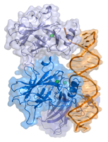
New research gives insight into how stems cells develop into other types of cells Thursday, 20 August 2009 Scientists have uncovered a vital link in the chain of events that gives stem cells their remarkable properties. Researchers from the Wellcome Trust Centre for Stem Cell Research at the University of Cambridge have pinpointed the final step in a complex process that gives embryonic stem cells their unique ability to develop into any of the different types of cells in the body (from liver cells to skin cells). Their findings, published today in the journal Cell, have important implications for efforts to harness the power of stem cells for medical applications. In the last few years, huge strides have been made in stem cell research. Scientists are now able to transform adult skin or brain cells into embryonic-like stem cells in the laboratory. Just like natural embryonic stem cells, these reprogrammed cells can make all the body's cell types. This extraordinary ability is known as pluripotency – 'having several potential outcomes'. It is the basis for the hope that stem cells will one day help fight illnesses like diabetes, Parkinson's or Alzheimer's disease. Despite such exciting developments, scientists still have only a very basic understanding of how cells become pluripotent. Dr Jose Silva, who led the Cambridge research with his colleague Dr Jennifer Nichols, says: "Exactly how pluripotency comes about is a mystery. If we want to create efficient, safe and reliable ways of generating these cells for medical applications, we need to understand the process; our research provides additional clues as to how it occurs. " The researchers studied how the rather poetically named protein Nanog helps give cells pluripotency. Nanog takes its name from the Celtic phrase 'Tir Nan Og', or 'land of the ever young'. It was identified as a key player in pluripotency in 2003, but until now its exact biological role remained unclear. "It was clear that Nanog was important, but we wanted to know how it works. Our research shows that this unique protein flips the last switch in a multi-step process that gives cells the very powerful property of pluripotency," Dr Silva said. Without Nanog, cells remain in a sort of half-way house. As a result, the embryo can't develop and attempts to re-programme adult cells fail. But Nanog does not work alone. It appears to be the conductor in charge of an orchestra of genes and proteins that must all play at the right time and in perfect harmony to create pluripotency. "The next challenge is to find out exactly how Nanog influences all these other molecules," Dr Silva added. Reference: Nanog Is the Gateway to the Pluripotent Ground State Jose Silva, Jennifer Nichols, Thorold W. Theunissen, Ge Guo, Anouk L. van Oosten, Ornella Barrandon, Jason Wray, Shinya Yamanaka, Ian Chambers, Austin Smith Cell, Volume 138, Issue 4, 722-737, 21 August 2009, doi:10.1016/j.cell.2009.07.039 ......... ZenMaster


For more on stem cells and cloning, go to CellNEWS at http://cellnews-blog.blogspot.com/ and http://www.geocities.com/giantfideli/index.html
Watching Stem Cells Repair the Human Brain

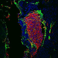
Tel Aviv University researcher shows viability of bone marrow stem cells with unique MRI tracking methods Thursday, 20 August 2009 There is no known cure for neurodegenerative diseases such as Huntington's, Alzheimer's and Parkinson's. But new hope, in the form of stem cells created from the patient's own bone marrow, can be found ― and literally seen ― in laboratories at Tel Aviv University. Dr. Yoram Cohen of Tel Aviv University. Credit: AFTAU.Dr. Yoram Cohen of TAU's School of Chemistry has recently proven the viability of these innovative stem cells, called mesenchymal stem cells, using in-vivo MRI. Dr. Cohen has been able to track their progress within the brain, and initial studies indicate they can identify unhealthy or damaged tissues, migrate to them, and potentially repair or halt cell degeneration. His findings have been reported in the journal Stem Cells. "By monitoring the motion of these cells, you get information about how viable they are, and how they can benefit the tissue," he explains. "We have been able to prove that these stem cells travel within the brain, and only travel where they are needed. They read the chemical signalling of the tissue, which indicate areas of stress. And then they go and try to repair the situation." Tracking live cells in the brain To test the capabilities of this innovative new stem cells, Dr. Cohen created a study to track the activity of the live cells within the brain using the in-vivo MRI at the Strauss Centre for Computational Neuro-Imaging. Watching the live, active cells has been central to establishing their viability as a therapy for neurodegenerative disease. Dr. Cohen and his team of researchers took magnetic iron oxide nanoparticles and used them to label the stem cells they tested. When injected into the brain, they could then be identified as clear black dots on an MRI picture. The stem cells were then injected into the brain of an animal that had an experimental model of Huntington's disease. These animals suffer from a similar neuropathology as the one seen in human Huntington's patients, and therefore serve as research tool for the disease. On MRI, it was possible to watch the stem cells migrating towards the diseased area of the brain. "Cells that go toward a certain position that needs to be rescued are the best indirect proof that they are live and viable," explains Dr. Cohen. "If they can migrate towards the target, they are alive and can read chemical signalling." An ethically viable stem cell This study is based on differentiated mesenchymal cells (MSC), which were discovered at Tel Aviv University. Bone marrow cells are transformed into NTFs-secreting stem cells, which can then be used to treat neurodegenerative diseases. This advance circumvents the ethical debate caused by the use of stem cells obtained from embryos. Although there is a drawback to using this particular type of stem cell ― the higher degree of difficulty involved in rendering them "neuron-like" ― the benefits are numerous. "Bone marrow-derived MSCs bypass ethical and production complications, and in the long run, the cells are less likely to be rejected because they come from the patients themselves. This means you don't need immunosuppressant therapy," says Dr. Cohen. Working towards a real-life therapy Dr. Cohen says the next step is to develop a real-life therapy for those suffering from neurodegenerative diseases. The ultimate goal is to repair neuronal cells and tissues. Stem cell therapy is thought to be the most promising future therapy to combat diseases such as Huntington's, Alzheimer's and Parkinson's diseases, and researchers may also be able to develop a therapy for stroke victims. If post-stroke cell degeneration can be stopped at an early stage, says Dr. Cohen, patients can live for many years with a good quality of life. ......... ZenMaster


For more on stem cells and cloning, go to CellNEWS at http://cellnews-blog.blogspot.com/ and http://www.geocities.com/giantfideli/index.html
Using Light to Manipulate Cell Movement

New technique expected to enhance understanding of how cancer spreads Thursday, 20 August 2009 One of the biggest challenges in scientists' quest to develop new and better treatments for cancer is gaining a better understanding of how and why cancer spreads. Recent breakthroughs have uncovered how different cellular proteins are turned 'on' or 'off' at the molecular level, but much remains to be understood about how protein signalling influences cell behaviour. A new technique developed by Klaus Hahn, Ph.D. and his colleagues uses light to manipulate the activity of a protein at precise times and places within a living cell, providing a new tool for scientists who study the fundamentals of protein function. Hahn is the Thurman Professor of Pharmacology at the University of North Carolina at Chapel Hill and a member of UNC Lineberger Comprehensive Cancer Center. In a paper published today in the journal Nature, Hahn described the technique, which uses light to control protein behaviour in cells and animals simply by shining light on the cells where they want the protein to be active.


Control of cell movement in living cells.A photo-activatable protein enables control of cell movement in living cells. Activation of Rac in the red circle led to localized cell protrusion and translocation of the kinase PAK to the cell edge (right hand image, Pak in red). Credit: Yi Wu, UNC-Chapel Hill.
"The technology has exciting applications in basic research – in many cases the same protein can be either cancer-producing or beneficial, depending on where in a cell it is activated. Now researchers can control where that happens and study this heretofore inaccessible level of cellular control," said Hahn. "Because we first tested this new technology on a protein that initiates cell movement, we can now use light to control where and how cells move. This is quite valuable in studies where cell movement is the focus of the research, including embryonic development, nerve regeneration and cancer metastasis," he added. The new technology is an advance over previous light-directed methods of cellular control that used toxic wavelengths of life, disrupted the cell membrane or could switch proteins 'on' but not 'off'. Reference: A genetically encoded photoactivatable Rac controls the motility of living cells Yi I. Wu, Daniel Frey, Oana I. Lungu, Angelika Jaehrig, Ilme Schlichting, Brian Kuhlman & Klaus M. Hahn Nature advance online publication 19 August 2009, doi:10.1038/nature08241 See also: Coordination of Rho GTPase activities during cell protrusion Matthias Machacek, Louis Hodgson, Christopher Welch, Hunter Elliott, Olivier Pertz, Perihan Nalbant, Amy Abell, Gary L. Johnson, Klaus M. Hahn & Gaudenz Danuser Nature advance online publication 19 August 2009, doi:10.1038/nature08242 ......... ZenMaster
For more on stem cells and cloning, go to CellNEWS at http://cellnews-blog.blogspot.com/ and http://www.geocities.com/giantfideli/index.html

Thursday, 13 August 2009

Technique Enables Efficient Gene Splicing in Human Embryonic Stem Cells

Technique Enables Efficient Gene Splicing in Human Embryonic Stem Cells Thursday, 13 August 2009 A novel technique allows researchers to efficiently and precisely modify or introduce genes into the genomes of human embryonic stem cells (ESCs) and induced pluripotent stem (iPS) cells, according to Whitehead scientists. The method uses proteins called zinc finger nucleases and is described in the August 13 issue of Nature Biotechnology. For years, scientists have easily swapped genes in and out of mouse ESC or iPS cell genomes, but have had a notoriously difficult time disrupting or inserting genes into their human equivalents. "It's not clear where this hurdle of genetic manipulation lies; it could be purely technical, but it could also be an inherent difference between human and mouse cells," says Dirk Hockemeyer. Hockemeyer and Frank Soldner are first authors on the article and postdoctoral researchers in Whitehead Member Rudolf Jaenisch’s lab. "Other people have genetically manipulated these human cells, but the process has been extremely laborious and extremely time consuming. Using the zinc finger nucleases makes the process very easy," says Hockemeyer. Earlier methods are so inefficient that fewer than 15 genes have been swapped into human ESCs since that cell type was discovered 10 years ago. By comparison, hundreds of genes have been introduced into the genomes of mouse ESCs. According to Jaenisch, this method could open a new phase in human genetics. "This is a proof of principle that zinc finger nucleases can be used to swap out many, many additional genes in human ESCs and iPS cells," says Jaenisch, who is also a professor of biology at MIT. "Now human ESC and iPS cell genetics can catch up to mouse genetics, which has had a 20-year head start." The inability to alter human ESC and iPS cells' genomes has hindered researchers from routinely creating specific cell types for modelling genetic diseases (e.g., the brain cells affected by Parkinson’s disease) and studying how embryonic stem cells mature into adult cells. (iPS cells are adult cells that have been reprogrammed to an embryonic-stem-cell-like state, so they have similar properties of ESCs: the ability to self-propagate and the ability to mature into any of an adult's approximately 220 cell types. iPS cells have the added benefit of possessing the same genes as the patient who donated the adult cells, thereby accurately reflecting that patient's specific genetic profile.) To substitute a gene in ESCs and iPS cells, Hockemeyer and Soldner adapted a recently developed technique to cut out one gene from the human ESCs and iPS cells and substitute it with another by putting two zinc finger nucleases and the replacement gene into the ESCs and iPS cells. Each zinc finger nuclease recognizes a particular sequence in a cell's DNA and then cuts through both strands of DNA at that site. The cell's DNA repair machinery recognizes that the DNA has been cut and tries to fix it using the replacement gene resulting in the desired alteration of the original gene. In addition to working so efficiently, the method can be tailored to precisely swap nearly any gene in the genome. "We can produce zinc finger nucleases that out of about three billion DNA base pairs can identify one specific site," says Soldner. "We also spent quite a bit of energy to see if the zinc finger nucleases cut somewhere other than the intended target site, and it was very unlikely." Reference: Efficient targeting of expressed and silent genes in human ESCs and iPSCs using zinc-finger nucleases Dirk Hockemeyer, Frank Soldner, Caroline Beard, Qing Gao, Maisam Mitalipova, Russell C. DeKelver, George E. Katibah, Ranier Amora, Elizabeth A. Boydston, Bryan Zeitler, Xiangdong Meng, Jeffrey C. Miller, Lei Zhang, Edward J. Rebar, Philip D. Gregory, Fyodor D. Urnov and Rudolf Jaenisch Nature Biotechnology, Published online: 13 August 2009, doi:10.1038/nbt.1562 ......... ZenMaster


For more on stem cells and cloning, go to CellNEWS at http://cellnews-blog.blogspot.com/ and http://www.geocities.com/giantfideli/index.html

Tuesday, 11 August 2009

Blood Cells Made from Embryonic Stem Cells

Scientists make multiple types of white blood cells directly from embryonic and adult stem cells Tuesday, 11 August 2009 In an advance that could help transform embryonic stem cells into a multipurpose medical tool, scientists at the University of Wisconsin-Madison have transformed these versatile cells into progenitors of white blood cells and into six types of mature white blood and immune cells. While clinical use is some years away, the new technique could produce cells with enormous potential for studying the development and treatment of disease. The technique works equally well with stem cells grown from an embryo and with adult pluripotent stem cells, which are derived from adult cells that have been converted until they resemble embryonic stem cells. If the adult cells came from people with certain bone marrow diseases, the new technique could produce blood cells with specific defects. It could also be used to grow specific varieties of immune cells that could target specific infections or tumours. The likely most immediate benefit is cells that can be used for safety screening of new drugs, says study leader Igor Slukvin, an assistant professor in the university's Department of Pathology and Laboratory Medicine. "Toxicity to the blood-forming system is a key limit on drug development, so these cells could be used for safety testing in any drug development," says Slukvin, who performs research at the National Primate Research Center in Madison. Bone marrow stem cells are already used to screen drugs, but the new technique promises to produce large quantities of cells in a dish that can be more exactly tailored to the task at hand, without requiring a constant supply of bone marrow cells from donors. The development of stem cells into mature, specialized cells is governed by trace amounts of biological signalling molecules, so Slukvin and colleagues Kyung-Dal Choi and Maxim Vodyanik exposed two types of highly versatile stem cells to various compounds. Eventually they found a recipe that would cause the cells to move through a process of progressive specialization into a variety of adult cells. Slukvin's study was published in the Journal of Clinical Investigation. The result included osteoclasts, cells that play a role in osteoporosis, and eosinophils, which are involved in allergy and asthma. Other adult cells included dendritic and Langerhans cells, which direct other immune cells to attack infections, and neutrophils, the most common type of white blood cell. "While we now can make almost all types of blood cells from embryonic and adult pluripotent stem cells, the next major challenge is to produce blood stem cells (called hematopoietic stem cells) that might be used in a bone marrow transplant," Slukvin says. This life-saving procedure can replace the entire blood-forming system in a patient with blood cancer, but more than one-third of patients cannot find a well-matched bone marrow donor and thus risk graft-versus-host disease, a sometimes-fatal attack on the patient by the transferred immune system. Compatibility problems should disappear if the blood-forming stem cells are based on the patient's own cells, Slukvin says. "Eventually, we want to make therapeutic cells that could be used instead of bone marrow transplants." In the interim, Slukvin expects the new technique to produce cells that model a variety of medical conditions. "We can take cells from patients with a disease of the blood system and explore the cause and treatment of that specific disease. We can generate blood cells which are normal or abnormal, and study the mechanisms and treatments of blood cancers," he says. Scientists now suspect that many cancers have their own stem cells, a long-lived malefactor that spawns cells that form the bulk of the tumour. "Cancer has these stem cells, and we need to target them for treatment. But when patients come to the clinic, they already have cancer, so the malignant transformation already started," says Slukvin. "By reprogramming blood cancer cells to pluripotent stem cells and differentiating these cells back to blood, we hope to generate cancer stem cells in a dish; that would be a good model for studying how these cells formed, to figure out what external factors make them go bad. This could be a crucial step in treating or preventing cancer." Reference: Generation of mature human myelomonocytic cells through expansion and differentiation of pluripotent stem cell–derived lin–CD34+CD43+CD45+ progenitors Kyung-Dal Choi1, Maxim A. Vodyanik2 and Igor I. Slukvin J. Clin. Invest. doi:10.1172/JCI38591 ......... ZenMaster


For more on stem cells and cloning, go to CellNEWS at http://cellnews-blog.blogspot.com/ and http://www.geocities.com/giantfideli/index.html

Monday, 10 August 2009

Simple, Cheap Complete Human Genome DNA Sequencing

Stanford professor sequences his entire genome at low cost, with small team Monday, 10 August 2009 The first few times that scientists mapped out all the DNA in a human being in 2001, each effort cost hundreds of millions of dollars and involved more than 250 people. Even last year, when the lowest reported cost was $250,000, genome sequencing still required almost 200 people. In a paper to be published online Aug. 9 by Nature Biotechnology, a Stanford University professor reports sequencing his entire genome for less than $50,000 and with a team of just two other people. In other words, a task that used to cost as much as a Boeing 747 airplane and required a team of people that would fill half the plane, now costs as much as a mid-priced luxury sedan and the personnel would fill only half of that car. "This is the first demonstration that you don't need a genome centre to sequence a human genome," said Stephen Quake, PhD, professor of bioengineering. "It's really democratizing the fruits of the genome revolution and saying that anybody can play in this game." There are at least two reasons why lowering the cost and effort required to sequence all the genetic information of individuals is important. The more examples scientists have of the whole human genetic code, the more they can discern about how specific genes and mutations result in the traits that make us all different, the diseases that plague us and our response to medicines. As that understanding increases and costs drop, doctors could then sequence their patients' genomes and provide "personalized medicine" in which prevention and treatment of disease would be informed by the patient's exact genetic profile. "This can now be done in one lab, with one machine, at a modest cost," said Quake, the Lee Otterson Professor in the School of Engineering and a member of Stanford's Cancer Center. "It's going to unleash an enormous amount of creativity and really broaden the field." Quake's genome, one of less than a dozen sequenced so far because of the cost and resources needed, is now available to researchers worldwide. Quake's colleagues at Stanford's School of Medicine have been looking through it and sometimes examining Quake himself, mining the data for interesting connections between what they can observe about him, his DNA and his family history. "Some of the doctors are starting to poke and prod me to see how they can couple my genome with medicine," he said. Simpler sequencing To sequence his genome, Quake's team used a commercially available, refrigerator-sized instrument called the Helicos Biosciences SMS Heliscope. Quake, who pioneered the underlying technology in 2003, is a co-founder of the Cambridge, Mass.-based company and chairs its scientific advisory board. The technology — the SMS in the instrument's name — is called single molecule sequencing. While many techniques require generating thousands of copies of a subject's DNA, the single molecule technique does not, reducing the cost and effort involved. Instead, the technique requires chopping the 3 billion or so fundamental units of DNA (called bases) into strands about 30 bases long. The four bases in DNA are adenine (abbreviated A), cytosine (C), guanine (G), and thymine (T). Each base of DNA matches with a specific other base: For example, T only matches with A. The machine captures each of the millions of strands on a specially treated glass plate, holds them there and washes successive waves of fluorescently labelled "letters" over the plate. As each complementary letter sticks next to a strand, the machine can read out the sequence of each strand. A video of the process can be seen on the Web. Assembling the strands back into a cohesive genome is then done by powerful computers, which compare it to the reference genomes that have been compiled before. The process is akin to assembling an enormous jigsaw puzzle by referring frequently to the picture on the box. The team said the sequencing process took about one month to complete. Still, several tricky problems had to be solved before the machine could reliably sequence a whole human genome. Quake worked with Norma Neff, a research manager in Quake's lab, and physics doctoral student Dmitry Pushkarev to write a sophisticated algorithm that would enable them to determine how accurate the process is. Overall, the genome is 95 percent complete, a rate comparable with other sequenced genomes, the team found. In the paper, the authors are careful to note that all genome-sequencing technologies, including the one they have demonstrated, have produced incomplete approximations of the actual genome. Still, it is enough to help produce genuine insights about a person's traits and health. A professor's personal revelations Quake's genome has already yielded a few interesting connections between his genetics and his health. One is that he carries a rare mutation associated with a heart disorder; the revelation, he said, sheds light on what members of his family have always wondered with regard to the health of prior generations. The good news, he said, is that he's also apparently genetically predisposed to respond well to common cholesterol-lowering statin medicines. Quake said the information has also forced him to take heed of that history. "If you know your uncle had something, you kind of discount that you can get it, but to see you've inherited the mutation for that is another matter altogether," he said. One amusing "revelation" is that Quake's code contains a form of a gene that has sometimes been associated with increased disagreeability, he said. The details of the code can be found on the Web. "Of course, you don't need my genome to tell you that," Quake acknowledged. "My wife could have told you that and certainly the dean could have as well." Reference: Single-molecule sequencing of an individual human genome Dmitry Pushkarev, Norma F Neff & Stephen R Quake Nature Biotechnology, Published online: 10 August 2009, doi:10.1038/nbt.1561 ......... ZenMaster


For more on stem cells and cloning, go to CellNEWS at http://cellnews-blog.blogspot.com/ and http://www.geocities.com/giantfideli/index.html

p53 Immortality Improves Cell Reprogramming

Tumour Suppressor Pulls Double Shift as Reprogramming Watchdog Monday, 10 August 2009 Tumour suppressor p53.A collaborative study by researchers at the Salk Institute for Biological Studies uncovered that the tumour suppressor p53, which made its name as “guardian of the genome,” not only stops cells that could become cancerous in their tracks but also controls somatic cell reprogramming. Although scientists have learned how to reprogram adult human cells such as skin cells into so-called induced pluripotent stem cells (iPSCs), the reprogramming efficiency is still woefully low. The Salk study, published in the Aug. 9 advance online edition of Nature, gives new insight into why only a few cells out of many can be persuaded to turn back the clock. “Although we have been able to reprogram specialized cells for a while now, there had been nothing known about the control mechanisms that prevent it from happening spontaneously in the body and why it has been so hard to change their fate in a Petri dish,” says Juan-Carlos Izpisúa Belmonte, Ph.D., a professors in the Gene Expression Laboratory, who worked closely with Geoffrey M. Wahl, Ph.D., also a professor in the Gene Expression Laboratory. Their findings bring iPSCs technology a step closer to fulfilling its promise as source of patient-specific stem cells but also force scientists to rethink the development of cancer. “There’s been a decade-old idea that cancer arises through the de-differentiation of fully committed and specialized cells but eventually it was discarded in favour of the currently fashionable cancer stem cell theory,” says Wahl. “Now, that we know that p53 prevents de-differentiation, I believe it is time to reconsider the possibility that reprogramming plays a role in the development of cancer since virtually all cancer cells lose p53 function in one way or another.” As mammalian embryos transition through a series of developmental stages, the choices of embryonic stem cells, which enjoy almost limitless prospects, are progressively limited till they eventually give rise to the roughly 200 cell types that make up our body and generally lack the ability to revert back to a less specialized stage. Although differentiation is generally irreversible, scientists have developed several methods to overcome the cells’ reluctance to be reprogrammed. The most widely used technology involves the forced expression of four transcription factors — Oct4, Sox2, Klf4, and c-Myc — in fully committed adult cells. “Unfortunately, Klf4 and c-Myc are oncogenes and adding them carries the risk of inducing cancer,” says Belmonte. Yet, despite the extra push provided by those powerful oncogenes, only a tiny fraction transmogrifies into iPSCs that look and act like embryonic stem cells, leading Belmonte to question whether what they were doing to get the cells to reprogram induced a response that stopped the cells from growing? A conversation with his next-door neighbour, cancer expert Wahl provided some fresh ideas that could be tested in the lab. “Normally, cells don’t reprogram so there must be a mechanism in place that prevents it,” says Wahl. “We knew that c-Myc and some of the other genes that are required for reprogramming activate the tumour suppressor p53 and we wondered whether it had any part in it.” Down-regulating p53 activity increases the reprogramming efficiency of adult somatic cells requiring only two reprogramming factors instead of the usual four. Credit: Courtesy of Dr. Juan-Carlos Belmonte, Salk Institute for Biological Studies.And sure enough, experiments by postdoctoral researchers and co-first authors Teruhisa Kawamura, Ph.D., and Jotaro Suzuki, Ph.D., revealed that adding the reprogramming factors c-Myc and Klf4, alone or in various combinations activated the p53 pathway. As a first-responder, the tumour suppressor p53 is called to action when cells experience stressful conditions. Depending on the situation, p53 then turns on genes that halt cell division to allow time for repairs or, when all rescue attempts prove futile, order the cell to stop dividing forever or to commit suicide. In cells genetically engineered to lack p53, reprogramming efficiency was at least 10-fold increased compared to control cells, demonstrating that p53 clearly played an important role in reigning in cells trying to revert back into a stem-like state. Because iPSCs generated with the full complement of reprogramming factors run the risk to turn malignant, Belmonte and his team wanted to know whether mouse cells lacking p53 could be reprogrammed using only two factors, Oct4 and Sox2. The cells readily converted into iPSCs and gave rise to healthy, full term mice that were able to reproduce passing the ultimate test for pluripotent embryonic stem cells. “This very successful collaboration is a prime example of what makes the Salk such a special place,” says Wahl. “Juan Carlos and I talk every day and we approach the same question from very different perspectives. He comes from a developmental biology perspective, while I come from the cancer side but when put together they can make for a great story.” About the Salk Institute for Biological Studies The Salk Institute for Biological Studies is one of the world's preeminent basic research institutions, where internationally renowned faculty probe fundamental life science questions in a unique, collaborative, and creative environment. Focused both on discovery and on mentoring future generations of researchers, Salk scientists make groundbreaking contributions to our understanding of cancer, aging, Alzheimer's, diabetes, and cardiovascular disorders by studying neuroscience, genetics, cell and plant biology, and related disciplines. Faculty achievements have been recognized with numerous honours, including Nobel Prizes and memberships in the National Academy of Sciences. Founded in 1960 by polio vaccine pioneer Jonas Salk, M.D., the Institute is an independent non-profit organization and architectural landmark. Comment: Now, five research teams, including Shinya Yamanaka's, have boosted their success rates by around a 100-fold by silencing the p53 pathway, which prevents mutations and preserves the sequence of the genome (see references below). Reference: Suppression of induced pluripotent stem cell generation by the p53–p21 pathway Hyenjong Hong, Kazutoshi Takahashi, Tomoko Ichisaka, Takashi Aoi, Osami Kanagawa, Masato Nakagawa, Keisuke Okita & Shinya Yamanaka Nature advance online publication 9 August 2009, doi:10.1038/nature08235 Immortalization eliminates a roadblock during cellular reprogramming into iPS cells Jochen Utikal, Jose M. Polo, Matthias Stadtfeld, Nimet Maherali, Warakorn Kulalert, Ryan M. Walsh, Adam Khalil, James G. Rheinwald & Konrad Hochedlinger Nature advance online publication 9 August 2009, doi:10.1038/nature08285 A p53-mediated DNA damage response limits reprogramming to ensure iPS cell genomic integrity Rosa M. Marión, Katerina Strati, Han Li, Matilde Murga, Raquel Blanco, Sagrario Ortega, Oscar Fernandez-Capetillo, Manuel Serrano & Maria A. Blasco Nature advance online publication 9 August 2009, doi:10.1038/nature08287 The Ink4/Arf locus is a barrier for iPS cell reprogramming Han Li, Manuel Collado, Aranzazu Villasante, Katerina Strati, Sagrario Ortega, Marta Cañamero, Maria A. Blasco & Manuel Serrano Nature advance online publication 9 August 2009, doi:10.1038/nature08290 Linking the p53 tumour suppressor pathway to somatic cell reprogramming Teruhisa Kawamura, Jotaro Suzuki, Yunyuan V. Wang, Sergio Menendez, Laura Batlle Morera, Angel Raya, Geoffrey M. Wahl & Juan Carlos Izpisúa Belmonte Nature advance online publication 9 August 2009, doi:10.1038/nature08311 See also: Immortality improves cell reprogramming Nature News Published online 9 August 2009, doi:10.1038/news.2009.809 ......... ZenMaster


For more on stem cells and cloning, go to CellNEWS at http://cellnews-blog.blogspot.com/ and http://www.geocities.com/giantfideli/index.html

Friday, 7 August 2009

Two Lines Account for Most Human Embryonic Stem Cell Research

Two Lines Account for Most Human Embryonic Stem Cell Research Friday, 07 August 2009 For the past eight years, scientists who wanted to use federal funds for research on human embryonic stem cells had to restrict their studies to 21 cell lines approved by the National Institutes of Health. But an analysis by a researcher at the Stanford University School of Medicine suggests that only two of those lines have been used routinely. "I was surprised by these results," said Christopher Scott, director of Stanford's Program on Stem Cells in Society. "I never imagined that we would find that three-fourths of the requests would be for the same two cell lines." On the one hand, the findings raise concerns about the reauthorization process of cell lines under way at the NIH — if these lines are now excluded from federal funding due to ethical considerations, researchers may abandon them, and their previous research, in favour of other lines. On the other, the findings draw attention to the possibility that these two lines may have abnormalities or characteristics that make them not as useful as newer lines. "Not only are scientists asking for these lines, they are publishing on them," said Scott, a senior research scholar at Stanford's Center for Biomedical Ethics. "They have become the reference standards against which new embryonic and iPS cell lines are being compared." (An iPS cell is an adult cell that has been induced to look and act like a human embryonic stem cell; comparing them with existing embryonic stem cell lines is important, as there is much debate about whether these iPS cells are functionally equivalent to human embryonic stem cells.) Scott collaborated with researchers from the Mayo Clinic and the University of Michigan to conduct the research, which will be published Aug. 7 in Nature Biotechnology. Together they analyzed the number and timing of requests placed by scientists for human embryonic stem cell lines housed at the two largest stem cell banks in the country: the National Stem Cell Bank at the WiCell Research Institute in Madison, Wisc., and the Harvard Stem Cell Institute in Massachusetts. Although the National Stem Cell Bank is meant to be the source of all NIH-approved lines, Scott and his colleagues found that at no time have all 21 lines been available for distribution; a maximum of 18 lines were available at the beginning of this year. Two cell lines, known as H1 and H9, made up the majority of requests — 941 out of 1,217, or 77 percent, since 1999. One other line, H7, was requested 111 times. In contrast, 13 of the previously approved lines were requested fewer than 10 times in the past decade. Research on the three most-requested lines from the NSCB is prevalent in the scientific literature: 83 percent of 534 peer-reviewed publications from 1999 to 2008 discussed research on H9, 61 percent on H1 and 24 percent used H7 (the numbers exceed 100 percent because many studies used more than one cell line). In contrast, fewer than 36 percent of the publications used any of the other NSCB-curated cell lines. Requests for cell lines from the Harvard Stem Cell Institute included a wider selection of lines, but even these were still relatively narrow, the researchers found. The majority of these cell lines were created by Harvard researcher Douglas Melton with private funds. Of the 17 cell lines available since 2004, 234 of 946 requests, or 25 percent, were for one of two lines: HUES1 and HUES9. Even though the Harvard stem cell lines increase the diversity available to researchers, their impact in the published research has so far been minimal: Only about 3 percent of the peer-reviewed articles included in Scott's study reported research on the two most popular Harvard lines. "It could be a first-mover advantage," said Scott of the researchers' bias toward just a few lines. "If one group publishes on a particular line, other groups want to replicate and extend that research." It's also possible, the authors theorize, that the complex thicket of federal and state restrictions on embryonic stem cell research simply made researchers skittish about branching off into new cell lines. "The trick will be to avoid this kind of situation with the NIH's new stem cell registry," said Scott, referring to the system that the agency will be establishing to ensure that only research on approved lines gets federal funding. Future policies should be designed, he said, to preserve researchers' ability to continue to work on these well-characterized lines while also encouraging them to plumb the new lines that are expected to become eligible for federal funding under the new regulations. "We're starting with a very scarce resource, and we have to figure out how to make it high quality." Reference: And then there were two: use of hESC lines Christopher Thomas Scott, Jennifer B McCormick & Jason Owen-Smith Nature Biotechnology 27, pp696 – 697, 2009, doi:10.1038/nbt0809-696 ......... ZenMaster


For more on stem cells and cloning, go to CellNEWS at http://cellnews-blog.blogspot.com/ and http://www.geocities.com/giantfideli/index.html

Thursday, 6 August 2009

What Makes Stem Cells Tick?

Researchers identify phosphorylated signalling proteins in human embryonic stem cells Thursday, 06 August 2009 Investigators at the Burnham Institute for Medical Research (Burnham) and The Scripps Research Institute (TSRI) have made the first comparative, large-scale phosphoproteomic analysis of human embryonic stem cells (hESCs) and their differentiated derivatives. The data may help stem cell researchers understand the mechanisms that determine whether stem cells divide or differentiate, what types of cells they become and how to control those complex mechanisms to facilitate development of new therapies. The study was published in the August 6 issue of the journal Cell Stem Cell. Protein phosphorylation, the biochemical process that modifies protein activities by adding a phosphate molecule, is central to cell signalling. Using sophisticated phosphoproteomic analyses, the team of Sheng Ding, Ph.D., associate professor at TSRI, Evan Y. Snyder, M.D., Ph.D., professor and director of Burnham's Stem Cell and Regenerative Biology program, and Laurence M. Brill, Ph.D., senior scientist at Burnham's Proteomics Facility, catalogued 2,546 phosphorylation sites on 1,602 phosphoproteins. Prior to this research, protein phosphorylation in hESCs was poorly understood. Identification of these phosphorylation sites provides insights into known and novel hESC signalling pathways and highlights signalling mechanisms that influence self-renewal and differentiation. "This research will be a big boost for stem cell scientists," said Dr. Brill. "The protein phosphorylation sites identified in this study are freely available to the broader research community, and researchers can use these data to study the cells in greater depth and determine how phosphorylation events determine a cell's fate." The team performed large-scale, phosphoproteomic analyses of hESCs and their differentiated derivatives using multi-dimensional liquid chromatography and tandem mass spectrometry. The researchers then used the phosphoproteomic data as a predictive tool to target a sample of the signalling pathways that were revealed by the phosphorylated proteins in hESCs, with follow-up experiments to confirm the relevance of these phosphoproteins and pathways to the cells. The study showed that many transcription regulators such as epigenetic and transcription factors, as well as a large number of kinases are phosphorylated in hESCs, suggesting that these proteins may play a key role in determining stem cell fate. Proteins in the JNK signalling pathway were also found to be phosphorylated in undifferentiated hESCs, which suggested that inhibition of JNK signalling may lead to differentiation, a result that was confirmed in hESC cultures. These methods were extremely useful to discover novel proteins relevant to the human embryonic stem cells. For example, the team found that phosphoproteins in receptor tyrosine kinase (RTK) signalling pathways were numerous in undifferentiated hESCs. Follow-up studies used this unexpected finding to show that multiple RTKs can support hESCs in their undifferentiated state. This research shows that phosphoproteomic data can be a powerful tool to broaden understanding of hESCs and how their ultimate fate is determined. With this knowledge, stem cell researchers may be able to develop more focused methods to control hESC differentiation and move closer to clinical therapies. The protein phosphorylation data is available on the Cell Stem Cell website, as well as on the PRIDE website. Another group from Utrecht, The Netherlands, has performed a similar study of phosphoproteins in hESCs (see the references). About Burnham Institute for Medical Research Burnham Institute for Medical Research is dedicated to revealing the fundamental molecular causes of disease and devising the innovative therapies of tomorrow. Burnham, with operations in California and Florida, is one of the fastest-growing research institutes in the country. The Institute ranks among the top-four institutions nationally for NIH grant funding and among the top-25 organizations worldwide for its research impact. Burnham utilizes a unique, collaborative approach to medical research and has established major research programs in cancer, neurodegeneration, diabetes, infectious and inflammatory and childhood diseases. The Institute is known for its world-class capabilities in stem cell research and drug discovery technologies. Burnham is a non-profit, public benefit corporation. About The Scripps Research Institute The Scripps Research Institute is one of the world's largest independent, non-profit biomedical research organizations, at the forefront of basic biomedical science that seeks to comprehend the most fundamental processes of life. Scripps Research is internationally recognized for its discoveries in immunology, molecular and cellular biology, chemistry, neurosciences, autoimmune, cardiovascular, and infectious diseases, and synthetic vaccine development. Established in its current configuration in 1961, it employs approximately 3,000 scientists, postdoctoral fellows, scientific and other technicians, doctoral degree graduate students, and administrative and technical support personnel. Scripps Research is headquartered in La Jolla, California. It also includes Scripps Florida, whose researchers focus on basic biomedical science, drug discovery, and technology development. Scripps Florida is located in Jupiter, Florida. References: Phosphoproteomic Analysis of Human Embryonic Stem Cells Laurence M. Brill, Wen Xiong, Ki-Bum Lee, Scott B. Ficarro, Andrew Crain , Yue Xu, Alexey Terskikh, Evan Y. Snyder, and Sheng Ding Cell Stem Cell, Volume 5, Issue 2, 204-213, 7 August 2009, doi:10.1016/j.stem.2009.06.002 Phosphorylation Dynamics during Early Differentiation of Human Embryonic Stem Cells Dennis Van Hoof, Javier Muñoz , Stefan R. Braam, Martijn W.H. Pinkse , Rune Linding , Albert J.R. Heck, Christine L. Mummery and Jeroen Krijgsveld Cell Stem Cell, Volume 5, Issue 2, 214-226, 7 August 2009, doi:10.1016/j.stem.2009.05.021 Unraveling the Human Embryonic Stem Cell Phosphoproteome Andrew P. Hutchins and Paul Robson Cell Stem Cell, Volume 5, Issue 2, 126-128, 7 August 2009, doi:10.1016/j.stem.2009.07.007 ......... ZenMaster


For more on stem cells and cloning, go to CellNEWS at http://cellnews-blog.blogspot.com/ and http://www.geocities.com/giantfideli/index.html

From Nerve Roots to Plant Roots

Research on hereditary spastic paraplegia yields surprises Thursday, 06 August 2009 Sprouting. Branching. Pruning. Neuroscientists have borrowed heavily from botanists to describe the way that neurons grow, but analogies between the growth of neurons and plants may be more than superficial. A new study from the National Institutes of Health and Harvard Medical School suggests that neurons and plant root cells may grow using a similar mechanism. The research also sheds light on the hereditary spastic paraplegias (HSP), a group of inherited neurological disorders in which some of the longest neurons in the body fail to grow and function properly. The genes behind HSP and their roles inside neurons are poorly understood. However, the study suggests that several forms of HSP share an underlying defect with each other – and with abnormal root hair development in a plant widely used for agricultural research. The strange implication is that the plant, Arabidopsis thaliana (mouse-ear cress), could prove useful for further research on HSP. "This study provides us with valuable new insights that will stimulate research toward therapies for hereditary spastic paraplegias," says Craig Blackstone, M.D., Ph.D., an investigator at NIH's National Institute of Neurological Disorders and Stroke (NINDS) and an HSP expert. Dr. Blackstone performed the study in collaboration with William Prinz, Ph.D., an investigator at the NIH's National Institute of Diabetes and Digestive and Kidney Diseases (NIDDK), and Tom Rapoport, Ph.D., a Howard Hughes Medical Institute investigator and a professor of cell biology at Harvard Medical School. HSP primarily affects corticospinal neurons, which extend projections called axons from the brain's cerebral cortex to the spinal cord. The longest corticospinal axons extend nearly all the way down the spinal cord – a distance up to about three feet – in order to control movement in the legs. In HSP, these long axons develop abnormally or they degenerate later in life, causing muscle stiffness and weakness in the legs. HSP exists in many forms in different families, and more than 40 genes have been implicated in the disease. In the new study, published in Cell, the researchers propose that defects in the shaping of a subcellular structure known as the endoplasmic reticulum (ER) are a common cause of HSP. The ER – named for its reticulated (or net-like) shape – is a cellular factory, where molecules such as proteins and lipids that are vital to cell growth are made and packaged for shipping to various cellular destinations. The researchers theorize that in several forms of HSP, the ER loses its complex shape and is unable to support the growth or maintenance of long corticospinal axons. Several years ago, other researchers showed that similar ER defects in Arabidopsis impair the growth of the plant's root hairs. These are wispy, microscopic projections that grow from the plant's individual root cells. The new study focuses on a gene called atlastin. This gene is defective in about 10 percent of HSP cases, and in previous research, Dr. Blackstone's group showed that it has a role in axon growth. The new study reveals that the atlastin protein is necessary for maintaining the shape of the ER in mammalian cells, and that an analogous protein called Sey1p performs the same function in baker's yeast. The researchers demonstrate that ER shaping defects have general relevance for HSP, by showing a connection between atlastin and a group of proteins known as the DP1 family. Years ago, Drs. Prinz and Rapoport reported that a yeast analogue of DP1 regulates the shape of the ER in yeast. Meanwhile, others researchers had independently reported that mutations in REEP1, a member of the DP1 family, cause 3 percent to 8 percent of HSP cases. The new study shows that atlastin interacts physically with DP1 in mammalian cells, and that Sey1p (the yeast atlastin) interacts with the DP1 analogue in yeast. Finally, Dr. Blackstone's study notes that Arabidopsis has an analogue of atlastin, called Root Hair Defective 3 (RHD3). Mutations affecting RHD3 cause the plant to grow short, wavy root hairs. If this connection between axon growth and root hair growth withstands further study, Arabidopsis could be a useful tool for investigating mechanisms of HSP. Arabidopsis is easy to raise in the lab, and the short root hairs of the RHD3 mutant are easy to observe, compared to the growth defects in atlastin-deficient neurons and yeast. Dr. Blackstone hopes to collaborate with other researchers to initiate a search for genes and compounds that correct root hair development in the RHD3 mutant, which might provide valuable therapeutic insights into HSP. Reference: A Class of Dynamin-Like GTPases Involved in the Generation of the Tubular ER Network Hu J, Shibata Y, Zhu P-P, Voss C, Rismanchi N, Prinz W, Rapoport TA, and Blackstone C. Cell, Volume 138, Issue 3, 549-561, 7 August 2009, doi:10.1016/j.cell.2009.05.025 ......... ZenMaster


For more on stem cells and cloning, go to CellNEWS at http://cellnews-blog.blogspot.com/ and http://www.geocities.com/giantfideli/index.html