Friday, 29 August 2008

Blood Created by Identifying Earliest Stem Cells

Blood Created by Identifying Earliest Stem Cells Friday, 29 August 2008 Johns Hopkins researchers have discovered the earliest form of human blood stem cells and deciphered the mechanism by which these embryonic stem cells replicate and grow. They also found a surprising biological marker that pinpoints these stem cells, which serve as the progenitors for red blood cells and lymphocytes. The biochemical marker, angiotensin-converting enzyme (ACE), is well known for its role in the regulation of blood pressure, blood vessel growth, and inflammation. ACE inhibitors are already widely used to treat hypertension and congestive heart failure. The findings are, the researchers say, likely to hold promise for developing new treatments for heart diseases, anaemia’s, leukaemia and other blood cancers, and autoimmune diseases because they show for the first time that ACE plays a fundamental role in the very early growth and development of human blood cells. "We figured out how to get the 'mother' of all blood stem cells with the right culture conditions," says Elias Zambidis, M.D., Ph.D., of the Institute of Cell Engineering at the Johns Hopkins University School of Medicine and the Division of Pediatric Oncology at the Sidney Kimmel Comprehensive Cancer Center at Johns Hopkins. "There is real hope that in the future we can grow billions of blood cells at will to treat blood-related disorders, and just as critically if not more so, we've got ACE as a 'new' old marker to guide our work," Zambidis adds. Researchers did not expect ACE to have a role in blood stem cells, he notes, "but were very pleasantly surprised to discover it as a beacon for finding the earliest blood stem cells known, as well as new ways to find and manipulate this marker to make them grow." The team's findings, published Aug. 26 in the online edition of the journal Blood, explain that these earliest stem cells marked by ACE, called hemangioblasts, first arise normally in the developing human foetus, when a woman is three or four weeks pregnant. Hemangioblasts can now be derived in unlimited supply experimentally from cultured human embryonic stem cells, which are the origin of all cell types in the body. These hemangioblasts go on to become either blood cells or endothelial cells, which form the inner lining of the heart, veins and arteries, and lymph vessels. The research grew out of Zambidis' interest in understanding the complex biological processes of blood development and the transformation of embryonic stem cells into the various types of cells that make up the human body. Hemangioblasts make the body's earliest form of blood in the foetal yolk sac, which nourishes a fertilized egg, and later in the foetal liver and bone marrow. However, because human embryonic cells disappear early in gestation, their role in the early production of blood could not be studied in humans because scientists had no way to identify these human progenitor blood stems cells to follow their development. The scientists suspected they existed in humans, however, because they have been found in mice and zebra fish. To find the blood stem cell, Zambidis' team grew human embryonic stem cells in culture and fed them growth factors over 20 days. Each time the cell colonies expanded, the researchers sampled individual cells, searching for ones capable of making both endothelial and blood cells, the hallmark of hemangioblasts. They plucked the newly discovered hemangioblasts from culture dishes, grew them in conditions that Zambidis and his team developed to speed replication, and tested cells for their ability to make endothelial and blood cells. Cells capable of making endothelial cells and all the elements of blood (platelets, and white and red cells) were specifically marked with ACE on their outer surface. The researchers found not only that ACE was a marker for hemangioblasts, but also turning off the enzyme that helps guide the cells' replication and maturation into either blood or endothelial cells. By treating the hemangioblasts with losartan, an ACE pathway-blocking agent routinely used to treat high blood pressure, dramatically increased the rate of blood cell production. The next step, Zambidis adds, is to test this research in animal models and show that "we can make lots and lots of blood cells from human stem cells for transfusions, regenerate new vascular trees for heart diseases, as well as create test tube factories for making transplantable blood cells that treat diseases.” “We are very far from treatment but this is a big step," Zambidis caution. If the new technique of mass-producing progenitor blood cells is proven to work in humans, it would allow patients getting bone marrow transplants to have their own stem cells creating the blood they need, significantly reducing rejection risk. The research reported today used federally approved embryonic stem cell lines, but other related research by the team comes from non-approved lines. The study was supported by grants from the National Institutes of Health and the Maryland Stem Cell Research Fund. Reference: Expression of ACE (CD143) identifies and regulates primitive hemangioblasts derived from human pluripotent stem cells Elias T Zambidis, Tea Soon Park, Wayne Yu, Ada Tam, Michal Levine, Xuan Yuan, Marina Pryzhkova, and Bruno Peault Blood, online August 26, 2008; DOI 10.1182/blood-2008-03-144766 ......... ZenMaster


For more on stem cells and cloning, go to CellNEWS at http://cellnews-blog.blogspot.com/ and http://www.geocities.com/giantfideli/index.html

How Blood Vessel Cells Form Tubes

How Blood Vessel Cells Form Tubes Friday, 29 August 2008 How do blood vessel cells understand that they should organise themselves in tubes and not in layers? A research group from Uppsala University shows for the first time that a special type of "instructor" molecule is needed to accomplish this. These findings, published in the scientific journal Blood, might be an important step towards using stem cells to build new organs. In order for a body to develop and function the cells in the body must be able to organise themselves in relation to each other. The way in which cells are arranged depends on the organ where they are located. Blood vessel cells need to form three-dimensional, tube-like structures that can transport blood. But how do blood vessel cells know that they should do that? An important part of the communication between cells and their environment is the use of growth factors. These are proteins that bind to receptors on the surface of the cell that receives the information. When the receptor in turn forms a complex with other proteins, on the inside of the cell, the read-out from the DNA can be altered. The information has "arrived". VEGF (vascular endothelial growth factor) is a family of closely related growth factors that control blood vessel cells throughout life. Blood vessel development in the foetus as well as later in life, for example during wound healing, is regulated by VEGF. In the present study the research group has examined how VEGF can instruct blood vessel cells to arrange themselves into a tube. The answer is that some variants of VEGF have the ability to attract another protein, an instructor molecule, which is joined together with VEGF and its receptor. The combination of instructor molecule, VEGF and receptor results in that a specific signal is sent inside the blood vessel cells, making them form a tube. Without the instructor molecule, the cells line up next to each other, in a layer. These results may become very useful. Today stem cells are used to create new cells, organs and even tissues, that in the future might be used to for transplantation instead of donated organs. If a patient's own stem cells are used the problem with organ rejection is avoided. But so far there has been a challenge to create three-dimensional structures from stem cells. “Our contribution can make it possible to create blood vessels from stem cells and to direct them to form a tube instead of a layer. Perhaps this knowledge can be transferred to the formation of other tube-like structures in the body, such as the lung and intestines. The perspectives for the future are very exciting,” says Lena Claesson-Welsh, who has led the study. ......... ZenMaster


For more on stem cells and cloning, go to CellNEWS at http://cellnews-blog.blogspot.com/ and http://www.geocities.com/giantfideli/index.html

Thursday, 28 August 2008

Vaccine Makes Cervical Cancer Control Feasible

Vaccine Makes Cervical Cancer Control in Developing World Feasible Thursday, 28 August 2008 Recent advances in cervical cancer prevention mean that controlling the disease in developing countries is becoming feasible for the first time, experts say. Developments such as highly effective vaccines against the human papilloma virus (HPV) and promising new screening tests provide an unprecedented opportunity to tackle the disease in poor countries. Pap smear screening has largely failed so far, because it is too expensive and too complicated to implement, experts said in a series of papers on the topic unveiled Thursday at the World Cancer Congress of the International Union against Cancer in Geneva. The papers are contained in a monograph published in the journal Vaccine. They present the best global thinking on cervical cancer prevention with vaccination and screening, as well as fresh regional and national research and insights to guide governments and donors in building plans. An independent collaboration of more than 180 leading experts, the monograph evaluates which strategies are most promising and likely to be most cost-effective and affordable and sets out various scenarios for programmes. Every year, about 500,000 women worldwide are diagnosed with cervical cancer and more than 250,000 die from the disease. It is the leading cancer in women in half the countries of the world and mostly affects relatively young and poor women. About 80%of cervical cancer deaths occur in developing countries. "Recent estimates indicate that if trends continue the way they are, developing countries will face a 75% increase in the number of cervical cancer cases because of growth and aging of the population in the next two decades. But it doesn't have to turn out that way," said the coordinator of the monograph, Professor Francesc Xavier Bosch of the Catalan Institute of Oncology in Barcelona, Spain. "The discovery of HPV as the cause of cervical cancer has shaken a field that was stagnating and we are now in a new era where developing countries no longer have to be left behind." The monograph presents the first broad analysis of the cost-effectiveness of introducing HPV vaccination and new screening methods into the hardest hit regions of the world – Asia-Pacific, Latin America and the Caribbean. The benefits varied, depending on the size and make-up of the population and the burden of cervical cancer in each country. Future monographs will address the situation in Africa, the Middle East and Eastern Europe. The experts determined that in the Asia-Pacific region, which accounts for more than half of the world's cervical cancer cases, vaccination would be cost-effective – even in the poorest countries – if the cost per vaccinated girl was US$10 - US$25. For Latin America and the Caribbean, the cost per vaccinated girl, including delivery and logistics costs, would have to be less than US$25 to be cost-effective for all countries. In the most developed populations in the region, vaccination would be cost saving if the cost per vaccinated girl is between US$25 and US$60, and cost-effective at higher prices. "Efforts are needed now to adapt the current price of the vaccines so they meet what individual countries can afford; the solution may be tiered pricing according to gross national income per capita and according to the scale of country efforts," Bosh said. Currently the vaccine's price in the private sector is about US$120 per dose, or US$360 per vaccinated girl. Many countries will need subsidies for some time. The monograph also presents updated evidence on the efficacy of various new screening alternatives compare to Pap smear testing. In addition, a discussion of innovative funds mechanisms for bringing HPV vaccination to poor countries, such as the sale of highly rated vaccine bonds to investors. New screening methods that are increasingly proving themselves in pilot studies provide viable alternatives to pap smears for the first time, the experts said. One such method, known as visual inspection with acetic acid, or VIA, involves painting the cervix with vinegar. It is an attractive alternative because it is cheap, seems to be very effective in detecting pre-cancerous lesions, entails only one visit and a simpler treatment that can be performed by nurses immediately and is less dependent on having a strong health infrastructure. A recent large study in India showed a significant reduction in cervical cancer cases and deaths in areas using VIA. Testing for HPV DNA is also a recent screening advance considered important for the developing world, especially as new rapid test kits being developed especially for poor countries are expected to be cheaper and easier to use. Studies have consistently shown that HPV testing works better than Pap smears as the primary screening test because it is better at picking up suspicious cases. "The models provide a useful roadmap for testing promising strategies in the field. More research is needed to determine an efficient combination of these new approaches and each country will have to decide which is best for them, but we are confident we have provided a valuable starting point for going forward," Bosch said. For the near future, the experts say, both vaccination and screening will be needed. However, in the beginning, many countries may have to continue to focus on screening alone until the vaccine becomes more affordable, the experts added. The price of the vaccine and the support for massive vaccination campaigns is one of the biggest barriers for the moment, but several other challenges lay ahead, the experts said. Those include generating the political support for an intervention whose payoff is two or more decades away, cultural acceptability of the vaccine and monitoring the circulating virus. Uncertainties that may affect the success of vaccination programmes include the duration of protection and whether booster shots might be needed, and whether the vaccines will be as effective in girls whose immune systems are suppressed by either malnutrition or other chronic infections such as HIV or malaria. To accelerate progress, advocates are launching a string of initiatives. At this week's conference in Geneva, three major advocacy groups – the International Union against Cancer, Cervical Cancer Action and PATH – announced an unprecedented coordination of public education campaigns. The groups plan to unveil a dossier of more than 400 letters, editorials and declarations from both developed and developing countries, global leaders, cancer control specialists and international organizations that call for improved cervical cancer prevention in the developing world. The International Union against Cancer also announced the launch of a series of professional training workshops on cervical cancer in developing countries, as well as funding for fellowships at leading institutions. Action is also being stepped up elsewhere. In June, the board of the GAVI Alliance, a partnership of public and private agencies focused on bringing vaccines to developing countries, included cervical cancer in its portfolio of priority diseases. The Alliance is now evaluating the introduction strategy and costs of HPV vaccines. In Africa, a network of first ladies was launched last month, with a meeting planned for next year, to further the cervical cancer agenda on the continent. Access to the Vaccine monograph will be free to developing countries and its contents will form the basis of a distant learning programme that includes tutoring for professional education. "This new era of cervical cancer presents many opportunities and challenges ahead. There is now realistic hope for controlling this disease where the toll is the highest and we have to seize this opportunity. We in the cervical cancer community will be stepping up all our efforts to help developing countries get this disease under control," said Isabel Mortara, executive director of the International Union against Cancer. ......... ZenMaster


For more on stem cells and cloning, go to CellNEWS at http://cellnews-blog.blogspot.com/ and http://www.geocities.com/giantfideli/index.html

Wednesday, 27 August 2008

Insulin-Producing Cells Created from Adult Pancreatic Cells

Insulin-Producing Cells Created from Adult Pancreatic Cells Wednesday, 27 August 2008 Howard Hughes Medical Institute researchers have converted adult pancreatic cells into insulin-producing beta cells in living mice. This is a first because the researchers directly changed the functional identity of adult cells without using embryonic stem cells or relying on techniques that reverse a cell’s genetic programming to its earliest stages. Douglas Melton at HSCI.At a meeting of the International Society for Stem Cell Research (ISSCR) in early June, Harvard Stem Cell Institute director Douglas Melton reported findings that strongly suggested this could be done. Using techniques akin to those that generated the first induced pluripotent stem (iPS) cells, a team spearheaded by Melton's postdoctoral associate Qiao Zhou turned digestive enzyme–producing pancreatic exocrine cells into insulin-secreting beta cells. The beta cells are rare to begin with in the pancreas and are in especially short supply in patients with diabetes, whereas the former comprise 95% of the cells of the pancreas. Therefore, converting one into the other is roughly equivalent to turning copper into gold. Remarkably, the investigators repurposed the adult cells quickly by using viruses to shuttle just three regulatory genes that triggered the remarkable developmental changes. It took only a brief blip of activity by the regulatory genes to imbue the cells with their new job descriptions, which they have retained for as long as nine months. The experiments, which are reported on August 27, 2008, in an advance online publication in the journal Nature, realize a long-time goal in regenerative medicine: To produce specialized repair cells directly from a pool of adult cells that are healthy, abundant and easily obtained. Until now, repair cells have been generated from embryonic stem cells or more recently from pluripotent stem cells created by fully reprogramming adult cells. “What this shows is that you can go directly from one type of adult cell to another, without going back to the beginning,” said Douglas A. Melton, a Howard Hughes Medical Institute (HHMI) investigator at Harvard University and co-director of the Harvard Stem Cell Institute. “You could say, for example, it's like turning a scientist into a lawyer without sending her all the way back to kindergarten.” In this case, the strategy was used in mice to convert exocrine cells, which compose 95 percent of the pancreas, to the relatively scarce beta cells that produce insulin. For more than a decade, Melton has studied how embryonic stem cells give rise to the pancreas and its insulin-producing beta cells, which are destroyed in patients with type 1 diabetes. Ultimately, his studies could lead to ways to generate new pancreatic beta cells that could be used as a treatment for diabetes. However, Melton cautioned that the new results are a proof of principle and do not have immediate medical applications. Exocrine cells are specialized to churn out an array of digestive enzymes. Although they, like all cells, carry the genes that enable insulin production, those genes have been silenced. Melton's experiments attempted to modify the genome of the exocrine cell to “awaken” certain genes and activate the insulin-producing features of beta cells. The concept of adult cell switching, or “lineage switching” as it is sometimes called, has been a major goal of regenerative medicine researchers. This approach has advantages because it avoids using stem cells derived from human embryos. With the advent of newer techniques that obviate the need for human embryonic cells, researchers have been racing to incorporate those ideas into their own work. In a major breakthrough in 2006, Japanese researcher Shinya Yamanaka and his colleagues made stem cells from adult mouse skin cells (fibroblasts) by inserting four specific genes that were active in mouse embryonic stem cells. Those genes, which code for transcription factors, reprogrammed the skin cells so they became pluripotent and therefore had the capacity to develop into any type of tissue. These “induced pluripotent stem cells” or iPS cells, could in theory be guided in the laboratory to become specialized cells that might repair damaged nerves, hearts, or other organs. Melton and postdoctoral fellow Qiao “Joe” Zhou, first author on the Nature paper, were encouraged by the revelation that a handful of transcription factor genes reactivated the embryonic program of adult skin cells. They wondered whether an equally small number of transcription factors could turn off the specialized functions of a given adult cell and turn on those needed to generate the target repair cell. Starting from a list containing all 1,100 transcription factors in mice, the HHMI scientists selected 200 that were active in cells that form the pancreas. They later narrowed that list to just 28 transcription factors that were most active in the region of the pancreas that contains beta cells. The researchers next used retroviruses to ferry genes for nine of the 28 transcription factors into the exocrine cells of live mice. Melton and Zhou were surprised to learn that, in fact, only three of the nine genes were necessary to turn exocrine cells into beta cells – an “extreme makeover,” as one of Melton's colleagues termed it. Those genes were Ngn3, Pdx1, and MafA. The manoeuvre converted about 20 percent of the exocrine cells to beta cells that produced insulin. This was enough to reduce blood sugar levels in diabetic mice. The expression of the three transcription factor genes disappeared less than two months after they were introduced with the virus – but the converted cells remained. While they believe that it will be possible to convert a wide range of adult cells to other cell types using a small number of regulatory genes, the scientists say a number of questions need to be explored. Among them: How closely related to the desired target cell does the donor cell need to be? What other types of cells can be converted to beta cells? And – since using viruses to ferry genes into human patients poses unacceptable risks — can the same outcome be accomplished with chemicals or other drugs? George Daley, an HHMI investigator and stem cell researcher at Children's Hospital Boston, commented: “Melton's work is going to inspire an explosion of experiments in directing the fate of tissues in one way or another in ways that may be more practical than having to reprogram them back to pluripotency.” Daley and colleagues reported recently they had converted cells from individuals with 10 degenerative diseases into stem cells that contained the same genetic errors. These newly created stem cells will allow researchers to reproduce human tissue formation in a Petri dish as it occurs in individuals with any of the diseases. Sir John Gurdon, the internationally renowned developmental biologist under whom Melton did his graduate work at Oxford University and the first scientist to successfully clone an adult animal – a frog, said: "What you really want is a missing cell type, one that is not functioning properly to be derived from something else. But you only want that cell type. So I think this is a really important step forward in exercising what people really wanted and showing how well it can work, by this gene over-expression procedure." Unlike the process involved in creating induced pluripotent stem cells (iPS), which have caused enormous excitement ever since their introduction two years ago by Japanese researcher Shinya Yamanaka, this direct reprogramming technique does not require turning adult cells into stem cells and then figuring out how to induce them to differentiate into a desired cell type. Both Melton and Daley emphasized, however, that direct reprogramming does not in any way eliminate the need for, or value of, work with iPS cells or human embryonic stem cells. "We need to attack problems from multiple angles," said Melton, stressing that his lab is using several approaches and will continue to work with iPS and hES cells. Reference: In vivo reprogramming of adult pancreatic exocrine cells to beta-cells Qiao Zhou, Juliana Brown, Andrew Kanarek, Jayaraj Rajagopal & Douglas A. Melton Nature advance online publication 27 August 2008, doi:10.1038/nature07314 See also: Disease-Specific Induced Pluripotent Stem Cells CellNEWS - Thursday, 07 August 2008 ......... ZenMaster


For more on stem cells and cloning, go to CellNEWS at http://cellnews-blog.blogspot.com/ and http://www.geocities.com/giantfideli/index.html

I Love the Olympic's! III

I Love the Olympic's! Tuesday, 19 August 2008


Dancing in the Water...

Dancing in the Water outside the WaterCube!


ZenMaster

I Love the Olympic's! II

I Love the Olympic's! Tuesday, 19 August 2008

With Swedish supporters...


ZenMaster

I Love the Olympic's! I

I Love the Olympic's! Saturday, 16 August 2008

I Love the Olympics... My friend in Beijing...


ZenMaster

Wednesday, 20 August 2008

Novel Method to Grow Human Embryonic Stem Cells

Uses no animal-based materials; could change how labs culture hESCs in the future Wednesday, 20 August 2008 The majority of researchers working with human embryonic stem cells (hESCs) – cells which produce any type of specialized adult cells in the human body – use animal-based materials for culturing the cells. But because these materials are animal-based, they could transmit viruses and other pathogens to the hESCs, making the cells unsuitable for medical use. Now, a stem-cell scientist at UC Riverside has devised a method of growing hESCs in the lab that uses no animal-derived materials – an important advance in the use of hESCs for future medical purposes. Because of their tremendous potential, hESCs are considered promising sources for future cell therapy to treat diseases such as Parkinson's disease and diabetes mellitus. Noboru Sato, an assistant professor of biochemistry, developed the new method, which is not only cleaner and easier to use than conventional methods of culturing hESCs but also results in hESCs whose pluripotency – the potential to differentiate into any of the specialized cells of the body such as neurons, cardiac muscles, and insulin-producing cells – is uncompromised. Currently in labs worldwide, many researchers grow hESCs on Matrigel-coated culture plates, Matrigel being the trade name for a gelatinous extract, taken from mouse tumour cells, that contains extracellular matrices (ECMs), made up of special proteins. The Matrigel coating provides the scaffolding to which the hESCs first attach and then grow in undifferentiated colonies before differentiating into specialized cells.


hESCs grown on Matrigel.Caption: hESCs grown on Matrigel in defined culture media. The mesh-like structure in the background is Matrigel. Credit: Sato lab, UC Riverside
"The development of animal-free coating methods for hESCs still remains a major challenge due to the complexity of ECMs and insufficient knowledge about how hESCs control cell-cell and cell-ECM interactions," explained Sato, who led the research project. His lab identified a specific signalling pathway, called Rho-Rock, which the hESCs use during colony formation and which plays an important role in physical interactions between hESCs. When the researchers blocked the pathway, they found, as expected, that the normal colony formation of hESCs was considerably impaired. They also found that the hESCs maintained their pluripotency. "Until now, it was generally assumed that the hESC colony formation was pivotal for maintaining pluripotency," Sato said. "But we show that pluripotency can be retained independent of close cell-cell contact."

Prue Talbot, the director of UCR's Stem Cell Center

of which Sato is a member, noted that Sato's discovery could affect the way embryonic stem cells are grown in the future. "His work is certainly an important step forward in both understanding signal transduction pathways in stem cells and in the development of an improved methodology for culturing stem cells," she said. In the study, Sato's group extensively screened various types of scaffold materials in combination with Y27632, a chemical compound that blocks the Rho-Rock pathway, and found that the Matrigel coating could be replaced with "poly-D-lysine," a chemically synthesized ECM. The major advantages of poly-D-lysine over Matrigel are that poly-D-lysine is completely animal-free, easy to handle, and its quality is consistent.


: hESCs grown on poly-D-lysine-coated plate.Caption: hESCs grown on poly-D-lysine-coated plate in defined culture media with Y27632. Credit: Sato lab, UC Riverside.
"We found that the growth of the hESCs under this novel culture condition was almost identical to the growth of hESCs on Matrigel-coated culture plates, with no compromise in pluripotency," Sato said. Having started his career as a physician in Japan, Sato began researching stem cell biology as a research fellow at The Rockefeller University, NY, one of the foremost research centres in the world. He accepted a faculty position in the Department of Biochemistry at UCR in 2006. Nicole Harb of UCR and Trevor K. Archer of the National Institute of Environmental Health Sciences (NIEHS), NC joined him in the research project. The research was a collaboration between UCR and NIEHS, and funded by UCR start-up funds to Sato and a grant to Archer from the National Institutes of Health. "Our research goal is to understand the basic mechanisms underlying unique biological functions of pluripotent stem cells, and to translate the obtained knowledge into future medical applications," Sato said. His group is now focusing on applying his technique to the latest stem cell technology, "induced pluripotent stem (iPS) cells," which are pluripotent stem cells artificially derived from adult cells without using embryos. "Our next step is to produce new animal-free iPS cell lines," Sato said. UCR's Office of Technology Commercialization has applied for a patent on Sato's discovery and is looking for industrial partners interested in further developing it. Reference: The Rho-Rock-Myosin Signaling Axis Determines Cell-Cell Integrity of Self-Renewing Pluripotent Stem Cells Nicole Harb, Trevor K. Archer, Noboru Sato PLoS ONE (2008), 3(8): e3001. doi:10.1371/journal.pone.0003001 ......... ZenMaster
For more on stem cells and cloning, go to CellNEWS at http://cellnews-blog.blogspot.com/ and http://www.geocities.com/giantfideli/index.html

Tuesday, 19 August 2008

Red Blood Cells Generated from Human Embryonic Stem Cells

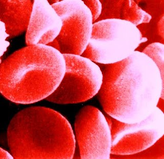
Could potentially be an inexhaustible source of "universal" blood for transfusion. Tuesday, 19 August 2008 Red blood cellsAdvanced Cell Technology, Inc. (ACT) reported that it is feasible to differentiate and mature human embryonic stem cells (hESCs) into functional oxygen-carrying red blood cells (RBCs) under conditions suitable for scale-up. The research by ACT and its collaborators at the Mayo Clinic and the University of Illinois, shows for the first time that the oxygen-carrying capacity of hESC-derived blood cells is comparable to normal transfusable RBCs, and that the cells respond to biochemical changes in a physiologically effective manner. It is published in advance online in the journal Blood. "Limitations in the supply of blood can have potentially life-threatening consequences for patients with massive blood loss," said Robert Lanza, M.D., Chief Scientific Officer at ACT, and senior author of the study. "Embryonic stem cells represent a new source of cells that can be propagated and expanded indefinitely, providing a potentially inexhaustible source of red blood cells for human therapy. We can currently generate 10 to 100 billion red blood cells from a single six-well plate of stem cells. The identification of a stem cell line with "O-"blood-type would permit the production of compatible "universal donor" blood. We also have work underway to generate reprogrammed (iPS) stem cells from individuals with universal-donor blood." The efficient and controlled differentiation of hESCs into homogeneous RBC populations has not been previously achieved. This paper describes for the first time the generation of RBCs from hESCs with oxygen-transporting capacity, and that the functional properties of these cells are similar to those of normal erythrocytes. Multiple stem cell lines were stimulated to undergo differentiation in vitro to form functional RBCs (blood types A, B, O, and both RhD+ and RhD-) on a large scale under conditions suitable for scale-up and clinical translation. Although alternative sources of progenitors for the generation of large-scale transfusable RBCs have been investigated, including cord blood, bone marrow and peripheral blood, it is clear that even after expansion and differentiation, these sources represent donor-limited sources of RBCs. Moreover, the low prevalence of O(-) type blood in the general population further intensifies the consequences of blood shortages for emergency situations and battlefield trauma care, where the need for blood typing can imposes serious delays in initiating transfusions Another critical issue for clinical utilization of hESC-derived RBCs is whether they can be enucleated in vitro. "We show that up to 65% of the blood cells underwent multiple maturation events that resulted in the extrusion of the nucleus," stated Shi-Jiang Lu, Ph.D., Director of Differentiation at ACT and first author of the paper. "They formed enucleated erythrocytes with a diameter of 6-8 mu-m, which is similar to normal red blood cells. We also showed that the cells could express adult beta-globin and respond normally to biochemical changes." "We believe this breakthrough could potentially benefit many Americans," stated William M. Caldwell, CEO and Chairman of ACT. "Although more work is required before this can move into the clinic, we are pleased with the rapid progress that is being made by our scientists and others. We are optimistic about the potential future role for stem cells as a donor-less source of blood for transfusion" About Advanced Cell Technology, Inc.: Advanced Cell Technology, Inc. is a biotechnology company applying cellular technology in the emerging field of regenerative medicine. Reference: Biological properties and enucleation of red blood cells from human embryonic stem cells Shi-Jiang Lu, Qiang Feng, Jennifer S. Park, Loyda Vida, Bao-Shiang Lee, Michael Strausbauch, Peter J. Wettstein, George R. Honig, and Robert Lanza Blood, online August 19, 2008; DOI 10.1182/blood-2008-05-157198 See also: ESCs Made to Blood Stem Cells CellNEWS - Monday, 28 April 2008 ......... ZenMaster


For more on stem cells and cloning, go to CellNEWS at http://cellnews-blog.blogspot.com/ and http://www.geocities.com/giantfideli/index.html
Limbs Saved by Menstrual Blood Stem Cells

Limbs Saved by Menstrual Blood Stem Cells Tuesday, 19 August 2008 Cells obtained from menstrual blood, termed 'endometrial regenerative cells' (ERCs) are capable of restoring blood flow in an animal model of advanced peripheral artery disease. A study published today in BioMed Central's open access Journal of Translational Medicine demonstrates that when circulation-blocked mice were treated with ERC injections, circulation and functionality were restored. Critical limb ischemia, an advanced form of peripheral artery disease, causes approximately 150,000 amputations per year in the US. Currently there are no medical or surgical interventions that are effective in the advanced stages of the disease. ERCs are cells taken from menstrual blood that are capable of forming into at least 9 different tissue types, including heart, liver and lung. Their discovery won the 'Medicine Research Award of the Year' award for BioMed Central's Research Awards in 2007. Dr. Michael Murphy, a vascular surgeon from Indiana University and lead author of this study has already performed clinical trials with adult stem cells for patients with peripheral artery disease. He stated: "The advantage of ERCs is that they can be used in an 'off the shelf' manner, meaning they can be delivered to the point of care, do not require matching, and are easily injectable without the need for complex equipment." The experiments were performed as collaboration between University of Western Ontario, Scripps Research Institute, Indiana University, and several other academic centres. The US publicly traded company Medistem Inc, who supported these studies, is currently developing the ERC cell population. "We are proud of assembling such a strong, clinically experienced team to contribute to these studies" said Dr. Thomas Ichim, CEO of Medistem. "Dr. Ewa Carrier and Suman Kambhampati are haematologists who use stem cells on a regular basis, Dr. Angle is a vascular surgeon, who like Dr. Murphy sees CLI on a daily basis, and Dr. Amit Patel has performed numerous cardiac stem cell clinical trials. With such a team that understands not only the science, but also the practical implementation, we feel we are well positioned to translate this discovery into a practical therapy in the near future." Reference: Allogenic Endometrial Regenerative Cells: An "Off the Shelf Solution" For Critical Limb Ischemia? Michael P Murphy, Hao Wang, Amit N Patel, Suman Kambhampati, Niren Angle, Kyle W Chan, Annette M Marleau, Andrew Pyszniak, Ewa Carrier, Thomas E Ichim and Neil H Riordan Journal of Translational Medicine (in press) ......... ZenMaster


For more on stem cells and cloning, go to CellNEWS at http://cellnews-blog.blogspot.com/ and http://www.geocities.com/giantfideli/index.html

Human Embryonic Stem Cells Induce Immune Response in Mice

Suggests that human therapy may face challenge, Stanford study shows Monday, 18 August 2008 Human embryonic stem cells trigger an immune response in mice, researchers from the Stanford University School of Medicine report. The finding suggests that the effectiveness of human therapies derived from the cells could be limited unless ways are found to dampen the rejection response. The researchers found the immune response in mice could be mitigated by the use of common anti-rejection medications. Overall, the work indicated that, contrary to previous suggestions, the immune system is not blind to the presence of foreign embryonic stem cells. “It’s getting harder and harder to believe that these cells are immunoprivileged,” said Joseph Wu, MD, PhD, assistant professor of cardiovascular medicine and of radiology. “In fact, the rejection of these cells confirms our suspicions that they do cause an immune response.” Embryonic stem cells form all cells in an embryo. Many researchers have suggested that these cells may receive a kind of “free pass” from the normally vigilant immune system in order to allow the growth of a foetus that contains both maternal and paternal genetic material. Such an immunological exemption could alleviate many concerns about using cells for therapy that don’t exactly match the recipient’s immune system — such as existing embryonic stem cell lines that are not directly derived from the recipient. “We all want to know what’s going to happen if you transplant these stem cells into a person,” said Mark Davis, MD, PhD, the Burt and Marion Avery Family Professor and professor of microbiology and immunology. But because unmodified embryonic stem cells can cause cancer, the researchers transplanted the cells into mice rather than people. Davis, who is also an investigator for the Howard Hughes Medical Institute, is a co-author of the paper, published Aug. 18 in the online early edition of the Proceedings of the National Academy of Sciences. Wu is the senior author of the research. Wu, Davis and their colleagues injected human embryonic stem cells into the leg muscles of mice with either normal or compromised immune systems. They followed the fate of the transplanted cells with a novel molecular imaging technique that can visualize whole, living animals. Previous studies of this type relied on microscopic examination of tissue samples from sacrificed animals, but this new approach allows researchers to watch the life or death of cells in real time. Although the cells died within about seven to 10 days in mice with functioning immune systems, they survived and proliferated in the immunocompromised mice. Repeated injections of cells into the immune-normal mice led to more rapid cell death, indicating that the immune system was becoming more efficient at recognizing and rejecting the cells. “The data is quite convincing,” said Wu. “Based on these results, we believe that transplanting these cells into humans would also cause an immune response.” It’s not known what triggers the immune system to attack the embryonic stem cells, but the scientists believe it may be a protein that begins to appear on the surface of the cells as they differentiate into more specialized tissues. Once the immune system has been primed to recognize the foreign molecules, it responds even more quickly to repeated invasion. “That’s the beauty of this kind of non-invasive imaging system,” said Wu. “It allows us to assess the response of one animal to a variety of conditions and gives us much more valuable information.” Because the aggressive reaction of the immune system somewhat mimics the way the body reacts to transplanted organs, the researchers wondered if common anti-rejection medications would increase cell survival. They found that a combination of two compounds — tacrolimus and sirolimus — allowed the cells to survive for up to 28 days in the mice with normal immune systems. Wu and his colleagues will continue to investigate whether different combinations can more effectively mitigate the immune response in mice. They also plan to conduct similar experiments in a mouse model that more closely approximates what would happen in humans. “A lot of research efforts are devoted to the basic science of stem cells,” said Davis. “This work is focused on the immediate practicalities of actually using these cells therapeutically.” Other Stanford authors include postdoctoral scholars Rutger-Jan Swijnenburg, MD; Johannes Govaert, MD; Feng Cao, MD, PhD, and Ahmad Sheikh, MD; as well as Sonja Schrepfer, MD, PhD, clinical instructor of cardiothoracic surgery; Katie Ransohoff, undergraduate; Andrew Connolly, MD, PhD, associate professor of pathology, and Robert Robbins, MD, professor and chair of cardiothoracic surgery. The work was supported by the National Institutes of Health, the Burroughs Wellcome Foundation, the California Institute of Regenerative Medicine, the Howard Hughes Medical Institute, the International Society for Heart & Lung Transplantation and a European Society for Organ Transplantation-Astellas Study and Research Grant. Astellas Pharma US, Inc. manufactures tacrolimis, which was used in this study. Stanford University Medical Center integrates research, medical education and patient care at its three institutions - Stanford University School of Medicine, Stanford Hospital & Clinics and Lucile Packard Children's Hospital. For more information, please visit the Office of Communication & Public Affairs site at http://mednews.stanford.edu/. ......... ZenMaster


For more on stem cells and cloning, go to CellNEWS at http://cellnews-blog.blogspot.com/ and http://www.geocities.com/giantfideli/index.html

Thursday, 14 August 2008

Universal Gene Signalling Mechanism Identified

Pathway is promising target for stem-cell therapies, anticancer strategies Thursday, 14 August 2008 A novel gene signalling mechanism that controls whether a stem cell develops into its destined tissue or fails to differentiate and becomes cancer has been identified by researchers in the multi-laboratory Molecular and Structural Neurobiology and Gene Therapy Program based at the University at Buffalo. The new pathway, identified as Integrative FGFR1 Signalling (INFS), presents a new and promising target for in vivo neural stem cell therapies and anticancer strategies. Michal K. Stachowiak, Ph.D., UB associate professor of pathology and anatomical sciences and head of the research program that identified this new signalling system, describes it as a universal "feed-forward-and-gate" signalling process. Its discovery puts to rest the idea that cell differentiation occurs out of a disorganized chaos of signals. In simple terms, "feed-forward-and-gate" involves two pathways working in tandem. One pathway "feeds forward" the classical cascade of signals initiated by diverse membrane receptors that activate sequence-specific transcription factors. In parallel, a separate pathway "counts" the signals and determines if enough "pros" versus "cons" have been received to open the gate and allow a coordinated signal to execute multi-gene developmental programs. This mechanism involves an unexpected behaviour of a known protein called Fibroblast Growth Factor Receptor-1 (FGFR1), which, instead of attaching to the cell surface, is transported to the nucleus as the universal feed-forward signal. Stachowiak and his wife Ewa Stachowiak, Ph.D., an instructor in pathology and anatomical sciences, have been working on the project for more than a decade. "I have been intrigued for years by the question of how a cell knows what to do," said Stachowiak. "It is exposed to a plethora of signals and many stimuli, and somehow it moves forward in the right direction most of the time. If it does not know what to do, the cell may continue to divide and become a cancer.” "Nature doesn't like chaos, so there had to be something logical, some process that tells the cell what to become, a pathway that integrates a variety of stimuli and comes up with a 'conclusion,'" he said. "INFS does that. It tells the cell 'Don't start anything until I tell you.' It also is called a safety mechanism. It's a bean counter — it counts the signals and 'averages' them until there is enough to open the gate." Until recently, explained Stachowiak, researchers were preoccupied with details of the individual classical signalling cascades, neglecting the fundamental question of how these signals can be integrated to command the multi-gene developmental programs. They were not interested in Stachowiak's findings. Meanwhile, young scientists at other institutions were contacting him to report they were arriving at similar conclusions, but were hesitant to go public and face the same criticism. Stachowiak and these researchers joined forces in 2005 and organized a session on their new theory of cell signalling at the American Society of Cell Biology meeting in San Francisco. "Scientists at the meeting finally accepted this new pathway," Stachowiak. "We were the first to have the guts to talk about it publicly. It took courage, and sticking to an important scientific principal — to report on nature as it is, rather than what was thought to be the situation — when our theory challenged the established scientific opinion. This unconventional, anti-doctrinaire approach has allowed us to explore areas and ideas that were overlooked by many scientists." Stachowiak and his UB group published several papers as the research progressed. The most recent publication, a review of the research to date titled "Integrative nuclear signaling in cell development — a role for FGF Receptor-1" appeared in the November 2007 issue of DNA and Cell Biology. The scientific community has shown great interest in the "feed-forward-and-gate" signalling process, which Stachowiak has discussed in several invited lectures, most recently at the University of Hannover, Germany, this June. Another paper is due to be published in the near future. "The INFS pathway offers a novel target for in vivo neural stem cell therapies and anticancer strategies," Stachowiak said. "It is a universal mechanism that can apply to many areas of biology. We have a key. Now we can move on to develop therapies." About University at Buffalo: The University at Buffalo is a premier research-intensive public university, a flagship institution in the State University of New York system and its largest and most comprehensive campus. UB's more than 28,000 students pursue their academic interests through more than 300 undergraduate, graduate and professional degree programs. The School of Medicine and Biomedical Sciences is one of the five schools comprising UB's Academic Health Center. Founded in 1846, the University at Buffalo is a member of the Association of American Universities. ......... ZenMaster


For more on stem cells and cloning, go to CellNEWS at http://cellnews-blog.blogspot.com/ and http://www.geocities.com/giantfideli/index.html

Wednesday, 13 August 2008

Casting a Security Net to Catch Harmful Bacteria

White Blood Cell Uses DNA 'Catapult' to Fight Infection 
Wednesday, 13 August 2008 

Swiss and US scientists have made a breakthrough in understanding how a type of white blood cell called the eosinophil may help the body to fight bacterial infections in the digestive tract, according to research published online this week in Nature Medicine. Hans-Uwe Simon, from the University of Bern, Switzerland, Gerald J.Gleich, M.D., from the University of Utah School of Medicine, and their colleagues discovered that bacteria can activate eosinophils to release mitochondrial DNA in a catapult-like fashion to create a net that captures and kills bacteria. 

“This is a fascinating finding,” says Gleich, professor of dermatology and internal medicine at the University of Utah and a co-author of the study. 

“The DNA is released out of the cell in less than a second.” 

Eosinophils, which comprise only 1 to 3 percent of human white blood cells, are known to be useful in the body’s defence mechanisms against parasites. However, their exact role in the immune system is not clear. Unlike other white blood cells, which are distributed throughout the body, eosinophils are found only in selected areas, including the digestive tract. Mitochondria – often referred to as the power plants of the cell – are components within cells that are thought to descend from ancient bacteria. Although most cellular DNA is contained in the nucleus, mitochondria have their own DNA. Previous research has shown that eosinophils secrete toxic granule proteins during parasite infections and that these granule proteins kill bacteria. 

Simon, Gleich, and their colleagues found that when eosinophils are stimulated by infection, such as E. coli, they rapidly secrete mitochondrial DNA. This DNA binds to the granule proteins and forms a net that is able to trap and kill bacteria. The researchers also found higher levels of eosinophils were linked to improved survival and lower numbers of bacteria in the blood of mice with widespread bacterial infections. 

 The toxic proteins released by eosinophils are not always helpful to the body, however, and can damage nearby tissues. The inflammation in some types of asthma and Crohn’s disease, a chronic inflammatory disease of the bowel, is attributed to eosinophils. In fact, Simon and his team first found evidence of these DNA-protein traps in tissue taken from the digestive tracts of people with Crohn’s disease. 

Earlier studies suggested another type of white blood cell – the neutrophil – also expels DNA and granule proteins to kill bacteria. However, this DNA comes from the nucleus and its release causes the neutrophil to die. The eosinophil is able to survive after expelling its mitochondrial DNA. The researchers hope to learn more about how eosinophils expel mitochondrial DNA. They speculate that the explosive mechanism might rely on stored energy, similar to the way plants release pollen into the air. 

“We don’t know how eosinophils are capable of catapulting mitochondrial DNA so quickly,” says Gleich. 

 Future investigation may focus on how this energy is generated and how this new knowledge can be applied to the treatment of bacterial infections and inflammatory diseases related to eosinophils. 

Reference: Catapult-like release of mitochondrial DNA by eosinophils contributes to antibacterial defense 
Shida Yousefi, Jeffrey A Gold, Nicola Andina, James J Lee, Ann M Kelly, Evelyne Kozlowski, Inès Schmid, Alex Straumann, Janine Reichenbach, Gerald J Gleich & Hans-Uwe Simon 
Nature Medicine, 10 August 2008, doi:10.1038/nm.1855 
......... 

ZenMaster


For more on stem cells and cloning, go to CellNEWS 
at http://cellnews-blog.blogspot.com/ 

Thursday, 7 August 2008

Complete Neanderthal Mitochondrial Genome Sequenced

Complete Neanderthal Mitochondrial Genome Sequenced 
Thursday, 07 August 2008 

A study reported in the August 8th issue of the journal Cell, a Cell Press publication, reveals the complete mitochondrial genome of a 38,000-year-old Neanderthal. The findings open a window into the Neanderthals past and helps answer lingering questions about our relationship to them. 

"For the first time, we've built a sequence from ancient DNA that is essentially without error," said Richard Green of Max-Planck Institute for Evolutionary Anthropology in Germany. 

 The key is that they sequenced the Neanderthal mitochondria — powerhouses of the cell with their own DNA including 13 protein-coding genes — nearly 35 times over. That impressive coverage allowed them to sort out those differences between the Neanderthal and human genomes resulting from damage to the degraded DNA extracted from ancient bone versus true evolutionary changes. 

 Although it is well established that Neanderthals are the hominid form most closely related to present-day humans, their exact relationship to us remains uncertain, according to the researchers. The notion that Neanderthals and humans may have "mixed" is still a matter of some controversy. Analysis of the new sequence confirms that the mitochondria of Neanderthal’s falls outside the variation found in humans today, offering no evidence of admixture between the two lineages although it remains a possibility. 

It also shows that the last common ancestor of Neanderthals and humans lived about 660,000 years ago, give or take 140,000 years. Of the 13 proteins encoded in the mitochondrial DNA, they found that one, known as subunit 2 of cytochrome c oxidase of the mitochondrial electron transport chain or COX2, had experienced a surprising number of amino acid substitutions in humans since the separation from Neanderthals. 

While the finding is intriguing, Green said, it is not yet clear what it means. 

"We also wanted to know about the history of the Neanderthal’s themselves," said Jeffrey Good, also of the Max-Planck Institute. 

For instance, the new sequence information revealed that the Neanderthal’s have fewer evolutionary changes overall, but a greater number that alter the amino acid building blocks of proteins. One straightforward interpretation of that finding is that the Neanderthal’s had a smaller population size than humans do, which makes natural selection less effective in removing mutations. That notion is consistent with arguments made by other scientists based upon the geological record, said co-author Johannes Krause. 

"Most argue there were a few thousand Neanderthals that roamed over Europe 40,000 years ago."

That smaller population might have been the result of the smaller size of Europe compared to Africa. The Neanderthals also would have had to deal with repeated glaciations, he noted. 

"It's still an open question for the future whether this small group of Neanderthals was a general feature, or was this caused by some bottleneck in their population size that happened late in the game?" Green said. 

Ultimately, they hope to get DNA sequence information for Neanderthals that predated the Ice Age, to look for a signature that their populations had been larger in the past. Technically, the Neanderthal mitochondrial genome presented in the new study is a useful forerunner for the sequencing of the complete Neanderthal nuclear genome, the researchers said, a feat that their team already has well underway and expected to be unveiled later this year. 

The complete Neanderthal nuclear genome sequence, together with comparison with the great apes DNA sequences, many hope will reveal the key genetic changes that propelled the evolution of human behaviour.

Reference: 
A Complete Neandertal Mitochondrial Genome Sequence Determined by High-Throughput Sequencing 
Richard E. Green, Anna-Sapfo Malaspinas, Johannes Krause, Adrian W. Briggs, Philip L.F. Johnson, Caroline Uhler, Matthias Meyer, Jeffrey M. Good, Tomislav Maricic, Udo Stenzel, Kay Prüfer, Michael Siebauer, Hernán A. Burbano, Michael Ronan, Jonathan M. Rothberg, Michael Egholm, Pavao Rudan, Dejana Brajković, Željko Kućan, Ivan GuÅ¡ić, MÃ¥rten Wikström, Liisa Laakkonen, Janet Kelso, Montgomery Slatkin, and Svante Pääbo 
Cell, Vol 134, 416-426, 08 August 2008 
......... 

ZenMaster


For more on stem cells and cloning, go to CellNEWS 
at http://cellnews-blog.blogspot.com/ 

Putting microRNAs on the Stem Cell Map

Putting microRNAs on the Stem Cell Map Thursday, 07 August 2008 Embryonic stem cells are always facing a choice — either to self-renew or begin morphing into another type of cell altogether. It is a tricky choice, governed by complex gene regulatory circuitry driven by a handful of key regulators known as "master transcription factors," proteins that switch gene expression on or off. In the past few years, scientists in the lab of Whitehead Member Richard Young and their colleagues have mapped out key parts of this regulatory circuitry, but the genes that produce the tiny snippets of RNA known as microRNAs have until now been a missing piece of the map. Since microRNAs are a second set of regulators that help to instruct stem cells whether to stay in that state, they play key roles in development. Young and colleagues have now discovered how microRNAs fit into the map of embryonic stem cell circuitry. With this map, the scientists have moved one step closer to understanding how adult cells can be reprogrammed to an embryonic state and then to other types of cells, and to understanding the role of microRNAs in cancer and other diseases. "By understanding how master transcription factors turn microRNAs on and off, we now see how these two groups of gene regulators work together to control the state of the cell," says Young, senior author on the study reported in the August 8 issue of Cell. "MicroRNAs are a special class of molecules because they not only contribute to cellular control but they play important roles in disease states such as cancer." Previous studies had shown that the microRNA machinery is important in maintaining embryonic stem cells in their embryonic state, but offered only partial views of how microRNA genes fit in with the overall gene regulation circuitry. To do so required mapping the sites in the genome from which microRNA genes start, explains Stuart Levine, co-lead author on the paper and postdoctoral scientist in Young's lab. "Knowing where genes start is essential to understanding their control," says Levine. "Based on our knowledge of microRNA gene start sites we were able to discover how these genes are controlled by the master transcription factors." The researchers first created genome-wide maps of human and mouse embryonic stem cells that pinpoint where transcription factors bind to DNA and launch gene expression. This pinpointed where four master transcription factors (known as Oct4, Sox2, Nanog and Tcf3) were occupying sites where microRNA genes start to be transcribed. They found that the four core transcription factors are interacting with two key sets of microRNA genes. One set of microRNA genes is actively expressed in embryonic stem cells. The other set is silenced in those cells by other gene regulatory proteins known as Polycomb proteins. These proteins repress genes that are key for later development, a role previously described by Young lab researchers and their colleagues. "We now have a list of what microRNAs are important in embryonic stem cells," says Alex Marson, co-lead author on the paper and an MD/PhD student in the Young lab. "This gives us clues of which microRNAs you might want to target to direct an embryonic stem cell into another type of cell. For example, you might be able to harness a microRNA to help drive an embryonic stem cell to become a neuron, aiding with neurodegenerative disease or spinal cord injury." Moreover, the results give scientists a better platform for analyzing microRNA gene expression in cancer and other diseases. "We and others are finding that the overall gene circuitry for embryonic stem cells and cancer cells is very similar," notes Marson. "Now that we have connected the circuitry to microRNAs, we can begin to compare microRNAs that are regulated in embryonic stem cells to those in cancer cells." Reference: Connecting microRNA Genes to the Core Transcriptional Regulatory Circuitry of Embryonic Stem Cells Alexander Marson, Stuart S. Levine, Megan F. Cole, Garrett M. Frampton, Tobias Brambrink, Sarah Johnstone, Matthew G. Guenther, Wendy K. Johnston, Marius Wernig, Jamie Newman, J.Mauro Calabrese, Lucas M. Dennis, Thomas L. Volkert, Sumeet Gupta, Jennifer Love, Nancy Hannett, Phillip A. Sharp, David P. Bartel, Rudolf Jaenisch, and Richard A. Young Cell, Vol 134, 521-533, 08 August 2008 ......... ZenMaster


For more on stem cells and cloning, go to CellNEWS at http://cellnews-blog.blogspot.com/ and http://www.geocities.com/giantfideli/index.html

iPS Without A Cancer-causing Virus Gene

Recipe for cell reprogramming adds a protein Thursday, 07 August 2008 A drug-like molecule called Wnt can be substituted for the cancer gene c-Myc, one of four genes added to adult cells to reprogram them to an embryonic stem cell like state, according to Whitehead researchers. Researchers hope that such embryonic stem cell like cells, known as induced pluripotent (iPS) cells, eventually may treat diseases such as Parkinson's disease and diabetes. Demonstrated in mice, the elimination of c-Myc represents an important step in creating iPS cells in a manner that in the future may be applied to human therapeutics. "This is a good sign for the possible replacement of the other three genes used to reprogram cells," says Ruth Foreman, a MD/PhD student in the lab of Whitehead Member Rudolf Jaenisch and a lead co-author on the paper, published online in Cell Stem Cell on August 6. The other lead co-authors are Alex Marson, an MD/PhD student in the labs of Jaenisch and Whitehead Member Richard Young, and Brett Chevalier, a research scientist in the Young lab. "iPS cells hold great potential for future medicine, but we must learn how to generate these cells in a manner that is safe for clinical therapies," says Young, who is also a professor of biology at Massachusetts Institute of Technology. "This advance in reprogramming is one key step toward that goal." Currently, iPS cells can be created by reprogramming adult cells through the use of viruses to transfer four genes (Oct4, Sox2, c-Myc and Klf4) into the cells' DNA. The activated genes then override the adult state and convert the cells to embryonic-like iPS cells. However, this method poses significant risks for potential use in humans. First, the viruses employed in the process, called retroviruses, are associated with cancer because they insert DNA anywhere in a cell's genome, thereby potentially triggering the expression of cancer-causing genes, or oncogenes. Second, c-Myc is a known oncogene whose over-expression can also cause cancer. For iPS cells to be employed to treat human diseases such as Parkinson's, researchers must find safe alternatives to reprogramming with retroviruses and oncogenes. Earlier research has shown that c-Myc is not strictly required for the generation of iPS cells. However, its absence makes the reprogramming process time-consuming and highly inefficient. To bypass these obstacles, the Whitehead researchers replaced c-Myc and its retrovirus with a naturally occurring signalling molecule called Wnt3a. When added to the fluid surrounding the cells being reprogrammed, Wnt3a promotes the conversion of adult cells into iPS cells. "We're not sure if the Wnt molecule is doing the same thing as c-Myc or complementing c-Myc's activity," says Chevalier. "But it does increase stem cell growth similar to c-Myc." "This is a good start toward using external cues instead of genetic manipulation to reprogram cells," says Marson. "But we still need to eliminate the need for retroviruses for the three other genes." Although the technique is promising in mouse cells, its potential applications in humans have not been studied, emphasizes Jaenisch, who is also a professor of biology at MIT. "Is the same pathway acting in the human system and can Wnt molecules be used to reprogram human cells?" he asks. "We don't know, but I think those are very important questions to investigate." Reference: Wnt stimulation substitutes for c-Myc in reprogramming somatic cells to induced pluripotent stem cells Alexander Marson, Ruth Foreman, Brett Chevalier, Michael Kahn, Richard A. Young, Rudolf Jaenisch Cell Stem Cell August 7, 2008 p. 132 (online August 6, 2007). See also: Genetic Modification-free Reprogramming to Induced Pluripotent Cells: Fantasy or Reality? Takashi Tada Cell Stem Cell, Vol 3, 121-122, 07 August 2008 ......... ZenMaster


For more on stem cells and cloning, go to CellNEWS at http://cellnews-blog.blogspot.com/ and http://www.geocities.com/giantfideli/index.html

Disease-Specific Induced Pluripotent Stem Cells

Scientists replicate diseases in the lab with new stem cell lines Thursday, 07 August 2008 A set of new stem cell lines will make it possible for researchers to explore ten different genetic disorders — including muscular dystrophy, juvenile diabetes, and Parkinson's disease — in a variety of cell and tissue types as they develop in laboratory cultures. Researchers led by Howard Hughes Medical Institute investigator George Q. Daley have converted cells from individuals with diseases into stem cells with the same genetic errors. These newly created stem cells will allow researchers to reproduce human tissue formation in a Petri dish as it occurs in individuals with any of the ten diseases, a vast improvement over current technology. Like all stem cells, these disease-specific stem cells grow indefinitely, and scientists can coax them into becoming a variety of cell types. Daley, who is at Children's Hospital Boston, worked with researchers from Harvard Medical School, Massachusetts General Hospital, and the University of Washington to create the disease-specific stem cell strains. The scientists will make the cell lines available to scientists worldwide through a core facility funded by the Harvard Stem Cell Institute. Daley and his colleagues published the details of the disease-specific stem cell lines in an advanced online publication of the journal Cell on August 7, 2008. "Researchers have long wanted to find a way to move a patient's disease into the test tube, to develop cells that could be cultured into the many tissues relevant to diseases of the blood, the brain and the heart, for example," he says. "Now, we have a way to do just that — to derive pluripotent cells from patients with disease, which means the cells can make any tissue and can grow forever. This enables us to model thousands of conditions using classical cell culture techniques." Daley's team has created disease-specific stem cell lines for Duchenne muscular dystrophy; Becker muscular dystrophy; juvenile-onset (type I) diabetes; Parkinson's disease; Huntington's disease; Down's syndrome; ADA severe combined immunodeficiency (a form of the disorder commonly known as "boy-in-the-bubble disease"); Shwachman-Bodian-Diamond syndrome (which causes bone marrow failure and a predisposition to leukaemia); Gaucher disease (an inherited metabolic disorder in which a fatty substance accumulates in several of the body's organs); and Lesch-Nyhan syndrome (an enzyme deficiency that causes a build-up of uric acid in body fluids). Many more cell lines are possible. For years, researchers have grown human cells in the laboratory in an attempt to mimic various genetic diseases, but the available techniques had significant shortcomings. Cells taken directly from affected patients typically have a limited lifespan when grown in laboratory dishes, restricting the types of studies for which they can be used. Researchers often turn to cells that have been modified to make them live in a dish forever, but altering cells to make them immortal changes their physiology and can cast doubt on a study's results. Recently, Daley's lab and others have demonstrated that adult cells can be converted to stem cells by introducing a set of genetic "reprogramming factors." To produce the disease-specific stem cells, Daley and his colleagues mixed cells from patients with the ten disorders with benign viruses to introduce the reprogramming factors into the cells. The resulting stem cells harboured the genetic diseases of the donors. Once the researchers isolated the disease-specific stem cells, they analyzed the genes and confirmed that the stem cells had the same disease-causing defects as the original donor cells. The researchers also made sure that the stem cells were pluripotent — able to differentiate into many different tissue types. Daley says that in many cases these new stem cell cultures will mimic human disease more reliably than animal models. Despite the vast genetic similarities between humans and mice, physiological differences invariably affect the course of disease in a mouse. In some cases, the genetic defect that produces a disorder in humans — such as Down's syndrome — does not cause the same symptoms in mice. Therefore, human cell cultures are an essential complement to research with animal models, Daley says. The most immediate application of the disease-specific stem cells will be to reproduce human diseases in culture to explore their development in different tissues, Daley says. The technique will even enable researchers to compare how the same disease varies among people, by generating disease-specific stem cell cultures from many individuals. The cells will also offer a proving ground for screening drugs to treat disease. Over the longer term, Daley expects the technique will be applied clinically. For example, it may allow scientists to develop therapies using a patient's own cells — reengineering the cells to correct a disease-causing defect then re-introducing them into the body. The Harvard Stem Cell Institute will make the stem cell lines available to the scientific community as quickly as possible, Daley says. The institute will also continue to work to generate cell lines for other diseases. Daley and his colleagues' techniques for reprogramming adult cells are readily available so other researchers can generate their own disease-specific stem cell lines. "Stem cells are quite finicky," Daley cautions. "They don't grow like weeds; they're more like orchids. You really have to tend to them." Therefore, he plans to collaborate with researchers at other institutions to help produce stem cell lines for the diseases they want to study.

The new iPS lines, developed from the cells of patients ranging in age from one month to 57-years-old and suffering from a range of conditions from Down Syndrome to Parkinson's disease, will be deposited in a new HSCI "core" facility being established at Massachusetts General Hospital (MGH), HSCI co-director Doug Melton announced yesterday. The operations of the iPS Core will be overseen by a faculty committee, which Daley will chair. "We wanted to produce a large number of disease models for ourselves, our collaborators, and the stem cell research community to accelerate research," Daley said. "The original embryonic stem cell lines are generic, and allow you to ask only basic questions. But these new lines are valuable tools for attacking the root causes of disease. Our work is just the beginning for studying thousands of diseases in a Petri dish," he said. Melton said that the HSCI iPS Core will serve as a repository for iPS cells produced by HSCI scientists. "The Core will also function as a technical laboratory to produce these disease- specific lines for use by scientists around the world," Melton said. He went on to say that "the suite of iPS cell lines reported by the Daley group marks an important achievement and a very significant advance for patients suffering from degenerative diseases. These disease-specific iPS cells are invaluable tools that will allow researchers to watch the development diseases in Petri dishes, outside of the patients. And we have good reason to believe that this will make it possible to find new treatments, and eventually drugs, to slow or even stop the course of a number of diseases. In years ahead, this report will be seen as opening the door to a new approach to develop therapies." "One of our goals in creating the NIH Director's Pioneer Award programs was to enable exceptionally creative scientists to move quickly in promising new directions, thereby speeding the intellectual and technical breakthroughs needed to address major challenges in biomedical or behavioural research," said National Institutes of Health Director Elias A. Zerhouni, M.D. "This is certainly the case for Drs. Daley and Hochedlinger, who deployed their Director's award resources to advance our ability to use induced pluripotent stem cells for disease-specific studies and drug development." Reference: Disease-Specific Induced Pluripotent Stem Cells In-Hyun Park, Natasha Arora, Hongguang Huo, Nimet Maherali, Tim Ahfeldt, Akiko Shimamura, M. William Lensch, Chad Cowan, Konrad Hochedlinger, and George Q. Daley Cell ......... ZenMaster


For more on stem cells and cloning, go to CellNEWS at http://cellnews-blog.blogspot.com/ and http://www.geocities.com/giantfideli/index.html