Wednesday 31 July 2013

Scientists Analyse Genetic Makeup of Human and Mouse Embryos in Unprecedented Detail

Single-cell RNA sequencing could lead to improved diagnoses of genetic diseases
Wednesday, 31 July 2013

UCLA scientists, in collaboration with teams in China, have used the powerful technology of single-cell RNA sequencing to track the genetic development of a human and a mouse embryo at an unprecedented level of accuracy. 

The technique could lead to earlier and more accurate diagnoses of genetic diseases, even when the embryo consists of only eight cells. 

The study was led by Guoping Fan, professor of human genetics and molecular biology and member of both the Jonsson Comprehensive Cancer Center and the Eli and Edythe Broad Center of Regenerative Medicine and Stem Cell Research. The findings were published in the online edition of the journal Nature and will appear later in the print edition.

Single-cell RNA sequencing allows researchers to determine the precise nature of the total gene transcripts, or all of the genes that are actively expressed in a particular cell.

"The advantages of this technique are twofold," Fan said.

"It is a much more comprehensive analysis than was achievable before and the technique requires a very minimal amount of sample material — just one cell."

Besides its implications for genetic diagnoses — such as improving scientists' ability to identify genetic mutations like BRCA1 and BRCA2, which predispose women to breast cancer and ovarian cancer, or genetic diseases that derive from protein dysfunction, such as sickle cell disease — the technology may also have important uses in reproductive medicine. 

The technique marks a major development in genetic diagnoses, which previously could not be conducted this early in embryonic development and required much larger amounts of biological material.

"Previous to this paper we did not know this much about early human development," said Kevin Huang, the study's co-first author and a postdoctoral scholar in Fan's laboratory.

"Now we can define what 'normal' looks like, so in the future we will have a baseline from which to compare possible genetic problems. This is our first comprehensive glance at what is normal."

With single-cell RNA sequencing, much more gene transcription was detected than before.

"The question we asked is, 'How does the gene network drive early development from one cell to two cells, two cells to four cells, and so on?'" Fan said.

"Using the genome data analysis methods developed by co-author Steve Horvath at UCLA, we have uncovered crucial gene networks and we can now predict possible future genetic disorders at the eight-cell stage."

Contact: Shaun Mason 

Reference:
Genetic programs in human and mouse early embryos revealed by single-cell RNA sequencing
Zhigang Xue, Kevin Huang, Chaochao Cai, Lingbo Cai, Chun-yan Jiang, Yun Feng, Zhenshan Liu, Qiao Zeng, Liming Cheng, Yi E. Sun, Jia-yin Liu, Steve Horvath & Guoping Fan
Nature (2013), doi:10.1038/nature12364
.........


For more on stem cells and cloning, go to CellNEWS at

Thursday 25 July 2013

Epilepsy in a Dish

Stem cell research reveals clues to disease's origins and possible treatment
Thursday, 25 July 2013

A new stem cell-based approach to studying epilepsy has yielded a surprising discovery about what causes one form of the disease, and may help in the search for better medicines to treat all kinds of seizure disorders.
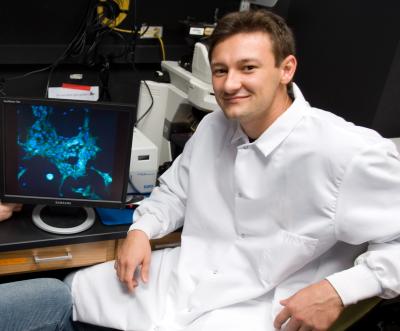
This diagram shows the process by which
scientists can take skin cells from patients
with epilepsy, convert them to stem cells, and
then create neurons (brain nerve cells) from
them. The induced neurons contain the same
genetic mutation(s) carried by the patients.
Credit: Parent lab, University of Michigan
Medical School. 
The findings, reported by a team of scientists from the University of Michigan Medical School and colleagues, use a technique that could be called "epilepsy in a dish".

By turning skin cells of epilepsy patients into stem cells, and then turning those stem cells into neurons, or brain nerve cells, the team created a miniature testing ground for epilepsy. They could even measure the signals that the cells were sending to one another, through tiny portals called sodium channels.

In neurons derived from the cells of children who have a severe, rare genetic form of epilepsy called Dravet syndrome, the researchers report abnormally high levels of sodium current activity. They saw spontaneous bursts of communication and "hyper excitability" that could potentially set off seizures. Neurons made from the skin cells of people without epilepsy showed none of this abnormal activity.

They report their results online in the Annals of Neurology, and have further work in progress to create induced pluripotent stem cell lines from the cells of patients with other genetic forms of epilepsy. The work is funded by the National Institutes of Health, the American Epilepsy Society, the Epilepsy Foundation and U-M.

The new findings differs from what other scientists have seen in mice – demonstrating the importance of studying cells made from human epilepsy patients. Because the cells came from patients, they contained the hallmark seen in most patients with Dravet syndrome: a new mutation in SCN1A, the gene that encodes the crucial sodium channel protein called Nav1.1. That mutation reduces the number of channels to half the normal number in patients' brains.

"With this technique, we can study cells that closely resemble the patient's own brain cells, without doing a brain biopsy," says senior author and team leader Jack M. Parent, M.D., professor of neurology at U-M and a researcher at the VA Ann Arbor Healthcare System.

"It appears that the cells are overcompensating for the loss of channels due to the mutation. These patient-specific induced neurons hold great promise for modelling seizure disorders, and potentially screening medications."

With the new paper, Parent, postdoctoral fellow Yu Liu, Ph.D. and their collaborators Lori Isom, Ph.D., professor of Pharmacology and of Molecular and Integrative Physiology at U-M, and Miriam Meisler, Ph.D., Distinguished University Professor of Human Genetics at U-M, report striking discoveries about what is happening at the cell level in the neurons of Dravet syndrome patients with a mutated SCN1A gene.

They also demonstrated that the effect is rooted in something that happens after function of the gene is reduced due to the mutation, though they don't yet know how or why the nerve cells overcompensate for partial loss of this channel.

And, they found that the neurons didn't show the tell-tale signs of hyper excitability in the first few weeks after they were made – consistent with the fact that children with Dravet syndrome often don't suffer their first seizures until they are several months old.

"In addition, reproduction of the hyperactivity of epileptic neurons in these cell cultures demonstrates that there is an intrinsic change in the neurons that does not depend on input from circuits in the brain," says co-author Meisler.

A platform for testing medications
Many Dravet patients don't respond to current epilepsy medications, making the search for new options urgent. Their lives are constantly under threat by the risk of SUDEP, sudden unexplained death in epilepsy – and they never outgrow their condition, which delays their development and often requires round-the-clock care.

"Working with patient families, and translating our sodium channel research to a paediatric disease, has made our basic science work much more immediate and critical," says Isom, who serves on the scientific advisory board of the Dravet Syndrome Foundation along with Meisler. Parent, who co-directs U-M's Comprehensive Epilepsy Program, was recently honoured by the foundation.

The team is now working toward screening specific compounds for seizure-calming potential in Dravet syndrome, by testing their impact on the cells in the "epilepsy in a dish" model. The National Institutes of Health has made a library of drugs that have been approved by the U.S. Food and Drug Administration available for researchers to use – potentially allowing older drugs to have a second life treating an entirely different disease from what they were initially intended.

Parent and his colleagues hope to identify drugs that affect certain aspects of sodium channels, to see if they can dampen the sodium currents and calm hyper excitability. The team is exploring new techniques that can make this process faster, using microelectrodes and calcium-sensitive dyes. They also hope to use the model to study potential drugs for non-genetic forms of epilepsy.

Having a U-M team that includes experts in induced pluripotent stem cell biology, sodium channel physiology and epilepsy genetics expertise helps the research progress, Parent notes.

"Epilepsy is a complicated brain network disease," he says.

"It takes team-based science to address it."

Patients as part of the research team
The U-M team's research wouldn't be possible without the participation of patients with Dravet syndrome and other genetic forms of epilepsy, and their parents.

More than 100 of them have joined the International Ion Channel Epilepsy Patient Registry, which is based at U-M and Miami Children's Hospital and co-funded by the Dravet Syndrome Foundation and the ICE Epilepsy Alliance. The researchers hope to be able to conduct clinical trials of potential drugs with participation by these patients and others.

Meanwhile, patients with other genetically based neurological diseases can also help U-M scientists discover more about their conditions, by taking part in other efforts to create induced neurons from skin cells. Parent and his team have worked with several other U-M faculty to create stem cell lines from skin cells provided by patients with other diseases including forms of ataxia and lysosomal storage disease.

Contact: Kara Gavin

Reference:
Dravet syndrome patient-derived neurons suggest a novel epilepsy mechanism
Liu, Y., Lopez-Santiago, L. F., Yuan, Y., Jones, J. M., Zhang, H., O'Malley, H. A., Patino, G. A., O'Brien, J. E., Rusconi, R., Gupta, A., Thompson, R. C., Natowicz, M. R., Meisler, M. H., Isom, L. L. and Parent, J. M.
Ann Neurol. (2013), doi: 10.1002/ana.23897
.........


For more on stem cells and cloning, go to CellNEWS at

Monday 22 July 2013

Common Stem Cell in Heart and Lung Development Explains Adaption for Life on Land

Common Stem Cell in Heart and Lung Development Explains Adaption for Life on Land
Monday, 22 July 2013

The evolution of adaptations for life on land have long puzzled biologists – are feathers descendants of dinosaur scales, how did arms and legs evolve from fins, and from what ancient fish organ did the lung evolve?

Biologists have known that the co-development of the cardiovascular and pulmonary systems is a recent evolutionary adaption to life outside of water, coupling the function of the heart with the gas exchange function of the lung. And, the lung is one of the most recent organs to have evolved in mammals and is arguably the most vital for terrestrial life.

The coordinated maturation of the cells of these two systems is illustrated during embryonic development, when the primitive lung progenitor cells protrude into the primitive cardiac progenitor cells as the two organs develop in parallel to form the cardiopulmonary circulation. However, little is known about the molecular cues guiding this simultaneous development, and how a common progenitor cell for both organs may influence the pathology of such related diseases as pulmonary hypertension.

Wnt2+ CPPs (green cells) populate multiple
cell lineages in the developing lung including
airway and vascular smooth muscle. The
smooth muscle of the branching airways
and large blood vessels are stained in red.
Credit: Edward E. Morrisey, Ph.D.,
Perelman School of Medicine, University
of Pennsylvania. 
In a new paper published this week online in Nature, a team from the Perelman School of Medicine, University of Pennsylvania, shows that the pulmonary vasculature, the blood vessels that connect the heart to the lung, develops even in the absence of the lung. Mice in which lung development is inhibited still have pulmonary blood vessels, which revealed to the researchers that cardiac progenitors, or stem cells, are essential for cardiopulmonary co-development.

The Penn team, led by Edward E. Morrisey, PhD, professor of Medicine and Cell and Developmental Biology and scientific director of the Penn Institute for Regenerative Medicine, identified a population of multi-potent CardioPulmonary mesoderm Progenitor cells they named CPPs. The CPPs can be distinguished from many other early embryonic cells by the expression of a well-studied signalling molecule Wnt2.

"We asked if these progenitor cells are capable of generating both heart and lung derivatives," says Morrisey.

"Our data show that Wnt2-positive cells exist prior to lung development and help coordinate lung and heart co-development by generating cell types in both tissues."

The issue of how the lung develops and connects to the cardiovascular system has intrigued the Morrisey lab for many years.

"It's pretty obvious to anyone who has looked at the anatomy of most terrestrial animals that the heart and lung are intimately linked. This is even reflected in clinical medicine where in many places, including the Perelman School of Medicine, the Division of Cardiovascular Medicine was once referred to as the Division of Cardiopulmonary Medicine," notes Morrisey.

The Morrisey lab began with a couple of simple questions: how do the lung and heart co-develop and what are the critical signals that regulate this process? The breakthrough in this work occurred when the team characterized the expression pattern of the Wnt2 gene.

"Wnt2 is expressed in a unique place in the early embryo – exactly in between the early heart and foregut tube, where the lung will arise from."

This allowed the researchers to create a model system in mice, whose cardiopulmonary anatomy is very similar to humans, and ask whether Wnt2-positive cells could coordinate heart and lung co-development.

Using cell lineage tracing analysis, they showed that Wnt2 cells generate single clones that, in turn, generate both heart and lung tissue, including cardiomyocytes and blood vessel cells such as vascular smooth muscle. Indeed, CPPs are capable of generating the vast majority of early embryonic cell types in the heart and lung. These studies also showed that the different cell lineages within the lung are related. For example, vascular smooth muscle and airway smooth muscle share a common progenitor cell in the lung.

The development of CPPs is regulated by the expression of another well-known protein called hedgehog, which is required for proper connection of the pulmonary vasculature to the heart. These studies show that hedgehog, which is also expressed by early lung progenitor cells, helps to promote CPPs to differentiate into the smooth muscle component of the pulmonary vasculature.

Together, these studies identify a novel population of multi-potent cardiopulmonary progenitors that coordinate heart and lung co-development, which is required for adaptation to terrestrial existence.

The finding that CPPs coordinate lung and heart co-development also has important implications for diseases that affect both organs, such as pulmonary hypertension. It is unclear whether pulmonary hypertension is primarily a lung disease or whether there are also intrinsic defects in the heart or cardiovascular system. The identification of CPPs could provide important insight into pulmonary hypertension and other diseases by identifying a common progenitor cell for both organs. Future studies will focus on whether CPPs exist in the adult cardiopulmonary system and whether they play a role in the response of the lung and heart to injury or disease.

Contact: Karen Kreeger

Reference:
Coordination of heart and lung co-development by a multipotent cardiopulmonary progenitor
Tien Peng, Ying Tian, Cornelis J. Boogerd, Min Min Lu, Rachel S. Kadzik, Kathleen M. Stewart, Sylvia M. Evans & Edward E. Morrisey
Nature, Published online 21 July 2013, doi:10.1038/nature12358
.........


For more on stem cells and cloning, go to CellNEWS at

Friday 19 July 2013

Scientists Discover More Versatile Approach to Creating Stem Cells

New method should hasten promise of regenerative medicine
Friday, 19 July 2013

Stem cells are key to the promise of regenerative medicine: the repair or replacement of injured tissues with custom grown substitutes. Essential to this process are induced pluripotent stem cells (iPSCs), which can be created from a patient's own tissues, thus eliminating the risk of immune rejection. However, Shinya Yamanaka's formula for iPSCs, for which he was awarded last year's Nobel Prize, uses a strict recipe that allows for limited variations in human cells, restricting their full potential for clinical application.

From left: Emmanuel Nivet Martinez and Juan
Carlos Belmonte. Seated: Ignacio Sancho
Martinez. Credit: Courtesy of the Salk Institute
for Biological Studies. 
Now, in this week's issue of Cell Stem Cell, the Salk Institute's Juan Carlos Izpisua Belmonte and his colleagues show that the recipe for iPSCs is far more versatile than originally thought. For the first time, they have replaced a gene once thought impossible to substitute, creating the potential for more flexible recipes that should speed the adoption of stem cells therapies.

Stem cells come in two types: embryonic stem cells (ESCs), which are immature cells that have never differentiated into specific cell types, and induced pluripotent stem cells, which are mature cells that have been reprogrammed back into an undifferentiated state. After the initial discovery in 2006 by Yamanaka that introducing four different genes into a mature cell could suffice for reprogramming the cell to pluripotency, most researchers adopted his recipe.

Izpisua Belmonte and his colleagues took a fresh approach and discovered that pluripotency (the stem cell's ability to differentiate into nearly any kind of adult cell) can also be accomplished by balancing the genes required for differentiation. These genes code for "lineage transcription factors," proteins that start a stem cell down the path to differentiate first into a particular cell lineage, or type, such as a blood cell versus a skin cell, and then finally into a specific cell, such as a white blood cell.

"Prior to this series of experiments, most researchers in the field started from the premise that they were trying to impose an 'embryonic-like' state on mature cells," says Izpisua Belmonte, who holds the Institute's Roger Guillemin Chair.

"Accordingly, major efforts had focused on the identification of factors that are typical of naturally occurring embryonic stem cells, which would allow or further enhance reprogramming."

The picture shows newly reprogrammed cells
expressing marks of pluripotency as identified
by fluorescence (NANOG in green, TRA-1-81
in red). Credit: Courtesy of the Salk Institute
for Biological Studies.
Despite these efforts, there seemed to be no way to determine through genetic identity alone that cells were pluripotent. Instead, pluripotency was routinely evaluated by functional assays. In other words, if it acts like a stem cell, it must be a stem cell.

That condition led the team to their key insight.

"Pluripotency does not seem to represent a discrete cellular entity but rather a functional state elicited by a balance between opposite differentiation forces," says Izpisua Belmonte.

Once they understood this, they realized the four extra genes weren't necessary for pluripotency. Instead, it could be achieved by altering the balance of "lineage specifiers," genes that were already in the cell that specified what type of adult tissue a cell might become.

"One of the implications of our findings is that stem cell identity is actually not fixed but rather an equilibrium that can be achieved by multiple different combinations of factors that are not necessarily typical of ESCs," says Ignacio Sancho-Martinez, one of the first authors of the paper and a postdoctoral researcher in Izpisua Belmonte's laboratory.

The group was able to show that more than seven additional genes can facilitate reprogramming to iPSCs. Most importantly, for the first time in human cells, they were able to replace a gene from the original recipe called Oct4, which had been replaced in mouse cells, but was still thought indispensable for the reprogramming of human cells. Their ability to replace it, as well as SOX2, another gene once thought essential that had never been replaced in combination with Oct4, demonstrated that stem cell development must be viewed in an entirely new way.

"It was generally assumed that development led to cell/tissue specification by 'opening' certain differentiation doors," says Emmanuel Nivet, a post-doctoral researcher in Izpisua Belmonte's laboratory and co-first author of the paper, along with Sancho-Martinez and Nuria Montserrat of the Center for Regenerative Medicine in Barcelona, Spain.

Instead, the successful substitution of both Oct4 and SOX2 shows the opposite.

"Pluripotency is like a room with all doors open, in which differentiation is accomplished by 'closing' doors," Nivet says.

"Inversely, reprogramming to pluripotency is accomplished by opening doors."

The team believes their work should help to overcome one of the major hurdles to the widespread adoption of stem cell therapies: the original four genes used to reprogram stem cells had been implicated in cancer.

"Recent studies in cancer, many of them done by my Salk colleagues, have shown molecular similarities between the proliferation of stem cells and cancer cells, so it is not surprising that oncogenes [genes linked to cancer] would be part of the iPSC recipe," says Izpisua Belmonte.

With this new method, which allows for a customized recipe, the team hopes to push therapeutic research forward.

"Since we have shown that it is possible to replace genes thought essential for reprogramming with several different genes that have not been previously involved in tumorigenesis, it is our hope that this study will enable iPSC research to more quickly translate into the clinic," says Izpisua Belmonte.

Contact: Kat Kearney 

Reference:
Reprogramming of human fibroblasts to pluripotency with lineage specifiers
Nuria Montserrat, Emmanuel Nivet, Ignacio Sancho-Martinez, Tomoaki Hishida, Sachin Kumar, Laia Miquel, Carme Cortina, Yuriko Hishida, Yun Xia, Concepcion Rodriguez Esteban and Juan Carlos Izpisua Belmonte
Cell Stem Cell, 18 July 2013, doi:10.1016/j.stem.2013.06.019
.........


For more on stem cells and cloning, go to CellNEWS at
http://cellnews-blog.blogspot.com/

Friday 12 July 2013

Nuclear Pore Complex: How to Build Your Gate

New approach solves large molecular puzzles
Friday, 12 July 2013

At first glance, this may look like the latest view
from a telescope, but it was taken by looking not
out at the vastness of space, but into a human
cell. What could pass for stars or distant galaxies
are actually gates to the genome that is locked up
inside the cell's nucleus, as imaged by a super-
resolution microscope. By combining images of
thousands of these gates, called nuclear pore
complexes, scientists at the European Molecular
Biology Laboratory (EMBL) in Heidelberg,
Germany, have solved a decade-old controversy
over how the pieces that form each pore's ring are
arranged. Credit: EMBL/A.Szymborska. 
It's a parent's nightmare: opening a Lego set and being faced with 500 pieces, but no instructions on how to assemble them into the majestic castle shown on the box. Thanks to a new approach by scientists at the European Molecular Biology Laboratory (EMBL) in Heidelberg, Germany, researchers studying large sets of molecules with vital roles inside our cells can now overcome a similar problem.

In a study published online today in Science, the scientists used super-resolution microscopy to solve a decade-long debate about the structure of the nuclear pore complex, which controls access to the genome by acting as a gate into the cell's nucleus.

Like the bewildered parent staring at the image on the box, scientists knew the gate's overall shape, from electron tomography studies. And thanks to techniques like X-ray crystallography and single particle electron microscopy, they knew that the ring which studs the nucleus' wall and controls what passes in and out is formed by sixteen or thirty-two copies of a Y-shaped building block. They even knew that each Y is formed by nine proteins. But how the Y’s are arranged to form a ring was up for debate.

"When we looked at our images, there was no question: they have to be lying head-to-tail around the hole" says Anna Szymborska, who carried out the work.

To figure out how the Y’s were arranged, the EMBL scientists used fluorescent tags to label a series of points along each of the Y's arms and tail, and analysed them under a super-resolution microscope. By combining images from thousands of nuclear pores, they were able to obtain measurements of where each of those points was, in relation to the pore's centre, with a precision of less than a nanometre – a millionth of a millimetre. The result was a rainbow of rings whose order and spacing meant the Y-shaped molecules in the nuclear pore must lie in an orderly circle around the opening, all with the same arm of the Y pointing toward the pore's centre.

Having resolved this decade-old controversy, the scientists intend to delve deeper into the mysteries of the nuclear pore – determining whether the circle of Y’s is arranged clockwise or anticlockwise, studying it at different stages of assembly, looking at other parts of the pore, and investigating it in three dimensions.

"There's been a lot of interest from other groups, so we'll soon be looking into a number of other molecular puzzles, like the different 'machines' that allow a cell to divide, which are also built from hundreds of pieces," says Jan Ellenberg, who led the work.

The work was carried out in collaboration with John Briggs' group at EMBL, who helped adapt the image averaging algorithms from electron microscopy to super-resolution microscopy, and Volker Cordes at the Max Planck Institute for Biophysical Chemistry in Göttingen, Germany, who provided antibodies and advice.

Contact: Sonia Furtado Neves

Reference:
Nuclear Pore Scaffold Structure Analyzed by Super-Resolution Microscopy and Particle Averaging
Szymborska, A., Marco, A., Daigle, N., Cordes, V.C., Briggs, J.A.G. & Ellenberg, J.
Science Express on 11 July 2013. DOI: 10.1126/science.1240672
..........


For more on stem cells and cloning, go to CellNEWS at

Thursday 11 July 2013

Researchers Create the Inner Ear from Stem Cells

Researchers Create the Inner Ear from Stem Cells
Thursday, 11 July 2013

Indiana University scientists have transformed mouse embryonic stem cells into key structures of the inner ear. The discovery provides new insights into the sensory organ's developmental process and sets the stage for laboratory models of disease, drug discovery and potential treatments for hearing loss and balance disorders.

A research team led by Eri Hashino, Ph.D., Ruth C. Holton Professor of Otolaryngology at Indiana University School of Medicine, reported that by using a three-dimensional cell culture method, they were able to coax stem cells to develop into inner-ear sensory epithelia - containing hair cells, supporting cells and neurons - that detect sound, head movements and gravity. The research was reportedly online Wednesday in the journal Nature.

Stem cell-derived sensory hair cells
Stem cell-derived sensory hair cells are in red
with hair bundles in green. Cellular nuclei are
shown in blue. Credit: IU Communications.
Previous attempts to "grow" inner-ear hair cells in standard cell culture systems have worked poorly in part because necessary cues to develop hair bundles - a hallmark of sensory hair cells and a structure critically important for detecting auditory or vestibular signals - are lacking in the flat cell-culture dish. But, Dr. Hashino said, the team determined that the cells needed to be suspended as aggregates in a specialized culture medium, which provided an environment more like that found in the body during early development.

The team mimicked the early development process with a precisely timed use of several small molecules that prompted the stem cells to differentiate, from one stage to the next, into precursors of the inner ear. But the three-dimensional suspension also provided important mechanical cues, such as the tension from the pull of cells on each other, said Karl R. Koehler, B.A., the paper's first author and a graduate student in the medical neuroscience graduate program at the IU School of Medicine.

"The three-dimensional culture allows the cells to self-organize into complex tissues using mechanical cues that are found during embryonic development," Koehler said.

"We were surprised to see that once stem cells are guided to become inner-ear precursors and placed in 3-D culture, these cells behave as if they knew not only how to become different cell types in the inner ear, but also how to self-organize into a pattern remarkably similar to the native inner ear," Dr. Hashino said.

Karl Koehler and Eri Hashino, Ph.D.
Karl Koehler and Eri Hashino, Ph.D. Credit:
IU Communications.
"Our initial goal was to make inner-ear precursors in culture, but when we did testing we found thousands of hair cells in a culture dish."

Electrophysiology testing further proved that those hair cells generated from stem cells were functional, and were the type that sense gravity and motion. Moreover, neurons like those that normally link the inner-ear cells to the brain had also developed in the cell culture and were connected to the hair cells.

Additional research is needed to determine how inner-ear cells involved in auditory sensing might be developed, as well as how these processes can be applied to develop human inner-ear cells, the researchers said.

However, the work opens a door to better understanding of the inner-ear development process as well as creation of models for new drug development or cellular therapy to treat inner-ear disorders, they said.

Contact: Eric Schoch

Reference:
Generation of inner ear sensory epithelia from pluripotent stem cells in 3D culture
Karl R. Koehler, Andrew M. Mikosz, Andrei I. Molosh, Dharmeshkumar Patel & Eri Hashino
Nature (2013), doi:10.1038/nature12298
.........


For more on stem cells and cloning, go to CellNEWS at

Wednesday 10 July 2013

Micro-particles Create Localized Control of Stem Cell Differentiation

Engineering stem cells
Wednesday, 10 July 2013

Before scientists and engineers can realize the dream of using stem cells to create replacements for worn out organs and battle damaged body parts, they'll have to develop ways to grow complex three-dimensional structures in large volumes and at costs that won't bankrupt health care systems.

Researchers are now reporting advances in these areas by using gelatine-based microparticles to deliver growth factors to specific areas of embryoid bodies, aggregates of differentiating stem cells. The localized delivery technique provides spatial control of cell differentiation within the cultures, potentially enabling the creation of complex three-dimensional tissues. The local control also dramatically reduces the amount of growth factor required, an important cost consideration for manufacturing stem cells for therapeutic applications.

Georgia Tech/Emory University Associate
Professor Todd McDevitt and graduate student
Anh Nguyen make microparticles to be used
for delivering growth factors to stem cells.
CreditGeorgia Tech Photo: Rob Felt.
The microparticle technique, which was demonstrated in pluripotent mouse embryonic cells, also offers better control over the kinetics of cell differentiation by delivering molecules that can either promote or inhibit the process. Based on research sponsored by the National Institutes of Health and the National Science Foundation, the developments were reported online July 1 in the journal Biomaterials and were presented at the 11th Annual International Society for Stem Cell Research meeting held in Boston June 12-15, 2013 .

"By trapping these growth factors within microparticle materials first, we are concentrating the signal they provide to the stem cells," said Todd McDevitt, an associate professor in the Wallace H. Coulter Department of Biomedical Engineering at Georgia Tech and Emory University.

"We can then put the microparticle materials physically inside the multicellular aggregate system that we use for differentiation of the stem cells. We have good evidence that this technique can work, and that we can use it to provide advantages in several different areas."

The differentiation of stem cells is largely controlled by external cues, including morphogenic growth factors, in the three-dimensional environment that surrounds the cells. Most stem cell researchers currently deliver the growth factors into liquid solutions surrounding the stem cell cultures with a goal of creating homogenous cultures of cells. Delivering the growth factors from microparticles, however, provides better control of the spatial and temporal presentation of the molecules that govern the growth and differentiation of the stem cells, potentially allowing formation of heterogeneous structures formed from different cells.

Groups of stem cells stick together as they develop, forming multicellular aggregates that form spheroids as they grow. The researchers took advantage of that by driving microparticles containing growth factor BMP4 or noggin – which inhibits BMP4 signalling – into layers of stem cells using centrifugation. When the cell aggregates formed, the microparticles became trapped inside.

The researchers used confocal imaging and flow cytometry to observe the differentiation process and found that growth factors in the microparticles directed the cells toward mesoderm and ectoderm tissues just as they do in solution-based techniques. But because the BMP4 and noggin molecules were directly in contact with the cells, much less growth factor was needed to spur the differentiation – approximately 12 times less than what would be required by conventional solution-based techniques.

"One of the major advantages, in a practical sense, is that we are using much less growth factor," said McDevitt, who is also director of the Stem Cell Engineering Center at Georgia Tech.

"From a bioprocessing standpoint, a lot of the cost involved in making stem cell products is related to the cost of the molecules that must be added to make the stem cells differentiate."

Beyond more focused signalling, the microparticles also provided a localized control not available through any other technique. That allowed the researchers to create spatial differences in the aggregates – a possible first step toward forming more complex structures with different tissue types such as vasculature and stromal cells.

Andres Bratt-Leal, a former graduate student
in the laboratory of Todd McDevitt, analyzes
stem cells using a microscope. Credit: Georgia
Tech Photo: Rob Felt.
"To build tissues, we need to be able to take stem cells and use them to make many different cell types which are grouped together in particular spatial patterns," explained Andres M. Bratt-Leal, the paper's first author and a former graduate student in McDevitt's lab.

"This spatial patterning is what gives tissues the ability to perform higher order functions."

After creating stem cell aggregates with microparticles containing different growth factors, the researchers observed a hemispherical organization of cells for several days, with the different cells remaining spatially segregated.

"We can see the microparticles had effects on one population that were different from the population that didn't have the particles," McDevitt said.

"This may allow us to emulate aspects of how development occurs. We can ask questions about how tissues are naturally patterned. With this material incorporation, we have the ability to better control the environment in which these cells develop."

The microparticles could also provide better control over the kinetics of cell differentiation. Including different amounts of molecules – one the growth factor and the other its antagonist – could vary the rate at which the stem cell differentiation proceeds.

While the research reported in this paper manipulated pluripotent mouse cells, the researchers have moved ahead in performing similar studies with human stem cells and achieved comparable types of results with the microparticle delivery approaches.

The developments not only help move stem cell technologies closer to the clinic, but also provide a new tool for research.

"Our findings will provide a significant new tool for tissue engineering, bioprocessing of stem cells and also for better studying early development processes such as axis formation in embryos," said Bratt-Leal.

"During development, particular tissues are formed by gradients of signalling molecules. We can now better mimic these signal gradients using our system."

Contact: John Toon

Reference:
.........


For more on stem cells and cloning, go to CellNEWS at
http://cellnews-blog.blogspot.com/

Tuesday 9 July 2013

First Child Born Following New Genome Embryo Screening

Validation studies underline accuracy of 'next generation sequencing' for embryo selection in IVF
Tuesday, 09 July 2013

The first birth has been achieved following the analysis of embryos using a new genome sequencing technique which promises to revolutionise embryo selection for IVF. The technique, which has never before been applied in the screening of embryos, is reported today at the annual meeting of ESHRE by Dr Dagan Wells of the NIHR Biomedical Research Centre at the University of Oxford, UK.

The analysis technique is known as "next generation sequencing", a powerful method capable of decoding entire genomes. Vast quantities of DNA data are produced from each sample tested, simultaneously revealing information on the inheritance of genetic disorders, chromosome abnormalities and mitochondrial mutations. Next generation sequencing (NGS) is already revolutionising many areas of genetic research and diagnostics, said Dr Wells, and, when applied to the assessment of embryos, will allow the concurrent analysis of serious inherited disorders and lethal chromosome abnormalities.

"Next generation sequencing provides an unprecedented insight into the biology of embryos," said Dr Wells.

The identification of an embryo destined to implant in the uterus and form a pregnancy remains the holy grail of IVF. On average, only around 30% of embryos currently selected for transfer actually implant. The reason for this high failure rate is unknown, but the prime suspects are unidentified genetic or chromosomal defects. Several genetic screening methods have been introduced over the past decade, but all have been shown to have drawbacks (and have not realised their potential) when tested in randomised clinical trials. This new NGS technique developed by Dr Wells and colleagues, however, seems to overcome the major drawbacks of current methods: 
  • Complete chromosome information can be produced revealing abnormalities often responsible for miscarriage;
  • Serious gene defects can be identified at the same time;
  • The analysis can be completed rapidly (around 16 hours), thus avoiding the need for embryo freezing while awaiting results;
  • The test could greatly reduce the costs of embryo screening, which is currently an expensive add-on to IVF.
The study described today was designed to test the accuracy and predictability of NGS in embryo selection. The validation was performed on multiple cells from cell-lines with known chromosome abnormalities, gene defects (cystic fibrosis) or mitochondrial DNA mutations.

Additionally, cells from 45 embryos, previously shown to be abnormal with another testing technique, were reanalysed by NGS in a blinded fashion. After high accuracy had been demonstrated, the method was applied clinically, with cells sampled from seven five-day-old embryos (blastocysts) produced by two couples undergoing IVF. The mothers were 35 and 39 years of age and one couple had a history of miscarriage.

NGS analysis in these two IVF patients identified three chromosomally healthy blastocysts in the first and two in the second; single embryo transfers based upon these results led to healthy pregnancies in both cases. The first pregnancy ended with the delivery of a healthy boy in June.

Dr Wells, who led the international research team behind the study, said:

"Many of the embryos produced during infertility treatments have no chance of becoming a baby because they carry lethal genetic abnormalities. Next generation sequencing improves our ability to detect these abnormalities and helps us identify the embryos with the best chances of producing a viable pregnancy. Potentially, this should lead to improved IVF success rates and a lower risk of miscarriage.”

"In the past few years, results from randomised clinical trials have suggested that most IVF patients would benefit from embryo chromosome screening, with some studies reporting a 50% boost in pregnancy rates. However, the costs of these genetic tests are relatively high, putting them beyond the reach of many patients. Next generation sequencing is a way which could make chromosome testing more widely available to a greater number of patients, improving access by cutting the costs. Our next step is a randomised clinical trial to reveal the true efficacy of this approach - and this will begin later this year."

Contact: Christine Bauquis
.........


For more on stem cells and cloning, go to CellNEWS at