Saturday 28 February 2009

Microenvironments Key to Future Stem Cell Therapies

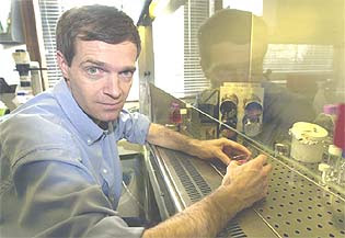
Unique microenvironment microarrays give new results Saturday, 28 February 2009 Medical researchers are praising adult stem cells and their more committed kin, progenitor cells, for their ability to produce different types of specialized cells. The potential of using these cells to repair or replace damaged tissue holds great promise for cancer therapies and regenerative medicine. However, the question that must first be answered is what determines the ultimate fate of a stem or progenitor cell? A team of researchers led by Berkeley Lab's Mark LaBarge and Mina Bissell appear to be well on the road to finding out. Mark LaBarge used micro patterning technology to imprint thousands of molecular combinations onto a glass microscope slide to mimic microenvironmental conditions within the human breast. Credit: Photo by Roy Kaltschmidt, Berkeley Lab Public Affairs.Working with unique microenvironment microarrays (MEArrays) of their own creation, LaBarge, Bissell, and their collaborators have shown that the ultimate fate of a stem or progenitor cell in a woman's breast – whether the cell develops normally or whether it turns cancerous – may depend upon signals from multiple microenvironments. "We found that adult human mammary stem and progenitor cells exhibit impressive plasticity in response to hundreds of unique combinatorial microenvironments," said LaBarge, a cell and molecular biologist in Berkeley Lab's Life Sciences Division. "Our results further suggest that rational modulation of the microenvironmental milieu can impose specific differentiation phenotypes on normal stem or progenitor cells, and perhaps even impose phenotypically normal behaviour on malignant cells during tissue genesis. All of this points to the rational manipulation of adult stem and progenitor cells as a promising pathway for beneficial therapies." Previous studies on how microenvironments affect the development of adult human stem or progenitor cells have been based on the behaviour of these cells in culture (in vitro) where they are exposed to a single molecular agent. However, when these cells are in an actual human being (in vivo) they are surrounded by a multitude of other cells plus a supporting network of fibrous and globular proteins called the extracellular matrix (ECM), as well as many other nearby molecules, all of which may be simultaneously sending them instructional signals. "With our MEArrays, we can use combinations of proteins from a select tissue to create multiple microenvironments on a single chip about two square centimetres in area," said LaBarge. "We think this approach will give us a much more realistic picture as to how stem and progenitor cells actually behave in vivo." "We have demonstrated that each discrete cell fate decision requires the integration of multiple pathways, and we have identified combinations of components in the human mammary microenvironment that impose distinct cell fates. These results are exciting because they indicate that we can test a large number of effectors and determine which ones to use to direct the fate of adult stem and progenitor cells. This give hope that one day - sooner rather than later - the information could be used for therapy," said Bissell, a Distinguished Scientist with Berkeley Lab's Life Sciences Division and one of the world's leading researchers on breast cancer. Collaborating with LaBarge and Bissell on this study were Jason Ruth, now at the University of Pennsylvania, Martha Stampfer of Berkeley Lab, Celeste Nelson, now with Princeton University, and Rene Villadsen, Agla Fridriksdottir and Ole Petersen, of the Panum Institute in Denmark. Human breast tissue harbours two types of epithelial cells: luminal - the cells that are able to produce milk and generally the ones that become cancerous; and myoepithelial – the cells that surround the luminal cells and push milk down the ducts to the nipples, but which rarely become cancerous. Like cells in other types of tissue these breast epithelial cells are spawned from stem and progenitor cells that despite being primitive - essentially a cellular blank slate - possess the exact same genome as their differentiated daughters. Once it was widely held that adult stem and progenitor cells intrinsically "know" when to self-renew and when to differentiate into one specific tissue cell or another based on pre-determined genetic programs. However, pioneering research by Bissell, in which it has been demonstrated that interactions between an epithelial breast cell and its ECM play a major role in determining whether that cell becomes cancerous, pointed the way to the idea that the ultimate fate of a stem or progenitor cell is heavily influenced by interactions with its neighbouring microenvironments.


Normal mammary gland.This is a section of a normal mammary gland that has been stained to find the terminal duct zones that are enriched for mammary stem cells (red) and to visualize a Notch signalling pathway (green). Impairing the Notch pathway caused malignant breast cancer cells to revert to a normal phenotype. Credit: Mark LaBarge.
"Adult stem cells are maintained inside a specialized microenvironment called a niche, whereas progenitor cells migrate to surrounding microenvironments that are distinct from the one around the niche," said LaBarge. "The ability of adult stem cells to self-maintain, as well as to give rise to progenitor cells that are targeted to become a specific tissue cell, indicates an ability to respond to changing microenvironmental demands, which would mean that a stem or progenitor cell is receiving instructional information from its surroundings." The fact that normal cells often lose their tissue-specific functions when placed in culture is further evidence of cell fate being tied in to signals from the microenvironment. However, proving such a hypothesis has been difficult in the past because the composition of cell microenvironments is extremely complex and requires a method by which a combination of carefully choreographed interactions can be observed. Given that experiments with human adult stem cell niches cannot be done in vivo and that scientists can only learn so much from mouse models, this means that cell culture studies must be done under as close as possible to in vivo conditions. "Our technology mimics actual in vivo conditions and enables us to perform highly parallel functional analysis of combinatorial microenvironments, and image analysis of 3-D organotypic cultures and micro patterned culture substrata," said LaBarge. "The 3-D capability is crucial because our studies show that orientation of the stem or progenitor cells with respect to the signalling molecules can be critical to what happens next." The MEArrays were fabricated using micro patterning technology originally adapted by co-author Nelson that LaBarge "tweaked." A robot imprinted arrays of 2,304 individual combinations of molecules onto a rubber-coated glass microscope slide (the rubber facilitates adsorption of the proteins onto the slide). An individual MEArray consisted of 192 unique combinatorial microenvironments replicated 12 times, with a plastic barrier running along the perimeter so that cell cultures could be placed on top. In addition to possible contributors to the stem cell niche, the microenvironments also comprised many ECM and signalling molecules that are expressed in the breast but had not been directly linked to stem cell function before. In all, adult mammary stem and progenitor cells were exposed to 8,000 different combinations of breast tissue protein and biological molecules. LaBarge, Bissell and their collaborators were able to distinguish between effects resulting from cell interactions with other cells and those resulting from cell interactions with the ECM or other signalling molecules. Both immortalized and primary human breast progenitors were analyzed with the MEArrays and the results were used in conjunction with physiologically relevant 3-D human breast cultures. This approach enabled the research team to identify conditions that induced cells to convert into normal breast cell types as well as conditions that kept the cells in their original, non-specialized state. One of the most intriguing results in this study was the suggestion that modulation of stem and progenitor cell differentiation pathways might be used to "normalize" malignant breast cells. "Normal and malignant mammary epithelial cells in 3-D cultures have distinct phenotypes," LaBarge said. "By impairing a signalling pathway known as Notch, we are able to revert malignant breast cancer cells to a normal phenotype." In previous studies, Bissell and her group had identified signalling pathways that could cause "phenotypic reversion" of breast cancer cells but this had never been tried before with stem cells. "The MEArray approach may be able to teach us how to direct stem cell function in a therapeutic setting and possibly to re-program non-stem cells to acquire other stem cell fates," said Bissell. While the MEArrays in this study were used to study adult stem and progenitor cells in breast tissue, the technique should also be applicable to any of the other 200 different types of tissue cells within other organs, LaBarge said. ......... ZenMaster
For more on stem cells and cloning, go to CellNEWS at http://cellnews-blog.blogspot.com/ and http://www.geocities.com/giantfideli/index.html

Researchers Piggyback to Safer Reprogrammed Stem Cells

Researchers Piggyback to Safer Reprogrammed Stem Cells Saturday, 28 February 2009 Austin Smith.Austin Smith and his research team at the Centre for Stem Cell Research in Cambridge have just published in the journal Development a new and safer way of generating pluripotent stem cells – the stem cells that can give rise to every tissue of the body. Rapid developments in stem cell research in recent years have provided a way for stem cell scientists to convert specialised cells, such as skin cells, into stem cells that can form numerous cell types of the body. Research into the creation of these 'reprogrammed' cells – so-called induced pluripotent stem (iPS) cells – is of vital importance because it could lead to new ways of creating human stem cells from adult tissues for the study and treatment of disease. But there is one key problem with the techniques currently used to generate such stem cells: they rely on potentially harmful viruses to deliver the reprogramming factors that change specialised cells into iPS cells. Now Austin Smith and his team report in the journal Development an approach that avoids the use of such viruses. They successfully persuaded partly specialised mouse cells, called Epi-stem cells, to reprogram into iPS cells using a single reprogramming factor called Klf4. Instead of relying on viruses to introduce Klf4 into the Epi-stem cells, they turned to a special type of DNA, called a transposable element, which can insert itself into an organism's DNA and carry a cargo with it, in this case Klf4. The transposable element Smith and colleagues used in their study is called Piggybac, which delivered a single copy of Klf4 into the Epi-stem cells, causing them to reprogram into iPS cells. The researchers then used an enzyme to cut the Klf4 out of Piggybac. In doing so, they discovered that the iPS cells could maintain themselves using their own Klf4 gene, which had been switched on during the reprogramming process. Once the Piggybac Klf4 is removed, they report, iPS cells can go on to create normal mice when introduced into newly developing mouse embryos and can give rise to the offspring of these mice by contributing to their reproductive cells. This is the most stringent test of the normality of iPS cells. As Professor Smith explains below, this is a significant advance in the field. "The paper we've published in Development, together with two other publications in Nature”, says Professor Smith, “is a significant technical development in the field as together these papers present a more reliable and precise method for generating iPS cells. The method allows for greater control over the genetic modification process and this is fully reversible once reprogramming is complete. Therefore, the final iPS cells carry no potentially damaging foreign DNA. Our findings published in Development show that this approach produces perfectly reprogrammed mouse cells. The Nature papers show that it can also work in human cells. These studies provide a new tool to help advance basic research into reprogramming and pave the way to the creation of human iPS cells suitable for biomedical applications." Reference: Klf4 reverts developmentally programmed restriction of ground state pluripotency Ge Guo, Jian Yang, Jennifer Nichols, John Simon Hall, Isobel Eyres, William Mansfield, and Austin Smith Development, 18 Feb 2009, doi: 10.1242/dev.030957 ......... ZenMaster


For more on stem cells and cloning, go to CellNEWS at http://cellnews-blog.blogspot.com/ and http://www.geocities.com/giantfideli/index.html

Self-digestion as a Means of Survival

How cells recognize and deal with protein waste Saturday, 28 February 2009 In times of starvation, cells tighten their belts: they start to digest their own proteins and cellular organs. The process - known as autophagy - takes place in special organelles called autophagosomes. It is a strategy that simple yeast cells have developed as a means of survival when times get tough, and in the course of evolution, it has become a kind of self-cleaning process. In mammalian cells, autophagosomes are also responsible for getting rid of misfolded proteins, damaged organelles or disease-causing bacteria. If this process malfunctions, it can result in infectious diseases, as well as cancer, Parkinson's or Alzheimer's disease. Biochemists at Frankfurt's Goethe University, working together with scientists from the University of Tromsø in Norway, the Weizmann Institute in Israel and the Tokyo Metropolitan Institute in Japan have just come up with an explanation as to how autophagosomes know exactly which proteins and organelles they should degrade. "Although autophagy has been known for more than 30 years, it is astonishing that no-one thought of looking for the receptors that make this process so selective" explains Prof. Ivan Dikic from the Institute of Biochemistry II and the Cluster of Excellence 'Macromolecular Complexes' in Frankfurt. He had a head start in this field, since over several years, he and his group have researched and now published their work on another self-cleaning process in the cell: the degradation of small proteins in the proteasome, which acts as a kind of molecular shredder. "We know that the molecules which are destined to be discarded are marked with the small protein ubiquitin and this is recognised by a receptor located at the gateway to the proteasome. It was natural to suggest a similar recognition mechanism for protein degradation by autophagosomes", says Dikic. Unlike the proteasome, which is a complex molecular machine, autophagosomes simply consist of a double membrane that floats around in the cytoplasm. Not unlike white blood cells, they can engulf larger proteins or even whole cell organelles. But since they have no enzymes with which they can digest their own cargo, they fuse with lysosomes. When a Yoshinori Ohsumi's group in Japan reported that they had discovered ubiquitin-like proteins (ATG8) on the outer surface of the autophagosome and gone on to prove that they were specific for autophagy, Dikic and his colleague Dr. Vladimir Kirkin immediately began their search for potential autophagy receptors that might bind to the family of ATG8 proteins. The team of international scientists report in the current issue of the renowned journal "Molecular Cell", that by employing methods from cell biology, biochemistry and mouse genetics, they have been able to identify a further protein, in addition to the known p62/SQSTM1 protein, that may act as a receptor. This is the protein NBR1, which has long been associated with cancer. Both proteins have a similar chain-like structure. At one end, they bind to the ubiquitin that marks the protein aggregates and organelles that are to be degraded. Next to the ubiquitin-binding site is a domain that binds to the ATG8 proteins found at the autophagosomal membrane. Here, the protein waste can dock onto the autophagosome and can then be wrapped up in the membrane. Vladimir Kirkin, who is now at Merck Serono in Darmstadt, is continuing these investigations with the long-term aim of developing new drugs. Dikic and his group are now concentrating on mitochondria - which are implicated in oxidative stress in cells - hoping to locate the receptors for autophagy on these important organelles. Reference: A Role for NBR1 in Autophagosomal Degradation of Ubiquitinated Substrates Vladimir Kirkin, Trond Lamark, Yu-Shin Sou, Geir Bjorkoy, Jennifer L. Nunn, Jack-Ansgar Bruun, Elena Shvets, David G. McEwan, Terje H. Clausen, Philipp Wild, Ivana Bilusic, Jean-Philippe Theurillat, Aud Overvatn, Tetsuro Ishii, Zvulun Elazar, Masaaki Komatsu,I van Dikic and Terje Johansen Molecular Cell, Volume 33, Issue 4, 505-516, 27 February 2009, doi:10.1016/j.molcel.2009.01.020 ......... ZenMaster


For more on stem cells and cloning, go to CellNEWS at http://cellnews-blog.blogspot.com/ and http://www.geocities.com/giantfideli/index.html

Friday 27 February 2009

Spun-sugar Fibres Spawn Sweet Technique for Nerve Repair

Spun-sugar Fibres Spawn Sweet Technique for Nerve Repair Friday, 27 February 2009 Researchers at Purdue University have developed a technique using spun-sugar filaments to create a scaffold of tiny synthetic tubes that might serve as conduits to regenerate nerves severed in accidents or blood vessels damaged by disease. The sugar filaments are coated with a corn-based degradable polymer, and then the sugar is dissolved in water, leaving behind bundles of hollow polymer tubes that mimic those found in nerves, said Riyi Shi, an associate professor in Purdue's Weldon School of Biomedical Engineering and Department of Basic Medical Sciences. The scaffold could be used to promote nerve regeneration by acting as a bridge placed between the ends of severed nerves, said biomedical engineering doctoral student Jianming Li, who is a member of Shi's research team that developed the technique.


Coated sugarstrands.Purdue researchers have developed a technique using sugar filaments spun like cotton candy and coated with a polymer to create a scaffold of tiny synthetic tubes that might serve as conduits to regenerate nerves severed in accidents or damaged by disease. The image on the left, taken with a scanning electron microscope and artificially coloured, shows the sugar strands in yellow and the polymer coating in blue. Images on the right, taken with the same instrument, show a side view of the tubes and tiny pores that are ideal for supplying nutrients to growing nerve cells and removing waste products from the cells. Credit: Weldon School of Biomedical Engineering, Department of Basic Medical Sciences and the Center for Paralysis Research, Purdue University.
The researchers are initially concentrating on the peripheral nerves found in the limbs and throughout the body because nerve regeneration is more complex in the spinal cord. About 800,000 peripheral nerve injuries are reported annually in the United States, with about 50,000 requiring surgery. The approach also might have applications in repairing blood vessels damaged by trauma and disease such as atherosclerosis and diabetes, Shi said. The new approach represents a potential alternative to the conventional surgical treatment, which uses a nerve "autograft" taken from the leg or other part of the body to repair the injured nerves. Researchers are trying to develop artificial scaffolds to replace the autografts because removing the donor nerve causes a lack of sensation in the portion of the body where it was removed. "The autograft is the lesser of two evils because you have to sacrifice a healthy nerve to repair a damaged segment," said Li, who led the research. New findings were published online in December and this month in the print edition of the journal Langmuir. The paper was written by Li, biomedical engineering doctoral student Todd A. Rickett and Shi. Rickett also is attending the Indiana University School of Medicine in an MD-Ph.D. program. Researchers from Cornell University published similar findings online Feb. 9 in the journal Soft Matter. Those findings focused on using the technique to create vascular networks for providing blood and nutrients to tissues and grafts.
Schwann cells (on left) growing on a tubule.Researchers at Purdue have developed a technique using sugar filaments spun like cotton candy and coated with a polymer to create a scaffold of tiny synthetic tubes that might serve as conduits to regenerate nerves severed in accidents or damaged by disease. These images, taken with fluorescent-dyed samples, show nerve-insulating cells called Schwann cells (on left) growing on a tubule, and a combination of Schwann cells and neurons aligned lengthwise along the tubes (on right). This alignment is critical for the fast growth of nerves. Credit: Weldon School of Biomedical Engineering, Department of Basic Medical Sciences, and the Center for Paralysis Research, Purdue University.
The synthetic scaffold resembles the structural assembly of natural nerves, which are made of thousands of small tubes bundled together. These tubes act as sheaths that house the conducting elements of the nerve cell. The first step in making the tubes is to spin sugar fibres from melted sucrose. "It's basically like making cotton candy," Li said. The sugar filaments were coated with a polymer called poly L-lactic acid. After the filaments were dissolved, hollow tubes of the polymer remained. The researchers then grew nerve-insulating cells called Schwann cells on these polymer tubes. These cells automatically aligned lengthwise along the tubes, as did nerve cells grown on top of the Schwann cells. This alignment is critical for the fast growth of nerves, Shi said. Nerve cells grew not only inside the hollow tubes but also around the outside of the tubes. "This finding is important because the increased surface area may accelerate the regeneration process following an accident," Li said. The scaffolds are designed specifically to regenerate a portion of a nerve cell called the axon, a long fibre attached to the cell body that transmits signals. Fast regeneration is essential to prevent the atrophy of muscles and organs connected to severed nerves. The researchers also discovered that the polymer tubes contain pores that are ideal for supplying nutrients to growing nerve cells and removing waste products from the cells. Images of the polymer-coated sugar strands were taken using a scanning electron microscope. Another instrument, called an atomic force microscope, was used to obtain images of the hollow tubes and pores in the walls of the tubules. Other images using fluorescent dyes revealed the nerve cell alignment along the tubes. The work was done using cell cultures in Petri-dishes, but ongoing work focuses on implanting the scaffolds in animals. The method for creating the scaffolds is relatively simple and inexpensive and does not require elaborate laboratory equipment, Shi said. "This is low-tech," he said. "We used the same kind of sugar found in candy and a cheap polymer to make samples of these scaffolds for a few dollars. The process easily lends itself to mass production. It is a unique idea, and the simplicity and efficiency of this technology distinguish it from other approaches for nerve repair." A provisional patent application on the material has been filed. Reference: Biomimetic Nerve Scaffolds with Aligned Intraluminal Microchannels: A "Sweet" Approach to Tissue Engineering Jianming Li, Todd A. Rickett and Riyi Shi Langmuir, 2009, 25 (3), pp 1813–1817, DOI: 10.1021/la803522f ......... ZenMaster
For more on stem cells and cloning, go to CellNEWS at http://cellnews-blog.blogspot.com/ and http://www.geocities.com/giantfideli/index.html

From Stem Cells to New Organs

Stanford and NYU scientists cross threshold in regenerative medicine Friday, 27 February 2009 By now, most people have read stories about how to "grow your own organs" using stem cells is just a breakthrough away. Despite the hype, this breakthrough has been elusive. A new report published in the March 2009 issue of The FASEB Journal brings bioengineered organs a step closer, as scientists from Stanford and New York University Langone Medical Center describe how they were able to use a "scaffolding" material extracted from the groin area of mice on which stem cells from blood, fat, and bone marrow grew. This advance clears two major hurdles to bioengineered replacement organs, namely a matrix on which stem cells can form a 3-dimensional organ and transplant rejection. "The ability to provide stem cells with a scaffold to grow and differentiate into mature cells could revolutionize the field of organ transplantation," said Geoffrey Gurtner, M.D., Associate Professor of Surgery at Stanford University and a senior researcher involved in the work. To make this advance, Gurtner and colleagues first had to demonstrate that expendable pieces of tissue (called "free flaps") could be sustained in the laboratory. To do this, they harvested a piece of tissue containing blood vessels, fat, and skin from the groin area of rats and used a bioreactor to provide nutrients and oxygen to keep it alive. Then, they seeded the extracted tissue with stem cells before it was implanted back into the animal. Once the tissue was back in the mice, the stem cells continued to grow on their own and the implant was not rejected. This suggests that if the stem cells had been coaxed into becoming an organ, the organ would have "taken hold" in the animal's body. In addition to engineering the stem cells to form a specific organ around the extracted tissue, they also could be engineered to express specific proteins which allows for even greater potential uses of this technology. "Myth has its lures, but so does modern science. The notion of using one tissue as the scaffold for another is as old as the Birth of Venus to the Book of Genesis," said Gerald Weissmann, M.D., Editor-in-Chief of The FASEB Journal. “Eve may or may not have been formed from Adam's rib, but these experiments show exactly how stem cell techniques can be used to turn one's own tissue into newly-formed, architecturally-sound organs." Reference: Tissue engineering using autologous microcirculatory beds as vascularized bioscaffolds Edward I. Chang, Robert G. Bonillas, Samyra El-ftesi, Eric I. Chang, Daniel J. Ceradini, Ivan N. Vial, Denise A. Chan, Joseph Michaels, V, and Geoffrey C. Gurtner. FASEB J. 2009 23: 906-915, doi: 10.1096/fj.08-114868. ......... ZenMaster


For more on stem cells and cloning, go to CellNEWS at http://cellnews-blog.blogspot.com/ and http://www.geocities.com/giantfideli/index.html
Wednesday 25 February 2009

Electrically Active Motor Neurons Made from iPS Cells

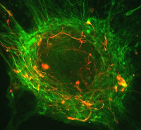
Discovery demonstrates the feasibility of using iPS-derived motor neurons to model and treat diseases Wednesday, 25 February 2009 Stem cells scientists at UCLA showed for the first time that human induced pluripotent stem (iPS) cells can be differentiated into electrically active motor neurons, a discovery that may aid in studying and treating neurological disorders. Additionally, the motor neurons derived from the iPS cells appeared to be similar in function and efficiency to those derived from human embryonic stem cells, although further testing needs to be done to confirm that. If the similarities are confirmed, the discovery may open the door for new treatments for neurological disorders using patient-specific cells. "It is clear from the literature that you can make at least immature versions of many different kinds of cells from human iPS cells," said William Lowry, a Broad Stem Cell Research Center scientist, an assistant professor of molecular, cell and developmental biology and senior author of the study. "But there is not a lot of data published describing the generation of fully functional cells from human iPS cells." Lowry and his team used skin fibroblasts and reprogrammed them back into an embryonic state, with the ability to differentiate into any cell type in the human body. They then took those cells and differentiated them into motor neurons. Neurons are the responsive cells in the nervous system that process and transmit information by electrochemical signalling. Motor neurons receive signals from the brain and spinal cord and regulate muscle contraction. The study demonstrates the feasibility of using iPS-derived motor neurons and their progenitors to replace damaged or dead motor neurons in patients with certain disorders. It also opens the possibility of studying motor neuron-related diseases in the laboratory to uncover their causes. Motor neurons are lost in many conditions, including spinal cord injury, Amyotrophic Lateral Sclerosis and Spinal Muscular Atrophy. "A primary objective of human embryonic stem cell and human iPS cell technology is to be able to generate relevant cell types to enable the repair of tissue damage and in vitro modelling of human disease processes," the study states. "Here, we demonstrate the successful generation of electrically active motor neurons from multiple human iPS cell lines and provide evidence that these neurons are molecularly and physiologically indistinguishable from motor neurons derived from human embryonic stem cells." "To our knowledge, our results present the first demonstration of the electrical activity of iPS-derived neurons and further suggest the feasibility of using these cells to explore how changes in motor neuron activity contributes to the degeneration of these cells underlying these disorders," the authors state. "These findings support the possibility that reprogrammed somatic cells might prove to be a viable alternative to embryo-derived cells in regenerative medicine," the authors note. When measuring the electrophysical properties of the iPS-derived neurons, the researchers found that the iPS cells followed a normal developmental progression to mature, electrically active neurons. "It seems possible that disease-specific somatic cells may be reprogrammed and utilized to model, and ultimately to treat a variety of human neurological disorders," says Miodrag Stojković, co-editor of the journal. Much may be learned from studying the iPS-derived motor neurons and comparing them to motor neurons derived from patients with neurological disorders to see how they differ. The next step for Lowry and his team is to combine the motor neurons with muscle cells to see if they can stimulate a response. If they do, researchers should be able to see the muscle cells contract. Reference: Directed differentiation of human induced pluripotent stem cells generates active motor neurons (p N/A) S Karumbayaram, BG Novitch, M Patterson, JA Umbach, L Richter, A Lindgren, AE Conway, AT Clark, SA Goldman, K Plath, M Wiedau-Pazos, HI Kornblum, WE Lowry Stem Cells, Online: Feb 23 2009, DOI: 10.1002/stem.31 ......... ZenMaster


For more on stem cells and cloning, go to CellNEWS at http://cellnews-blog.blogspot.com/ and http://www.geocities.com/giantfideli/index.html

Monday 23 February 2009

Photo’s of Human Embryonic Stem Cells

CIRM Announces Winners of the 2008 Image Contest Monday, 23 February 2009 Neurosphere sending out processes. Photo by the courtesy of CIRM.The twelve winning images from the first California Institute for Regenerative Medicine stem cell photo contest range from a whirling galaxy of developing nerve cells to a moonscape of retinal pigment epithelial cells, both derived from human embryonic stem cells. Other images show a lightning bolt of a motor neuron axon shooting through a sky of blue and green nerve cells, a sunburst ball of neural progenitor cells and paisley colonies of human embryonic stem cells. Together, this collection of images from CIRM-funded researchers shows the diversity and the beauty of stem cell research. Human embryonic stem cells differentiating into neurons. Photo by the courtesy of CIRM.“When scientists read about a research project that has taken a creative route to arrive at an enlightening finding, it is not unusual for them to exalt it as ‘a beautiful piece of science,’” said Alan Trouson, CIRM president. “With these images we show clearly that such exaltations can have two meanings.” Winners of the contest received $100 and a professional enlargement of their entry. The contest winners join a growing collection of stem cell images on the CIRM Flickr site. Images on the site include embryonic stem cell lines created by CIRM grantees and tissues derived from those cells. These more mature cell types, such as cells of the nervous system, eye, liver or heart, are being used to study the origin of diseases and learn how to reverse the effects of those disorders. Three neurons and human embryonic stem cells. Photo by the courtesy of CIRM. Don Gibbons, chief communications officer, said the collection of images is part of an ongoing effort to keep Californians informed about the agency’s work. The effort includes a stem cell basics primer about stem cell research, summaries of CIRM-funded research findings and videos about the basics of stem cell research and CIRM news at the agency’s YouTube site. “Together, these materials make it easier for Californians to stay abreast of stem cell developments in the state,” Gibbons said. The image contest winners include: Neuron derived from neural stem cells. Photo by the courtesy of CIRM.- Juan Carlos Izpisua Belmonte, PhD, Salk Institute for Biological Studies - Bruce Conklin, MD, the Gladstone Institute of Cardiovascular Disease - Brian Cummings, PhD, University of California, Irvine - Guoping Fan, PhD, University of California, Los Angeles - Susan Fisher, PhD, University of California, San Francisco (two images) - Fred H. Gage, PhD, Salk Institute for Biological Studies - Anirvan Ghosh, PhD, University of California, San Diego - David Hinton, MD, University of Southern California - Paul Knoepfler, PhD, University of California, Davis - Martin Pera, PhD, University of Southern California - Prue Talbot, PhD, University of California, Riverside

All pictures by the courtesy of CIRM.
About CIRM: The California Institute for Regenerative Medicine (CIRM) was established in 2004 with the passage of Proposition 71, the California Stem Cell Research and Cures Act. The state-wide ballot measure, which provided $3 billion in funding for stem cell research at California universities and research institutions, was overwhelmingly approved by voters, and called for the establishment of an entity to make grants and provide loans for stem cell research, research facilities, and other vital research opportunities. To date, the CIRM governing board has approved 279 research and facility grants totalling more than $693 million, making CIRM the largest source of funding for human stem cell research in the world. For more information, please visit www.cirm.ca.gov. ......... ZenMaster
For more on stem cells and cloning, go to CellNEWS at http://cellnews-blog.blogspot.com/ and http://www.geocities.com/giantfideli/index.html

Human Embryonic Stem Cells: A New Model for Lou Gehrig's Disease

Motor neurons derived from embryonic stem cells mimic the progress of familial ALS Monday, 23 February 2009 Amyotrophic lateral sclerosis (ALS), also known as Lou Gehrig's disease, is a devastating condition in which motor neuron degeneration causes progressive loss of movement and muscle tone, leading to death. Overcoming the limited success of previous models, a report published in Disease Models & Mechanisms (DMM), describes how neurons can be derived from human stem cells, and engineered to mimic inherited ALS. Researchers at the University of California Los Angeles developed an optimized protocol to generate motor neurons from human embryonic stem cells (ES cells), which express normal or mutant forms of the SOD-1 gene, which is linked to inherited, familial ALS. Resulting cells exhibit hallmark characteristics of motor nerve cells, and neurons expressing mutant SOD-1 display abnormalities typical of ALS. Defects included shortened cell projections and a reduced life span compared to cells containing the normal SOD-1 gene. This human cell-derived model of ALS provides a new method of studying this disease and testing novel therapeutics. This is especially helpful as only one drug is approved to help slow ALS progression, and animal models currently used in drug development have had limited success. Additionally, this research may aid other gene-linked neurodegenerative diseases, as they too may benefit from studies in a human cell-derived model. Reference: Human embryonic stem cell-derived motor neurons expressing SOD1 mutants exhibit typical signs of motor neuron degeneration linked to ALS Saravanan Karumbayaram, Theresa K. Kelly, Andres A. Paucar, Anne J. T. Roe, Joy A. Umbach, Andrew Charles, Steven A. Goldman, Harley I. Kornblum, and Martina Wiedau-Pazos Dis. Model. Mech. published online before print February 23, 2009, doi:10.1242/dmm.002113 ......... ZenMaster


For more on stem cells and cloning, go to CellNEWS at http://cellnews-blog.blogspot.com/ and http://www.geocities.com/giantfideli/index.html

Wednesday 18 February 2009

Stem Cell Research Recommendations for the New US Administration

Stem Cell Research Recommendations for the New US Administration Wednesday, 18 February 2009 US President Barack Obama will soon issue an executive order lifting an eight-year ban on embryonic stem cell research imposed by his predecessor, President George W. Bush, a senior adviser said on Sunday. "We're going to be doing something on that soon, I think. The president is considering that right now," Presidential adviser David Axelrod said on "Fox News Sunday”. Obama vowed to reverse Bush's ban during his presidential campaign and in his inaugural address last month promised to return science to its proper place in the United States. Furthermore, the US Food and Drug Administration last month cleared the way for the first trial to see if human embryonic stem cells could treat people safely. In January, Baker Institute fellows Neal Lane and Kirstin Matthews released policy recommendations on stem cell research for the Obama administration. They recommend that stem cell research be allowed to expand in a responsible, thoughtful and ethical manner and that a federal stem cell policy be developed. They encourage the administration to expand federal funding for human embryonic stem cells and charge the National Institutes of Health (NIH) with oversight. Lane is a senior fellow in science and technology policy. He also served President Clinton as science adviser and is the former director of the National Science Foundation. Matthews is a fellow in science and technology policy. Matthews and Lane's policy recommendations for the Obama administration:

  • Support research on all types of human stem cells, including embryonic, adult, nuclear-transfer-derived (also known as therapeutic cloning) and induced-pluripotent-derived.
  • Authorize federal funding of human embryonic stem cell research on lines derived according to strict ethical guidelines, regardless of the date the cell lines were derived or created.
  • Remove the
Dickey Amendment (which severely limits the NIH funding of embryonic research) from the Department of Health and Human Services appropriation bills.
  • Ban any effort to clone a human being, regardless of the source of funding.
  • Create an Embryonic Stem Cells Research Oversight (ESCRO) board within the NIH to review controversial research and recommend policy for the agency.
  • Continue the President's Council on Bioethics.
  • References: Human Embryonic Stem Cell Research: Recommendations for the Next Administration Neal Lane and Kirstin Matthews, Rice University Stem Cell Research: A Science and Policy Overview Kirstin R. Matthews, Rice University ......... ZenMaster
    For more on stem cells and cloning, go to CellNEWS at http://cellnews-blog.blogspot.com/ and http://www.geocities.com/giantfideli/index.html
    Tuesday 17 February 2009

    An Inexhaustible Source of Neural Cells

    Researchers in Bonn demonstrate that embryonic stem cells may be derived from brain stem cells Tuesday, 17 February 2009 Research scientists at University of Bonn have succeeded in deriving so-called brain stem cells from human embryonic stem cells. These can not only be conserved almost indefinitely in culture, but can also serve as an inexhaustible source of diverse types of neural cell. The scientists have also shown that these neural cells are capable of synaptic integration in the brain. Their results have been published in the latest edition of the Proceedings of the National Academy of Sciences. For years, stem cell research appeared to be divided between two worlds: on the one hand, were the embryonic stem cells – omnipotent, with unlimited development potential, and on the other, were the so-called somatic stem cells, which were obtainable from adult tissue, but have only limited potential for self-renewal and development. Scientists in Bonn have now succeeded in combining these two worlds: they have derived brain stem cells of almost unlimited self-renewal capacity and conservation potential from human embryonic stem cells. Using these stable cell lines, they were then able to obtain a continual in vitro supply of diverse types of human neural cell including, for example, those which fail with Parkinson’s disease. Using the new cells, researchers are now also able to reduce their requirements for embryonic stem cells, which have hitherto been indispensable as basic material for every individual cell creation process. "The new cells, in contrast, serve as an inexhaustible source: they provide a supply of human neural cells over periods of months and years without demanding any recourse to supplementary embryonic stem cells", declares Professor Dr. Oliver Brüstle, head of the research team at the Institute for Reconstructive Neurobiology at Bonn University. Using animal experiments, the researchers in Bonn provided direct proof that these artificially derived neural cells will also function. Transplanted into the brain of a mouse, these cells made contact with the recipient brain and were subsequently able both to send and receive signals. "This is the first direct evidence that neural cells derived from human stem cells are capable of synaptic integration in the brain", declares Dr. Philipp Koch, the original author of the study. The scientists in Bonn are now also hoping to exploit this inexhaustible cell source to study neurodegenerative diseases and possible active agents directly in human neural cells. Brüstle and his team were the first scientists in Germany to receive permission to import human embryonic stem cells. They had played a major role in the public discussion of this hot topic. "Our current results make it clear how smoothly research into embryonic and somatic stem cells can be combined, and at the same time, how important this is", Brüstle stresses. Reference: A rosette-type, self-renewing human ES cell-derived neural stem cell with potential for in vitro instruction and synaptic integration Koch, P., Opitz, T., Steinbeck, J., Ladewig, J., Brüstle, O. PNAS February 10, 2009, 106 (6), doi: 10.1073/pnas.0808387106 ......... ZenMaster


    For more on stem cells and cloning, go to CellNEWS at http://cellnews-blog.blogspot.com/ and http://www.geocities.com/giantfideli/index.html

    Tuesday 10 February 2009

    Nerve-muscle ‘Connectome’ Diversity Mapped

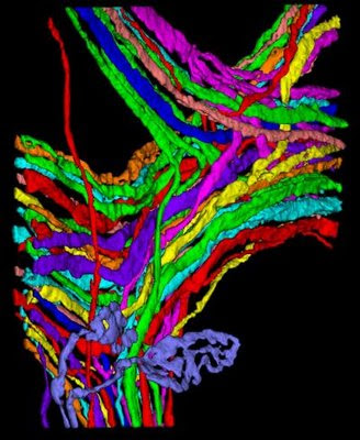
    Reconstruction of the complete set of motor axons innervating a muscle reveals extensive wiring variability Tuesday, 10 February 2009 Genetics may play a surprisingly small role in determining the precise wiring of the mammalian nervous system, according to painstaking mapping of every neuron projecting to a small muscle mice use to move their ears. These first-ever mammalian "connectomes," or complete neural circuit diagrams, reveal that neural wiring can vary widely even in paired tissues on the left and right sides of the same animal. Scientists at Harvard University and the Massachusetts Institute of Technology describe the work this week in the journal PLoS Biology, accompanied by vivid images depicting neurons that are strikingly treelike, but also tremendously varied.

    Axons in the connectomes of the left and right interscutularis muscles.Axons in the connectomes of the left and right interscutularis muscles of a 1-month old animal were colour-coded based on the rank-order of their motor unit sizes (the number of muscle fibres innervated by each axon) in each connectome. Thus, each axon and its contra-lateral counterpart can be identified in the connectomes, and subsequently their morphologies can be compared. Credit: Ju Lu, Harvard University.
    "We had expected to find a great degree of neural symmetry in the same mouse's two interscutularis muscles, but this isn't even close to true," says Jeff W. Lichtman, professor of molecular and cellular biology in Harvard's Faculty of Arts and Sciences. "It looks like the mammalian nervous system may be a bit like a football game," he adds. "Even when the rules are the same, every single outcome is unique." Curiously, the connectome of the mouse interscutularis — a muscle also found in dogs, rats, and other mammals that readily move their ears — reveals that some of its neurons are as much as 25 percent longer than is necessary. This casts doubt on a longstanding assumption among neuroscientists that neural wiring length is generally minimized to conserve space, energy, and resources.
    Detail of complete wiring diagram.In order to understand how each nerve cell integrates into the functional organization of a neural circuit, it is necessary to obtain the complete wiring diagram (connectome) of the circuit by tracing out all the neural processes in the sample. We used confocal laser scanning microscopy to image all the axons that innervate a small ear muscle in transgenic mice that express fluorescent protein in motor neurons. Shown here is one of the reconstructed image stacks containing branches of ~ 10 axons. Each axon was traced out in a semi-automated image processing program and rendered in 3D with a distinct colour. This image stack is representative of the hundreds of image stacks from which the entire connectome was reconstructed. Credit: Ju Lu, Harvard University.
    "This well-known hypothesis that wiring length should be minimized has been in the scientific literature for decades," says Ju Lu, a postdoctoral researcher in molecular and cellular biology at Harvard. "It's very surprising, frankly, to find so much excess wiring in the mammalian nervous system." Lichtman and Lu's work represents only the second connectome to date, following one for the worm
    Caenorhabditis elegans. While their task initially appeared manageable — the entire interscutularis muscle is but a few millimetres in length — teasing out the muscle's tangle of about 15 intricately branched and intertwined axons proved fiendishly complex. "It's a bit like taking a giant plate of spaghetti and, without unravelling it, trying to figure out which strand goes where," says Lu. "Except in this case, each strand of spaghetti has up to 37 branches."
    The entire connectome.The entire connectome was obtained by montage of hundreds of image stacks individually reconstructed. Each axon was pseudo-coloured to indicate its trajectory and the position of its neuromuscular terminals. The connectome gives the anatomical underpinning of the graded tensions elicited by motor neurons according to Henneman’s size principle. Credit: Ju Lu, Harvard University.

    Working with mice containing a gene that causes motor neurons to fluoresce, Lichtman and Lu used an automated microscope to gather tens of thousands of images. These images were analyzed with semi-automated tracing tools, although the need for frequent corrections and manual editing by Lu slowed the pace of the mapping to a scant half-millimetre per hour. Connectomes from a mouse's two interscutularis muscles depict dramatically different neural circuitry even within mirror-image tissues from the same animal. "Comparison of each neuron and its counterpart on the opposite side of the animal revealed that each connectome was unique," Lichtman says, "demonstrating wiring diagrams that differ substantially in form, even within a common genetic background." Lichtman says the research suggests the mammalian nervous system is in some ways unexpectedly primitive, its freeform structure lacking the regimentation seen in insects and worms. But, he adds, this seeming randomness may be advantageous. "This may explain why humans and other mammals can quickly adapt their behaviours to a changing environment," Lichtman says. "We may be less perfected in our genetic evolution, but our flexible neural wiring may allow us to undergo behavioural evolution at a very rapid rate." Such variation in the nervous system, he adds, could help explain why different humans, each equipped with the same neural building blocks, excel at tasks ranging from dancing to mathematical computations, and from crossword puzzles to bowling. Reference: The Interscutularis Muscle Connectome Ju Lu, Juan Carlos Tapia, Olivia L. White, Jeff W. Lichtman PLoS Biol 7(2): e1000032 doi:10.1371/journal.pbio.1000032 ......... ZenMaster


    For more on stem cells and cloning, go to CellNEWS at http://cellnews-blog.blogspot.com/ and http://www.geocities.com/giantfideli/index.html

    Friday 6 February 2009

    Are We Selling Personalized Medicine Before Its Time?

    Limitations of genetic screening reported in Public Library of Science study Friday, 06 February 2009 We may be a long way off from using genetics to reliably gauge our risks for specific diseases, say researchers at the University of Pittsburgh Schools of the Health Sciences in a study published on Feb. 5 in the online journal PLoS Genetics. Yet, many companies currently offer personalized genetic testing for diseases like cancer, heart disease and diabetes, and tout the ability of DNA testing to predict future health risks. "The rapid discovery of new genetic risk factors is giving us vitally important insights into human health, but a strong association between these factors and disease risk may not reliably predict which health issues a specific individual will face in the future," said Daniel E. Weeks, Ph.D., senior author and professor of human genetics and biostatistics at the University of Pittsburgh Graduate School of Public Health. "Our study indicates that even though we can paint a picture of our genetic makeup with current tests, this may not be enough to help us understand our individual risk for disease." The study focused on single nucleotide polymorphisms, or SNPs – variations in short DNA sequences that have been linked to the presence of particular diseases, and that exist in the millions in the human genome. A number of companies currently offer individualized estimates for disease risks based on genome-wide SNP genotyping. These tests typically scan 500,000 to 1 million SNPs, searching for only a handful associated with a specific disease. Dr. Weeks and colleagues focused their study on diseases for which there are strongly associated genetic variants: age-related macular degeneration, type 2 diabetes, prostate cancer, cardiovascular disease and Crohn's disease. They found that a strong genetic association did not guarantee they could accurately discriminate between actual disease cases and controls in both mathematical models and real-world examples. Part of the problem may be a statistical one. To provide meaningful insights, a test for disease risk needs to accurately identify positive cases and, at the same time, provide a low false positive rate. One of the challenges with current approaches to genetic testing is that they are based on a very small number of common variants, "making it likely that you will identify people at high risk who may not be at risk at all," said Dr. Weeks. "With such a small pool of variants, it's difficult to develop a very meaningful test for predicting disease risk." In addition, he said, few health care providers have adequate genetics training to make sense of the risk calculations now commercially offered and to advise their patients accordingly. Dr. Weeks suggests the need for longitudinal studies to define true risk and to understand how genetic susceptibility may interact with known environmental and lifestyle risk factors. "With more study, our hope is that genetic testing will benefit people and encourage positive lifestyle changes and guide clinical decisions. In the meantime, we need to take a step back and proceed with caution and allow the insights gained from these new association findings to be used to explore the basic biological causes of disease," he said. ......... ZenMaster


    For more on stem cells and cloning, go to CellNEWS at http://cellnews-blog.blogspot.com/ and http://www.geocities.com/giantfideli/index.html

    Thursday 5 February 2009

    Single Factor Converts Adult Stem Cells into Embryonic-like Stem Cells

    Single Factor Converts Adult Stem Cells into Embryonic-like Stem Cells Thursday, 05 February 2009 The simple recipe scientists earlier discovered for making adult stem cells behave like embryonic-like stem cells just got even simpler. A new report in the February 6th issue of the journal Cell shows for the first time that neural stem cells taken from adult mice can take on the characteristics of embryonic stem cells with the addition of a single transcription factor. Transcription factors are genes that control the activity of other genes. The discovery follows a 2006 report also in the journal Cell that showed that the introduction of four ingredients could transform differentiated cells taken from adult mice into "induced pluripotent stem cells" (iPS) with the physical, growth, and genetic characteristics typical of embryonic stem cells. Pluripotent refers to the ability to differentiate into most other cell types. The same recipe was later shown to work with human skin cells as well. Subsequent studies found that the four-ingredient recipe could in some cases be pared down to just two or three essential ingredients, said Hans Schöler of the Max Planck Institute for Molecular Biomedicine in Germany. "Now we've come down to just one that is sufficient. In terms of the biology, it's really quite amazing." The discovery sheds light on centuries-old questions about what distinguishes the embryonic stem cells that give rise to egg and sperm from other body cells, Schöler said. It might also have implications for the use of reprogrammed stem cells for replacing cells lost to disease or injury. Other researchers led by Shinya Yamanaka showed that adult cells could be reprogrammed by adding four factors – specifically Oct4, Sox2, Klf4, and c-Myc. Recently, Schöler and his colleagues demonstrated that Oct4 and Klf4 are sufficient to induce pluripotency in neural stem cells. By omitting Klf4 in the new study, they have now established that Oct4 is the "driving force" behind the conversion of the neural stem cells into iPS cells. The lone transcription factor is not only essential, but it is also sufficient to make neural stem cells pluripotent. Those cells, which Schöler's team calls "1F iPS" can differentiate into all three germ layers. Those primary germ layers in embryos eventually give rise to all the body's tissues and organs. Not only can those cells efficiently differentiate into neural stem cells, heart muscle cells, and germ cells, they show, but they are also capable of forming tumours when injected under the skin of nude mice. Those tumours, or teratomas, contain tissue representing all three germ layers. When injected into mouse embryos, the 1F iPS cells also found their way into the animals' developing organs and were able to be transmitted through the germ line to the next generation, they report. The results show that adult stem cells can be made pluripotent without c-Myc and Klf4, both of which are "bona fide" oncogenes that can help turn normal cells into cancer cells, Schöler said. Limiting the number of factors is also a bonus because it means fewer genes must be inserted into the genome, where they can potentially have detrimental effects. "Strikingly, Oct4 alone is sufficient to induce pluripotency in neural stem cells, which demonstrates its crucial role in the process of reprogramming…" the researchers concluded. "Future studies will show whether other sources of neural stem or progenitor cell populations such as mouse or human bone marrow-derived mesenchymal stem cells or dental pulp can be reprogrammed to iPS cells and whether expression of Oct4 can be induced by non-retroviral means, a prerequisite for the generation of iPS cells of therapeutic value." ......... ZenMaster


    For more on stem cells and cloning, go to CellNEWS at http://cellnews-blog.blogspot.com/ and http://www.geocities.com/giantfideli/index.html

    The Nonsense in Our Genes

    Is 1 in 200 human genes superfluous? Thursday, 05 February 2009 A study of the genetic code of more than 1,000 people, published in the American Journal of Human Genetics, has found that at least one in 200 human genes can be inactivated in apparently healthy people. The findings suggest that, though these genetic mutations can be harmful, they generally have little effect on the individual and could occasionally even be beneficial in evolutionary terms. The study also found that individuals carry on average 46 of these inactivating mutations. 1 in 200 of our human genes can be inactivated with no detectable effect on our health. A study by Wellcome Trust Sanger Institute scientists raises new questions about the effects of gene loss on our wellbeing and evolution. The study, published today in The American Journal of Human Genetics, explores single letter changes in our genetic code that affect the ability of genes to produce proteins. The researchers' findings suggest that such mutations, while sometimes harmful, generally have little consequence for the individual and may occasionally even be beneficial in evolutionary terms. The team studied variations in the genetic code of more than 1000 people from around the world. They focused their work on single-letter changes (called SNPs) that disrupt proteins, leading to versions that are either shorter or completely absent. One might intuitively expect that such a change - called a nonsense-SNP - would be harmful to the person. "We knew that these mutations existed and that many have been associated with genetic diseases, but we were amazed to find that they were so common in the general population," said Bryndis Yngvadottir, lead author on the study.

    Frequency of nonsense SNPs in the study sample.


    "We found that 167 genes could be inactivated by nonsense mutations, and that individuals carry on average at least 46 such variations. For 99 of the genes, both copies could be lost in adults living a normal existence." Human DNA contains approximately 20,000 genes: the total of 99 genes with nonsense-SNPs means that at least 1 in 200 genes is dispensable. Some harmful nonsense-SNPs were also present among the 167 genes studied: 8 are listed in the Human Gene Mutation Database which catalogues disease-causing mutations. While the researchers found that inactivating genes was, on the whole, slightly harmful, there were exceptions. In East Asia, but not in other places, it seems to have been advantageous to lose the MAGEE2 gene. "There is a theory that 'less is more' where genes are concerned" explained the study's coordinator, Chris Tyler-Smith, "and we already knew of a couple of examples of advantageous gene loss. But this is the first large-scale investigation of its significance for recent human evolution.” "The MAGEE2 gene is an interesting new example, although we have absolutely no idea what this gene does, or why some people are better off without it. However, our study suggests that overall, gene loss has not been a major evolutionary force: our genome does not seem to be in a hurry to get rid of these 'superfluous' genes." "Certain types of genes tend to be lost preferentially. We found the biggest decrease in the genes that contribute to our sense of smell. Perhaps early humans didn't like smelly partners, and so when humans started to live together in big groups it helped their chances of finding true love if they couldn't smell their partner too strongly," speculated Bryndis Yngvadottir. Genetic variation in nonsense-SNP numbers was significant: participants in the survey had between 29 and 65 of these mutations each and varied on average by 24 genes as a consequence. 18 of the 169 nonsense-SNPs investigated are also present in the Craig Venter genome published last year. About: The Wellcome Trust Sanger Institute, which receives the majority of its funding from the Wellcome Trust, was founded in 1992 as the focus for UK sequencing efforts. The Institute is responsible for the completion of the sequence of approximately one-third of the human genome as well as genomes of model organisms such as mouse and zebrafish, and more than 90 pathogen genomes. In October 2005, new funding was awarded by the Wellcome Trust to enable the Institute to build on its world-class scientific achievements and exploit the wealth of genome data now available to answer important questions about health and disease. These programmes are built around a Faculty of more than 30 senior researchers. The Wellcome Trust Sanger Institute is based in Hinxton, Cambridge, UK. The Wellcome Trust is the largest charity in the UK. It funds innovative biomedical research, in the UK and internationally, spending over £600 million each year to support the brightest scientists with the best ideas. The Wellcome Trust supports public debate about biomedical research and its impact on health and wellbeing. Reference: A Genome-wide Survey of the Prevalence and Evolutionary Forces Acting on Human Nonsense SNPs Bryndis Yngvadottir , Yali Xue , Steve Searle , Sarah Hunt , Marcos Delgado , Jonathan Morrison , Pamela Whittaker , Panos Deloukas and Chris Tyler-Smith The American Journal of Human Genetics, 05 February 2009, 10.1016/j.ajhg.2009.01.008 ......... ZenMaster


    For more on stem cells and cloning, go to CellNEWS at http://cellnews-blog.blogspot.com/ and http://www.geocities.com/giantfideli/index.html

    Wednesday 4 February 2009

    New Chemical Keeps Stem Cells Young

    New Chemical Keeps Stem Cells Young Wednesday, 04 February 2009 Scientists at the University of Bath and University of Leeds have discovered a chemical that stops stem cells from turning into other cell types, allowing researchers to use these cells to develop new medical treatments more easily. Stem cells have the ability to develop into many other cell types in the body, and scientists believe they have huge potential to treat diseases or injuries that do not currently have a cure. Professor Melanie Welham Credit: Nic Delves-Broughton, University of Bath.Professor Melanie Welham's team at the University of Bath's Department of Pharmacy & Pharmacology, collaborating with Professor Adam Nelson at the University of Leeds, have discovered a chemical that can be added to embryonic stem cells grown in the lab, allowing them to multiply without changing into other cell types. This breakthrough will help scientists produce large stocks of cells that are needed for developing new medical therapies. Professor Welham, who is co-director of the University of Bath's Centre for Regenerative Medicine, explained: "Stem cells have great potential for treating spinal injuries and diseases like type I diabetes because they can change into a range of specialised cell types including nerve or pancreatic cells, which could be used to repair damaged tissues.” "Unfortunately, when you grow stem cells in the lab, they can spontaneously develop into specialised cells, making it difficult to grow large enough stocks to use for medical research.” "We've identified a chemical that will put this process on hold for several weeks so that we can grow large numbers of them in their unspecialised state. This is reversible, so when you take it away from the cells, they still have the ability to change into specialised cells." Professor Adam Nelson's team, at the Astbury Centre for Structural Molecular Biology, made more than 50 chemical compounds that were tested for activity in the stem cells. The researchers found that the chemicals worked by blocking glycogen synthase kinase 3 (GSK-3), that can control when the stem cell switches to a more specialised cell type. Professor Nelson, who is Director of the Astbury Centre at the University of Leeds, said: "This research is a great example of how small molecules can be used as tools to understand biological mechanisms." Reference: Involvement of GSK-3 in Regulation of Murine Embryonic Stem Cell Self-Renewal Revealed by a Series of Bisindolylmaleimides Heather K. Bone,Teresa Damiano,Stephen Bartlett,Alexis Perry,Julie Letchford,Yolanda Sanchez Ripoll,Adam S. Nelson andMelanie J. Welham Chemistry & Biology, Volume 16, Issue 1, 15-27, doi:10.1016/j.chembiol.2008.11.003 ......... ZenMaster


    For more on stem cells and cloning, go to CellNEWS at http://cellnews-blog.blogspot.com/ and http://www.geocities.com/giantfideli/index.html