Monday 30 June 2008

Adult Stem Cells Reprogrammed in The Brain

Adult Stem Cells Reprogrammed in Their Natural Environment Monday, 30 June 2008 In recent years, stem cell researchers have become very adept at manipulating the fate of adult stem cells cultured in the lab. Now, researchers at the Salk Institute for Biological Studies achieved the same feat with adult neural stem cells still in place in the brain. They successfully coaxed mouse brain stem cells bound to join the neuronal network to differentiate into support cells instead. The discovery, which is published ahead of print on Nature Neuroscience's website, not only attests to the versatility of neural stem cells but also opens up new directions for the treatment of neurological diseases, such as multiple sclerosis, stroke and epilepsy that not only affect neuronal cells but also disrupt the functioning of glial support cells. "We have known that the birth and death of adult stem cells in the brain could be influenced be experience, but we were surprised that a single gene could change the fate of stem cells in the brain," says the study's lead author, Fred H. Gage, Ph.D., a professor in the Laboratory for Genetics and the Vi and John Adler Chair for Research on Age-Related Neurodegenerative Diseases. Throughout life, adult neural stem cells generate new brain cells in two small areas of mammalian brains: the olfactory bulb, which processes odours, and the dentate gyrus, the central part of the hippocampus, which is involved in the formation of memories and learning. After these stem cells divide, their progenitors have to choose between several options – remaining a stem cell, turning into a nerve cell, also called a neuron, or becoming part of the brain's support network, which includes astrocytes and oligodendrocytes. Astrocytes are star-shaped glia cells that hold neurons in place, nourish them, and digest parts of dead neurons. Oligodendrocytes are specialized cells that wrap tightly around axons, the long, hair-like extensions of nerve cell that carry messages from one neuron to the next. They form a fatty insulation layer, known as myelin, whose job it is to speed up electrical signals travelling along axons. When pampered and sheltered in a petri dish, adult neural stem cells can be nudged to differentiate into any kind of brain cell but within their natural environment in the brain career options of neural stem cells are thought to be mostly limited to neurons. "When we grow stem cells in the lab, we add lots of growth factors resulting in artificial conditions, which might not tell us a lot about the in vivo situation," explains first author Sebastian Jessberger, M.D., formerly a post-doctoral researcher in Gage's lab and now an assistant professor at the Institute of Cell Biology at the Swiss Federal Institute of Technology in Zurich. "As a result we don't know much about the actual plasticity of neural stem cells within their adult brain niche."


TOP: Throughout life, adult neural stem cells generate new brain cells in the dentate gyrus, the central part of the hippocampus, which is involved in the formation of memories and learning (shown in white). BOTTOM: Over expression of a single gene changed the fate of neural stem cells bound to join the neuronal network in the brain. Instead they differentiated into glial support cells (shown in green). Credit: Courtesy of Dr. Sebastian Jessberger, Swiss Federal Institute of Technology in Zurich
To test whether stem cells in their adult brain environment can still veer off the beaten path and change their fate, Jessberger used retroviruses to genetically manipulate neural stem cells and their progeny in the dentate gyrus of laboratory mice. Under normal conditions, the majority of newborn cells differentiated into neurons. When he introduced the Ascl1, a transcription factor which had previously been shown to be involved in the generation of oligodendrocytes and inhibitory neurons, he successfully redirected the fate of newborn cells from a neuronal to an oligodendrocytic lineage. "It was quite surprising that stem cells in the adult brain maintain their fate plasticity and that a single gene was enough to reprogram these cells," says Jessberger. "We can now potentially tailor the fate of stem cells to treat certain conditions such as multiple sclerosis." In patients with multiple sclerosis, the immune system attacks oligodendrocytes, which leads to the thinning of the myelin layer affecting the neurons' ability to efficiently conduct electrical signals. Being able to direct neural stem cells to differentiate into oligodendrocytes may alleviate the symptoms. About Salk Institute for Biological Studies: The Salk Institute for Biological Studies in La Jolla, California, is an independent non-profit organization dedicated to fundamental discoveries in the life sciences, the improvement of human health and the training of future generations of researchers. Jonas Salk, M.D., whose polio vaccine all but eradicated the crippling disease poliomyelitis in 1955, opened the Institute in 1965 with a gift of land from the City of San Diego and the financial support of the March of Dimes. Reference: Directed differentiation of hippocampal stem/progenitor cells in the adult brain Sebastian Jessberger, Nicolas Toni, Gregory D Clemenson Jr, Jasodhara Ray & Fred H Gage Nature Neuroscience, Published online: 29 June 2008, doi:10.1038/nn.2148 ......... ZenMaster
For more on stem cells and cloning, go to CellNEWS at http://cellnews-blog.blogspot.com/ and http://www.geocities.com/giantfideli/index.html

Saturday 28 June 2008

Artificial Polio Virus Could Make Safe New Vaccine

‘Wimpy’ viruses could become a method to produce new and better vaccines Saturday, 28 June 2008 A team of molecular biologists and computer scientists at Stony Brook University has designed and synthesized a new class of weakened polioviruses. They used their synthesizing method with computer software to systematically re-code the poliovirus genome. In doing so, the team is the first to demonstrate that a synthetic weakened virus can immunize an animal. These results show promise in the creation of new attenuated (‘live virus’) anti-viral vaccines and are reported in the June 27 issue of Science. Six years ago, Eckard Wimmer, Ph.D., Distinguished Professor, Department of Molecular Genetics and Microbiology at Stony Brook University, and colleagues synthesized and generated poliovirus, the first artificial synthesis of any virus. Dr. Wimmer and other scientists within the Department built on that finding in their recent work. “Synthesizing the wild-type poliovirus was an essential and important first step toward our current research,” says Dr. Wimmer, noting that the new method involves impeding the synthesis of viral proteins, a new approach to developing attenuated vaccines. This type of vaccine is created by mutating the virus so it cannot cause disease. Generally, attenuated vaccines are easy to administer, inexpensive, and sometimes offer the best protection against disease. “As all viruses depend on their host’s cellular machinery to produce their proteins, targeting the synthesis of viral proteins by the host may be universally applicable to creating weakened strains of other viruses,” says Steffen Mueller, Ph.D., Senior Author and Research Assistant Professor of Molecular Genetics and Microbiology, referring to the implications of the research results. Because of the redundancy of the genetic code, there are an unimaginably large number of ways to encode any given protein. For poliovirus proteins, there are more possible encodings (10 to the 442 power) than atoms in the universe. Using a powerful computer algorithm, the team found particular re-codings of the genome predicted to weaken the virus. The researchers made hundreds of small mutations in the genome that perfectly preserved the viral proteins but changed the way those proteins were encoded by RNA (ribonucleic acid), so that pairs of amino acids were added by transfer RNAs (tRNAs) that rarely work together in normal proteins. They call the process “Synthetic Attenuated Virus Engineering,” or “SAVE.” The resulting virus contains completely authentic, wild-type poliovirus proteins. However, each of the hundreds of mutations causes a tiny defect by creating an obstacle – a genetic “speed bump” – in translating the genetic code into a protein. “Translation of this unusual genome into viral proteins was inefficient, and the most highly re-coded virus was weakened to the point where it was unable to infect cells,” says J. Robert Coleman, Lead Author and a graduate student in Molecular Genetics and Microbiology. The reduced translational efficiency of these chimeric viruses reduced their ability to cause disease. The team injected mice with the re-coded polioviruses. Most mice showed no signs of disease but did produce anti-polio antibodies. These mice were then immune against infection by the normal, fully virulent poliovirus. “Ultimately we created a wimpy poliovirus that can be customized and does not cause disease unless given at high doses,” explains Bruce Futcher, Ph.D., Co-author and Professor of Molecular Genetics and Microbiology. “These viruses are still far from suitable vaccines for humans, but there is a lot of potential for this approach,” continues Dr. Futcher. “A virus modified using ‘SAVE’ might act as a vaccine by providing immunity against the normal virus.” The inclusion of computer programming essential to developing these synthetic polioviruses featured the work of Steven Skiena, Ph.D., Professor of Computer Science. Dr. Skiena, in collaboration with his graduate student Dimitris Papamichail, developed the sequence design algorithm. “Sophisticated computer algorithms are necessary to design the hundreds of changes to sufficiently cripple the virus for our ‘death by a thousand cuts’ approach,” summarizes Dr. Skiena. “Because of the large number of changes, the weakened virus can never mutate back to wild-type.” The research team hopes this “death by a thousand cuts” virus mutation strategy can be applicable to attenuating many kinds of viruses. They are looking into applications with other viruses. References: Virus Attenuation by Genome-Scale Changes in Codon Pair Bias J. Robert Coleman, Dimitris Papamichail, Steven Skiena, Bruce Futcher, Eckard Wimmer, Steffen Mueller Science 27 June 2008, Vol. 320. no. 5884, pp. 1784 – 1787, DOI: 10.1126/science.1155761 Dangerous Virus Made from Mail-order Kits CellNEWS - Thursday, 11 July 2002 Pentagon Behind The Creation of The "Synthetic" Poliovirus CellNEWS - Friday, 12 July 2002 ......... ZenMaster


For more on stem cells and cloning, go to CellNEWS at http://cellnews-blog.blogspot.com/ and http://www.geocities.com/giantfideli/index.html

Thursday 26 June 2008

Ronin Controls Embryonic Stem Cells

Ronin Controls Embryonic Stem Cells Thursday, 26 June 2008 Like the master-less samurai for whom it is named, the protein Ronin chooses an independent path, maintaining embryonic stem cells in their undifferentiated state and playing essential roles in genesis of embryos and their development, said Baylor College of Medicine researchers who reported on this novel cellular regulator in the current issue of the journal Cell. Three proteins – Oct4, Sox2 and Nanog – had previously been considered the "master" regulators of embryonic stem cells. "Ronin could be as important as these three," said Dr. Thomas Zwaka, assistant professor in the Stem Cells and Regenerative Medicine (STaR) Center at BCM. In fact, he said, if the action of Oct4, considered the most important, is reduced in embryonic stem cells, Ronin can compensate for the loss. Embryonic stem cells are pluripotent, meaning they have the potential for becoming all other kinds of cells in the body. They are also capable of self-renewal. Oct4, Sox2 and Nanog were previously thought the major method by which embryonic stem cells remained in their pristine state. Now, Ronin represents a different and parallel pathway to achieve the same result. Ronin is also expressed in early embryonic development of mice. If it is not present, the embryos die, said Zwaka. It is also found in mature oocytes or egg cells. "Ronin is a potent transcription repressor," he said. In fact, it prevents the action of genes that promote the differentiation of cells into the various tissues and organs of the body. "It does it more effectively than the other three factors together," he said. It silences the differentiation genes epigenetically through specific chemical mechanisms that modify histones, the chief packaging proteins for DNA. He and his colleagues found Ronin as a follow-up to an earlier study that showed a component of the cell death system called caspase-3 actually cleaved and reduced the amount of Nanog protein. This caused the embryonic stem cells to stop self-renewal and begin differentiation into other kinds of cells. Zwaka and his colleagues searched for other proteins affected by the caspase and found Ronin, which was previously unknown. The finding prompts other questions. Can Ronin be used to reprogram differentiated cells into those that more closely resemble embryonic stem cells? What is the significance of the portion of Ronin that resembles a "jumping gene" or transposon called P element transposase, usually found in the genomes of fruit flies? Ronin is also found in areas of the brain such as the hippocampus and the Purkinje cells of the cerebellum. "What role does it play in the brain?" asked Zwaka. Reference: Ronin Is Essential for Embryogenesis and the Pluripotency of Mouse Embryonic Stem Cells Marion Dejosez, Joshua S. Krumenacker, Laura Jo Zitur, Marco Passeri, Li-Fang Chu, Zhou Songyang, James A. Thomson, and Thomas P. Zwaka Cell, Vol 133, 1162-1174, 27 June 2008 ......... ZenMaster


For more on stem cells and cloning, go to CellNEWS at http://cellnews-blog.blogspot.com/ and http://www.geocities.com/giantfideli/index.html

Should Great Apes be Granted Human Rights?

Should Great Apes be Granted Human Rights? Thursday, 26 June 2008 Reuters report that "Spain Give Great Apes Human Rights". What do you think? Are great apes equal to humans, or should humans instead be considered equal to great apes?

And what problem would it pose for the medical community? ......... ZenMaster


For more on stem cells and cloning, go to CellNEWS at http://cellnews-blog.blogspot.com/ and http://www.geocities.com/giantfideli/index.html

Spain Give Great Apes Human Rights

Spain Give Great Apes Human Rights Thursday, 26 June 2008 Reuters report that a Spanish parliamentary committee voiced its support on Wednesday for the rights of great apes to life and freedom in what will apparently be the first time any national legislature has called for such rights for non-humans. The parliament’s environmental committee approved resolutions urging Spain to comply with the Great Apes Project, devised by scientists and philosophers who say our closest genetic relatives deserve rights hitherto limited to humans. Philosophers Peter Singer and Paola Cavalieri founded the Great Apes Project in 1993, arguing that "non-human hominids" like chimpanzees, gorillas, orang-utans and bonobos should enjoy the right to life, freedom and not to be tortured. "This is a historic day in the struggle for animal rights and in defence of our evolutionary comrades, which will doubtless go down in the history of humanity," said Pedro Pozas, Spanish director of the Great Apes Project. The new resolutions have cross-party or majority support and are expected to become law and the government is now committed to update the statute book within a year to outlaw harmful experiments on apes in Spain. Keeping apes for circuses, television commercials or filming will also be forbidden and breaking the new laws will become an offence under Spain's penal code. Keeping an estimated 315 apes in Spanish zoos will not be illegal, but supporters of the bill say conditions will need to improve drastically in 70 percent of establishments to comply with the new law. See also: The Rights of Apes – and Humans by Peter Singer Published by Project Syndicate ......... ZenMaster


For more on stem cells and cloning, go to CellNEWS at http://cellnews-blog.blogspot.com/ and http://www.geocities.com/giantfideli/index.html

Wednesday 25 June 2008

Genetically Modified ESCs Improved Brain Function

Nerve cells derived from stem cells and transplanted into mice may lead to improved brain treatments Wednesday, 25 June 2008 Scientists at the Burnham Institute for Medical Research have, for the first time, genetically programmed embryonic stem cells (ESCs) to become nerve cells when transplanted into the brain, according to a study published today in The Journal of Neuroscience. The research, an important step toward developing new treatments for stroke, Alzheimer's, Parkinson's and other neurological conditions showed that mice afflicted by stroke showed tangible therapeutic improvement following transplantation of these cells. None of the mice formed tumours, which had been a major setback in prior attempts at stem cell transplantation. The team was led by Stuart A. Lipton, M.D., Ph.D., professor and director of the Del E. Webb Neuroscience, Aging, and Stem Cell Research Center at Burnham. Dr. Lipton is also a clinical neurologist who treats patients with these disorders. Collaborators included investigators from The Scripps Research Institute. "We found that we could create new nerve cells from stem cells, transplant them effectively and make a positive difference in the behaviour of the mice," said Dr. Lipton. "These findings could potentially lead to new treatments for stroke and neurodegenerative diseases such as Parkinson's disease." Conditions such as stroke, Alzheimer's, Parkinson's and Huntington's disease destroy brain cells, causing speech and memory loss and other debilitating consequences. In theory, transplanting neuronal brain cells could restore at least some brain function, just as heart transplants restore blood flow. Prior to this research, creating pure neuronal cells from ESCs had been problematic as the cells did not always differentiate into neurons. Sometimes they became glial cells, which lack many of the neurons' desirable properties. Even when the neuronal cells were created successfully, they often died in the brain following transplant — a process called programmed cell death or apoptosis. In addition, the cells would sometimes become tumours. Dr. Lipton solved these problems by inducing ESCs to express a protein, discovered in his laboratory called myocyte enhancer factor 2C (MEF2C). MEF2C is a transcription factor that turns on specific genes which then drive stem cells to become nerve cells. Using MEF2C, the researchers created colonies of pure neuronal progenitor cells, a stage of development that occurs before becoming a nerve cell, with no tumours. These cells were then transplanted into the brain and later became adult nerve cells. MEF2C also protected the cells from apoptosis once inside the brain. "To move forward with stem cell-based therapies, we need to have a reliable source of nerve cells that can be easily grown, differentiate in the way that we want them to and remain viable after transplantation," said Dr. Lipton. "MEF2C helps this process first by turning on the genes that, when expressed, make stem cells into nerve cells. It then turns on other genes that keep those new nerve cells from dying. As a result, we were able to produce neuronal progenitor cells that differentiate into a virtually pure population of neurons and survive inside the brain." The next step was to determine whether the transplanted neural progenitor cells became nerve cells that integrated into the existing network of nerve cells in the brain. Performing intricate electrical studies, Dr. Lipton's investigative team showed that the new nerve cells, derived from the stem cells, could send and receive proper electrical signals to the rest of the brain. They then determined if the new cells could provide cognitive benefits to the stroke-afflicted mice. The team executed a battery of neurobehavioral tests and found that the mice that received the transplants showed significant behavioural improvements, although their performance did not reach that of the non-stroke control mice. These results suggest that MEF2C expression in the transplanted cells was a significant factor in reducing the stroke-induced deficits. The work was supported in part by National Institutes of Health (NIH) grants and a Senior Scholar Award in Aging Research from the Ellison Medical Foundation. About Burnham Institute for Medical Research Burnham Institute for Medical Research is dedicated to revealing the fundamental molecular causes of disease and devising the innovative therapies of tomorrow. Burnham is one of the fastest growing research institutes in the country with operations in California and Florida. The Institute ranks among the top four institutions nationally for NIH grant funding and among the top 25 organizations worldwide for its research impact. Burnham utilizes a unique, collaborative approach to medical research and has established major research programs in cancer, neurodegeneration, diabetes, infectious and inflammatory and childhood diseases. The Institute is known for its world-class capabilities in stem cell research and drug discovery technologies. Burnham is a non-profit, public benefit corporation. ......... ZenMaster


For more on stem cells and cloning, go to CellNEWS at http://cellnews-blog.blogspot.com/ and http://www.geocities.com/giantfideli/index.html
Our Genome Changes Over Lifetime

May explain many 'late-onset' diseases, Johns Hopkins experts say Tuesday, 24 June 2008 Researchers at Johns Hopkins have found that epigenetic marks on DNA Рchemical marks other than the DNA sequence Рdo indeed change over a person's lifetime, and that the degree of change is similar among family members. Reporting in the June 25 issue of the Journal of the American Medical Association, the team suggests that overall genome health is heritable and that epigenetic changes occurring over one's lifetime may explain why disease susceptibility increases with age. "We're beginning to see that epigenetics stands at the centre of modern medicine because epigenetic changes, unlike DNA sequence which is the same in every cell, can occur as a result of dietary and other environmental exposure," says Andrew P. Feinberg, M.D., M.P.H, a professor of molecular biology and genetics and director of the Epigenetics Center at the Johns Hopkins School of Medicine. "Epigenetics might very well play a role in diseases like diabetes, autism and cancer." If epigenetics does contribute to such diseases through interaction with environment or aging, says Feinberg, a person's epigenetic marks would change over time. So his team embarked on an international collaboration to see if that was true. They focused on methylation-one particular type of epigenetic mark, where chemical methyl groups are attached to DNA. "Inappropriate methylation levels can contribute to disease-too much might turn necessary genes off, too little might turn genes on at the wrong time or in the wrong cell," says Vilmundur Gudnason, MD, PhD, professor of cardiovascular genetics at the University of Iceland director of the Icelandic Heart Association's Heart Preventive Clinic and Research Institute. "Methylation levels can vary subtly from one person to the next, so the best way to get a handle on significant changes is to study the same individuals over time." The researchers used DNA samples collected from people involved in the AGES Reykjavik Study (formerly the Reykjavik Heart Study). Within the study, about 600 people provided DNA samples in 1991, and again between 2002 and 2005. Of these, the research team measured the total amount of DNA methylation in each of 111 samples and compared total methylation from DNA collected in 2002 to 2005 to that person's DNA collected in 1991. They found that in almost one-third of individuals, methylation changed over that 11-year span, but not all in the same direction. Some individuals gained total methylation in their DNA, while others lost. "What we saw was a detectable change over time, which showed us proof of the principle that an individual's epigenetics does change with age," says M. Daniele Fallin, Ph.D., an associate professor of epidemiology at the Johns Hopkins Bloomberg School of Public Health. "What we still didn't know was why or how, but we thought 'maybe this, too, is something that's heritable' and could explain why certain families are more susceptible to certain diseases." The team then measured total methylation changes in a different set of DNA samples collected from Utah residents of northern and western European descent. These DNA samples were collected over a 16-year span from 126 individuals from two- and three-generation families. Similar to the Icelandic population, the Utah family members also showed varied methylation changes over time. But they found that family members tended to have the same kind of change Рif one individual lost methylation over time, they saw similar loss in other family members. "We still haven't concretely figured out what this means for health and disease, but as an epidemiologist, I think this is very interesting, since epigenetic changes could be an important link between environment, aging and genetic risk for disease," Fallin says. The research was funded by the National Institutes of Health, Swedish Cancer Foundation, Icelandic Parliament, Huntsman General Clinical Research Center, W. M. Keck Foundation, George S. and Delores Dor̩ Eccles Foundation, Fulbright Foundation and the Icelandic Student Innovation Fund. Reference: Intra-individual Change Over Time in DNA Methylation With Familial Clustering Hans T. Bjornsson, Martin I. Sigurdsson, M. Daniele Fallin, Rafael A. Irizarry, Thor Aspelund, Hengmi Cui, Wenqiang Yu, Michael A. Rongione, Tomas J. Ekstr̦m, Tamara B. Harris, Lenore J. Launer, Gudny Eiriksdottir, Mark F. Leppert, Carmen Sapienza, Vilmundur Gudnason, Andrew P. Feinberg JAMA, 2008, 299(24) 2877-2883. ......... ZenMaster


For more on stem cells and cloning, go to CellNEWS at http://cellnews-blog.blogspot.com/ and http://www.geocities.com/giantfideli/index.html

Monday 23 June 2008

Scientists Begin Untangling Genetics behind Dog Breeds

Discovery offers potential benefits for dogs and their owners Monday, 23 June 2008 What makes a pointer point, a sheep dog herd, and a retriever retrieve? Why do Yorkshire terriers live longer than Great Danes? And how can a tiny Chihuahua possibly be related to a Great Dane? Dogs vary in size, shape, colour, coat length and behaviour more than any other animal and until now, this variance has largely been unexplained. Now, scientists have developed a method to identify the genetic basis for this diversity that may have far-reaching benefits for dogs and their owners. In the cover story of today’s edition of the science journal Genetics, research reveals locations in a dog's DNA that contain genes that scientists believe contribute to differences in body and skull shape, weight, fur colour and length – and possibly even behaviour, trainability and longevity. "This exciting breakthrough, made possible by working with leaders in canine genetics, is helping us piece together the canine genome puzzle which will ultimately translate into potential benefit for dogs and their owners," said study co-author Paul G. Jones, PhD, a Mars Veterinary™ genetics researcher at the Waltham® Centre for Pet Nutrition – part of Mars® Incorporated, a world leader in pet care that has been studying canine genetic science for the past eight years. "By applying this research approach, we may be able to decipher how genes contribute to physical or behavioural traits that affect many breeds." Dogs originally derived from the wolf more than 15,000 years ago – a blink of the eye in evolutionary terms. Selective breeding produced dogs with physical and behavioural traits that were well suited to the needs or desires of their human owners, such as herding or hunting ability, coat colour and body and skull shape and size. This resulted in the massive variance seen among the more than 350 distinct breeds that make up today's dog population. Until now, the genetic drivers of this diversity have intrigued scientists who have been trying to explain how and why the difference in physical and behavioural traits in dogs changed so rapidly from its wolf origins. An international team of researchers, which included scientists at the National Human Genome Research Institute, the University of Utah, Sundowners Kennels in Gilroy, California and Mars' Waltham Center for Pet Nutrition in the United Kingdom, studied simple genetic markers known as Single Nucleotide Polymorphisms, or SNPs, to find places in the dog genome that correlate with breed traits. Because many traits are "stereotyped" – or fixed within breeds – researchers can zero in on these "hot spots" to see what specific genes are in the area that might contribute to differences in traits. The research used 13,000 dog DNA samples provided by Mars Veterinary, which holds one of the most comprehensive canine DNA banks in the world. This collection has been built up with the help of pet owners who have consented to their pets providing cheek swabs and blood samples for the database. Mars' DNA bank allowed the study to cover most of the American Kennel Club recognized breeds that span a wide variety of physical and behavioural traits and differences in longevity. "With further refinement and additional data, this method could be used to tailor products that may benefit the health of pets," Jones said. "Pet owners and veterinarians may be able to develop better care regimes based on this knowledge. In addition, genetic information about behavioural traits, such as trainability and temperament, could also help veterinarians identify the most lifestyle-appropriate pet for an owner." This research may also have implications for human health, as dogs suffer from many of the same diseases that we do. Reference: Single-Nucleotide-Polymorphism-Based Association Mapping of Dog Stereotypes Paul Jones, Kevin Chase, Alan Martin, Pluis Davern, Elaine A. Ostrander and Karl G. Lark Genetics, Vol. 179, 1033-1044, June 2008, doi:10.1534/genetics.108.087866 ......... ZenMaster


For more on stem cells and cloning, go to CellNEWS at http://cellnews-blog.blogspot.com/ and http://www.geocities.com/giantfideli/index.html

Sunday 22 June 2008

New Source of Heart Stem Cells Discovered

Finding offers hope of recapitulating developmental events to regenerate tissue Sunday, 22 June 2008 Researchers at Children's Hospital Boston are continuing to document the heart's earliest origins. Now, they have pinpointed a new, previously unrecognized group of stem cells that give rise to cardiomyocytes, or heart muscle cells. These stem cells, located in the surface of the heart, or epicardium, advance the hope of being able to regenerate injured heart tissue. This finding, published online by the journal Nature on June 22, comes on the heels of parallel cardiac stem cell discoveries in 2006, at both Children's and Massachusetts General Hospital. Then, the Children's team found that a specific stem cell or progenitor, marked by expression of a gene called Nkx2-5, forms many components of the heart: heart muscle cells, vascular smooth muscle cells, and the endothelial cells lining blood vessels in the heart's left-sided chambers. The team at MGH found a related progenitor, marked by expression of the Isl1 gene, which produces these same cell-types in the right-sided heart chambers. Now, researchers at Children's have shown that heart muscle cells can also be derived from a third type of cardiac progenitor, located within the epicardium and identifiable through its expression of a gene called Wt1. "There's a lot of interest in finding places to obtain new cardiomyocytes, because in heart failure, you lose cardiomyocytes, so the only way to reverse heart failure is to make more of these cells," said William Pu, MD, a paediatric cardiologist at Children's who was the study's senior investigator. Although epicardial cells are known to give rise to smooth muscle and endothelial cells during coronary vessel formation, nobody previously thought that epicardial cells might turn into cardiomyocytes. "I couldn't believe it at first, myself," said Bin Zhou, MD, a research fellow in Pu's laboratory and the study's first author. The results were independently corroborated by researchers from the University of California, San Diego. Using a different genetic marker, Tbx18, the UCSD team also showed that cardiomyocytes can be derived from the epicardium, and their study will be published in the same issue of Nature. Pu and Zhou showed that a specific population of cells in the epicardium, marked by Wt1 expression, not only differentiated into cardiomyocytes, but also smooth muscle cells, endothelial cells and fibroblasts (found in connective tissue). "If you're going to regenerate a tissue, you need to regenerate the whole tissue, not just the cardiomyocytes," said Pu. "This progenitor population contains all the potential to regenerate multiple tissue types within the heart." In recent years, the scientific literature has described many progenitors for cardiomyocytes, Pu added, but the markers used frequently did not play a direct role in heart development. For example, Sca-1 and c-Kit are markers that most stem cells express throughout the body, with no cardiac or developmental specificity. "I think our best chance of getting a cell to do what we want is to modify what it was designed to do," Pu elaborated. "Some of these other progenitors were isolated in the adult heart, but we don't know what they do in the normal heart, and what they're related to in the embryo. However, we clearly know what progenitors expressing Wt1, Nkx2-5, and Isl1 do in the foetus: they can make fibroblasts, blood vessels, and cardiomyocytes. Therefore we think we have a good shot, in the adult heart, of recapitulating these events." Pu considers his and Zhou's discovery to be a fortunate accident. They were trying, instead, to study a different gene, GATA4, by deleting it in the epicardium. "The tool we created for that experiment irreversibly marks the cells involved, so you can see where their descendants are headed in normal development," Pu explained. "Unexpectedly, we saw that these epicardial cells were becoming cardiomyocytes — it was a lucky observation."


Pu and Zhou tagged the Wt-1 expressing epicardial cells with a fluorescent red protein, and then allowed the cells to differentiate. The image shows a descendent cardiomyocyte (green) that carries the same red marker, and another cell that arose from different origins. (The blue stain indicates cell nuclei). Credit: Bin Zhou, MD (Children's Hospital Boston).
Using an enzyme called Cre recombinase, Pu and Zhou labelled epicardial cells in live mouse embryos with red fluorescent protein (RFP). Each time the Wt1 gene in these cells was activated, RFP lit up. Since the marker is inherited by descendants of the Wt1-expressing cell, the researchers could identify these descendants by looking for RFP. "If the marker shows up in a cardiomyocyte, then I know that cardiomyocyte came from the Cre-expressing progenitor," said Pu. At the moment, scientists are still trying to figure out whether and how the Wt1-expressing progenitors relate to the progenitors reported in 2006. "What we think is that very early on, our particular progenitor expresses Nkx2-5 and Isl1, but quickly loses expression of both and starts expressing Wt1," said Pu. "Think of a lineage hierarchy with Nkx2-5 and Isl1 at the top, and Wt1 as a branch. These two lineages separate pretty early, before the heart is present in the embryo. However, the Wt1-expressing progenitor may retain some of the developmental capabilities of the progenitors expressing Nkx2-5 and Isl1." Pu and Zhou now want to know whether the epicardium in an adult mouse could be induced to make cardiomyocytes. "If so, obviously this would be much more translatable to human studies," Pu said. Other ongoing questions are whether this newly-discovered progenitor is truly multipotent (able to turn into all other cell types), how multipotency is controlled, and whether this can be used therapeutically to benefit adults with heart failure. The fact that the Wt1-expressing progenitors can also differentiate into fibroblasts in the developing heart suggests that they can contribute to scar formation in the adult heart after injury, Pu added. "But if we can turn the progenitors away from making scars, and instead turn them towards making cardiomyocytes, that would be pretty exciting." Put another way, Pu and Zhou would love to learn what controls a progenitor's choice — to become a fibroblast, a smooth muscle cell, or a cardiomyocyte — and develop ways of "biasing progenitors to make the choice or choices we want," says Pu. "We still don't know how we can manipulate these progenitors, and there's no way to predict which ones will be useful. But I think having more choices is good, because then hopefully one of them will work." Reference: Epicardial progenitors contribute to the cardiomyocyte lineage in the developing heart Bin Zhou, Qing Ma, Satish Rajagopal1, Sean M. Wu, Ibrahim Domian, José Rivera-Feliciano, Dawei Jiang, Alexander von Gise, Sadakatsu Ikeda1, Kenneth R. Chien & William T. Pu Nature advance online publication 22 June 2008, doi:10.1038/nature07060 ......... ZenMaster
For more on stem cells and cloning, go to CellNEWS at http://cellnews-blog.blogspot.com/ and http://www.geocities.com/giantfideli/index.html

Wednesday 18 June 2008

Human Ovulation Moment Caught on Camera

Human Ovulation Moment Caught on Camera 
Wednesday, 18 June 2008 

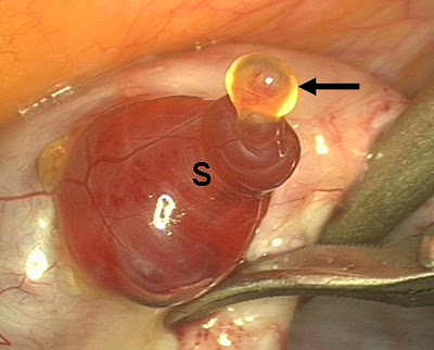
 Fertile women release one or more eggs every month, but until now, only animal ovulation has been recorded in detail. Now, gynaecologist Dr Jacques Donnez spotted the release of a human egg in progress, during a routine hysterectomy. He could film the process in close-up, when the egg was emerging from the ovary. Human eggs are produced by follicles, fluid-filled sacs on the side of the ovary, which, around the time of ovulation, produce a reddish protrusion seen in the pictures. The egg comes from the end of this, surrounded by a jelly-like substance containing supportive cells. The egg itself is only the size of a full-stop, and the whole ovary, which contains many immature eggs, just a couple of inches long. Dr Donnez, from the Catholic University of Louvain, told New Scientist that the pictures would help scientists understand the mechanisms involved in ovulation. He said that some theories had suggested an "explosive" release for the egg, but the ovulation he witnessed took 15 minutes to complete, so the event was progressive.

These images are the first time the event of ovulation in humans has been captured in clear detail. The yellow blob is a protruding egg cell, surrounded by supportive cumulus cells (at the black arrow). The reddish part is the follicle (S), and the pale pink tissue is part of the ovary (F).

Ovulation takes place on the surface of the ovarian tissue.

The egg, surrounded by supportive cumulus cells, is shown emerging from the follicle on the ovary.


After the release from the follicle, the egg travels down the Fallopian tube where it can be fertilised. 
Credit: The above pictures was kindly provided by Prof. Jacques Donnez, at Université Catholique de Louvain, Brussels, Belgium, and Eric Steinmehl, managing editor at “Fertility and Sterility” and the American Society for Reproductive Medicine, Birmingham, Alabama. 

Reference: 
Laparoscopic observation of spontaneous human ovulation 
Jean-Christophe Lousse, M.D., Jacques Donnez, M.D., Ph.D. 
Fertility and Sterility, published online 28 April 2008, doi:10.1016/j.fertnstert.2007.12.049

See also: 
How much is a human egg worth? 
CellNEWS - Wednesday, 21 February 2007 
Most egg cells in a female body die naturally by programmed cell death 
CellNEWS - Tuesday, 24 July 2007 
Back to the question: How much is a human egg worth? 
CellNEWS - Tuesday, 09 October 2007 
.........


ZenMaster


For more on stem cells and cloning, go to CellNEWS at 
http://cellnews-blog.blogspot.com/

Benefits of Broad Genetic Testing Still Limited

Benefits of Broad Genetic Testing Still Limited Wednesday, 18 June 2008 Five years ago, scientists finished mapping the complete human genetic code, but genetic assessment of disease risk remains in its infancy, according to the June issue of Mayo Clinic Women’s HealthSource. Scientists know that people share essentially the same genetic makeup, differing only by one-tenth of 1 percent. By studying that small variation, they hope to explain why one person is healthy and another sick and which treatments are best suited for each individual. Scientists have identified “simple” genetic disorders caused by mutation of a single gene, for example, sickle cell anaemia. Much more research is necessary on common diseases such as cancer, heart disease and diabetes, which are likely caused by a combination of genetic changes along with environmental influences. Already, some private companies are providing DNA analysis to the public to assess disease risk. A few also offer genetic counselling and disease prevention and screening advice. Experts caution that current tests don’t identify all genetic variants that combine to predict vulnerability or resistance to a disease. And even then, an accurate test might not provide useful information. For example, most women have an estimated 10 percent lifetime risk of breast cancer. Some medical providers question the practicality of informing a woman her personal risk may be slightly higher. In most cases, screening and prevention options aren’t changed. Though not as exciting as new technology, a careful analysis of the extended family medical history can provide a form of genetic risk assessment that is just as useful as newer tests. See also: Human Genetic Variation CellNEWS - Wednesday, 20 February 2008 ......... ZenMaster


For more on stem cells and cloning, go to CellNEWS at http://cellnews-blog.blogspot.com/ and http://www.geocities.com/giantfideli/index.html

Key Developmental Pathway Activates Lung Stem Cells

Pathway could hold promise for lung tissue repair Wednesday, 18 June 2008 Researchers from the University of Pennsylvania School of Medicine found that the activation of a molecular pathway important in stem cell and developmental biology leads to an increase in lung stem cells. Harnessing this knowledge could help develop therapies for lung-tissue repair after injury or disease. The investigators published their findings online last week in advance of print publication in Nature Genetics. "The current findings show that increased activity of the Wnt pathway leads to expansion of a type of lung stem cell called bronchioalveolar stem cells," says senior author Edward Morrisey, Ph.D., Associate Professor of Medicine and Cell and Developmental Biology. "This information will give us a more extensive basic understanding of Wnt signalling in adult tissue repair in the lung and other tissues and also start to help us determine whether pharmacological activation or inhibition of this pathway can be utilized for treatments," explains Morrisey, who is also the Scientific Director of the Penn Institute for Regenerative Medicine. Activation of the Wnt signalling pathway leads to expansion, or increase in number, of bronchioalveolar stem cells in the lung. A protein called GATA6 inhibits Wnt signalling by directly regulating the expression of another protein in the Wnt pathway called frizzled 2 (Fzd2). Wnt signalling is a major pathway in stem cell biology. The finding that GATA6 negatively regulates Wnt signalling and that GATA6 has been shown to play important roles in embryonic stem cell replication and differentiation suggests that these two pathways are linked not only in lung stem cells but in other tissues where they play important roles including the heart, gut, and pancreas. "We were surprised by the robust activation of Wnt signalling after loss of GATA6 expression in the lung," says Morrisey. "Such a robust activation is rarely observed." Wnt signalling can be pharmacologically modulated with compounds, including lithium, already approved by the FDA. Use of such compounds, both known and newly identified through ongoing screens, could allow for forced expansion and differentiation of key stem cell populations in the lung and other tissues for adult tissue repair after injury or disease. Future directions of the Morrisey lab include not only a more extensive basic understanding of Wnt signalling in adult-tissue repair in the lung and other tissues, but also starting to determine whether pharmacological activation or inhibition of this pathway can really be utilized for treatments. Reference: A Gata6-Wnt pathway required for epithelial stem cell development and airway regeneration Yuzhen Zhang, Ashley M Goss, Ethan David Cohen, Rachel Kadzik, John J Lepore, Karthika Muthukumaraswamy, Jifu Yang, Francesco J DeMayo, Jeffrey A Whitsett, Michael S Parmacek & Edward E Morrisey Nature Genetics, 8 June 2008, doi:10.1038/ng.157 ......... ZenMaster


For more on stem cells and cloning, go to CellNEWS at http://cellnews-blog.blogspot.com/ and http://www.geocities.com/giantfideli/index.html

Tuesday 17 June 2008

Adult Stem Cells Aid Broken Bone

UNC study lays groundwork for potential treatments Tuesday, 17 June 2008 In an approach that could become a new treatment for the 10 to 20 percent of people whose broken bones fail to heal, researchers at the University of North Carolina at Chapel Hill have shown that transplantation of adult stem cells can improve healing of fractures. Adult stem cells are specialized cells with the ability to regenerate tissue in response to damage. However, many patients lack sufficient numbers of these cells and thus cannot heal properly. Researchers have used adult stem cells in a few cases to improve fracture healing, but further studies were needed to show that this method was truly effective and safe before it can be pursued as a new treatment. Now scientists at UNC have provided the scientific foundation for future clinical trials of this approach by demonstrating in animal models that these cells can be used to repair broken bones. "This finding is critical to patients who lack the proper healing process and to individuals prone to broken bones, such as those with osteoporosis and the rare genetic condition known as brittle bone disease," said Dr. Anna Spagnoli, associate professor of paediatrics and biomedical engineering in the UNC School of Medicine and senior author on the study. The study, presented Monday, June 16 at the annual Endocrine Society meeting in San Francisco by the first author, Froilan Granero-Molto, Ph.D., post-doctoral associate researcher in UNC's paediatrics department, is the first to visualize the action of transplanted adult stem cells as they mend fractures in mice. During normal fracture healing, stem cells migrate to the site of the break, forming the cartilage and bone needed to fuse the broken bones back together. But in more than 600,000 Americans a year, this process does not occur as it should and these bones stay broken. The result can be long periods of immobilization, pain, bone deformities and even death. Current therapies, such as multiple surgeries with bone autografts and artificial prosthetic materials, often are not enough to cure these patients. "Man-made materials do not address the normal bone's function, and recurrent fractures, wear and toxicity are a real problem," Spagnoli said. "There is clearly a need to develop alternative therapies to enhance fracture healing in patients with bone union failure." Kicking stem cells into repair mode is one of the objectives of a new branch of medicine called regenerative medicine. With a little prodding, stem cells in human bone marrow – called mesenchymal stem cells – can turn into bone, cartilage, fat, muscle and blood vessel cells. "The beauty of regenerative medicine is that we are helping the body improve its innate ability to regenerate healthy tissue on its own, rather than introducing manmade materials to try to patch up a broken bone," Spagnoli said. Granero-Molto and other colleagues led by Spagnoli demonstrated this approach by transplanting adult stem cells in mice with fractures of the tibia, the long bone of the leg. The cells were taken from the bone marrow of mice that produce luciferase, the same molecule that allows fireflies to glow. In addition to possessing the ability to glow, the cells were engineered to express a molecule called insulin-like growth factor 1 (IGF-1). IGF-1 is a potent bone regenerator necessary for bones to grow both in size and strength. The researchers transplanted the cells through a simple intravenous injection and then placed the mice into a dark box so they could track the glowing stem cells as they migrated within the rodent. They found that these cells were specifically attracted to the fracture site, and that a particular molecule called CXCR4 – which acts as a homing signal – was necessary for the migration. Using a computerized tomography (CT or CAT) scan, the researchers showed that the stem cells not only migrated to the site of the fracture, but also improved healing there by increasing the bone and cartilage that bridged the bone gap. The bone at the fracture site in the treated mice was about three times stronger than that of untreated controls. If scientists can duplicate the results of this animal study in humans, it may lead to a new treatment for the millions of people who suffer fractures that do not heal properly, Spagnoli said. Once a physician determines that the bone has not healed, they could obtain adult stem cells from the person's bone marrow in a minimally invasive procedure and transplant them at the same time the patient is receiving a bone graft. Spagnoli said adult stem cells may pose fewer problems than embryonic stem cells, since they are not associated with the ethical controversy that surrounds the latter. Also, they may avoid the problem of rejection by the immune system, since the patient's own cells can be used. ......... ZenMaster


For more on stem cells and cloning, go to CellNEWS at http://cellnews-blog.blogspot.com/ and http://www.geocities.com/giantfideli/index.html

Stem Cell Researchers Give Old Muscle New Vigour

Stem Cell Researchers Give Old Muscle New Vigour Tuesday, 17 June 2008 Old muscle got a shot of youthful vigour in a stem cell experiment by bioengineers at the University of California, Berkeley, setting the path for research on new treatments for age-related degenerative conditions such as muscle atrophy or Alzheimer's and Parkinson's diseases. In a new study to be published June 15 in an advanced online issue of the journal Nature, researchers identified two key regulatory pathways that control how well adult stem cells repair and replace damaged tissue. They then tweaked how those stem cells reacted to those biochemical signals to revive the ability of muscle tissue in old mice to repair itself nearly as well as the muscle in the mice's much younger counterparts. Irina Conboy, an assistant professor of bioengineering and an investigator at the Berkeley Stem Cell Center and at the California Institute for Quantitative Biosciences (QB3), led the research team conducting this study.


Shown above is muscle tissue from a young mouse. On the right side are healthy, new cells created to replace damaged tissue. This ability to regenerate new cells diminishes with age. Credit: Photo courtesy of Morgan Carlson and Irina Conboy, UC Berkeley
Because the findings relate to adult stem cells that reside in existing tissue, this approach to rejuvenating degenerating muscle eliminates the ethical and medical complications associated with transplanting tissues grown from embryonic stem cells. "We are one step closer to having a point of intervention where we can rejuvenate the body's own stem cells so we don't have to suffer from some of the debilitating diseases associated with aging," said the study's lead author, Morgan Carlson, a recent Ph.D. graduate of Conboy's lab. The researchers focused on the interplay of two competing molecular pathways that control the stem cells, which sit next to the mature, differentiated cells that make up our working body parts. When the mature cells are damaged or wear out, the stem cells are called into action to begin the process of rebuilding. "We don't realize it, but as we grow our bodies are constantly being remodelled," said Conboy. "We are constantly falling apart, but we don't notice it much when we're young because we're always being restored. As we age, our stem cells are prevented, through chemical signals, from doing their jobs."
Shown here is muscle tissue from an old mouse. After UC Berkeley researchers manipulated the biochemical response of adult stem cells in the old tissue, the muscle was able to repair itself from damage almost as well as muscle from young mice. Credit: Photo courtesy of Morgan Carlson and Irina Conboy, UC Berkeley
The good news, the researchers said, is that the stem cells in old tissue are still ready and able to perform their regenerative function if they receive the appropriate chemical signals. Studies have shown that when old tissue is placed in an environment of young blood, the stem cells behave as if they are young again. "Conversely, we have found in a study published last year that even young stem cells rapidly age when placed among blood and tissue from old mice," said Carlson, who will stay on at UC Berkeley to expand his work on stem cell engineering either as a QB3 fellow or a postdoctoral researcher.
Adult stem cells have a receptor called Notch that, when activated, tells them that it is time to grow and divide, the researchers said. But stem cells also have a receptor for the protein TGF-beta that sets off a chain reaction activating the molecule pSmad3 and ultimately producing cyclin-dependent kinase (CDK) inhibitors, which regulate the cell's ability to divide. "Interestingly, activated Notch competes with activated pSmad3 for binding to the regulatory regions of the same CDK inhibitors in the stem cell," said Conboy. "We found that Notch is capable of physically kicking off pSmad3 from the promoters for the CDK inhibitors within the stem cell's nucleus, which tells us that a precise manipulation of the balance of these pathways would allow the ability to control stem cell responses." Notch and TGF-beta are well known in molecular biology, but Conboy's lab is the first to connect them to the process of aging, and the first to show that they act in opposition to each other within the nucleus of the adult stem cell.
As muscle tissue ages, it loses its ability to adequately repair itself from damage. Instead of creating healthy, new cells to replace damaged ones, the old muscle tissue is left with fibroblasts and scar tissue, as shown here. Credit: Photo courtesy of Morgan Carlson and Irina Conboy, UC Berkeley.
Aging and the inevitable march towards death are, in part, due to the progressive decline of Notch and the increased levels of TGF-beta, producing a one-two punch to the stem cell's capacity to effectively rebuild the body, the researchers said. "What we discovered is the interplay between two pathways - one an aging pathway, and the other a youthful pathway," said Conboy. But what would happen if researchers blocked the adult stem cells in old tissues from reacting to those TGF-beta signals? The researchers put that question to the test in a living organism by comparing the muscle regeneration capacity of old, 2-year-old mice, comparable in age to a 75- to 80-year-old human, with that of 2-month-old mice, similar in age to a 20- to 25-year-old human. For a group of the old mice, the researchers disabled the "aging pathway" that tells stem cells to stop dividing by using an established method of RNA interference that reduced levels of pSmad3. The researchers then examined the muscle of the different groups of mice one to five days after injury to compare how well the tissue repaired itself. As expected, the researchers found that muscle tissue in the young mice easily replaced damaged cells with new, healthy cells. In contrast, the areas of damaged muscle in the control group of old mice were characterized by fibroblasts and scar tissue. However, muscles in the old mice whose stem cell "aging pathway" had been dampened showed levels of cellular regeneration that were comparable to their much younger peers, and that were 3 to 4 times greater than those of the group of "untreated" old mice. The researchers cautioned that shutting down the TGF-beta/pSmad3 pathway altogether by turning off the gene that controls it could lead to many health problems. The ability to suppress cell division is critical in controlling the development of tumours, for instance. "When we are young, there is an optimal balance between Notch and TGF-beta," said Conboy. "We need to find out what the levels of these chemicals are in the young so we can calibrate the system when we're older. If we can do that, we could rejuvenate tissue repair for a very long time." The researchers also warn against interpreting this research as the cure-all for aging. "We're not at a point where we're ready to inject ourselves with TGF-beta antibodies and call it a day," said Carlson. "There are multiple mechanisms involved in how our body functions. We know that TGF-beta is involved in one aspect of aging, but we don't know where it fits in the global scheme of aging." In addition to their work on adult stem cells, Carlson and Conboy have also discovered that human embryonic stem cells can actually neutralize the effects of aging. Conboy received funding last year from the California Institute for Regenerative Medicine (CIRM) to pursue this line of research. Reference: Imbalance between pSmad3 and Notch induces CDK inhibitors in old muscle stem cells Morgan E. Carlson, Michael Hsu & Irina M. Conboy Nature advance online publication 15 June 2008, doi:10.1038/nature07034 .........
ZenMaster
For more on stem cells and cloning, go to CellNEWS at http://cellnews-blog.blogspot.com/ and http://www.geocities.com/giantfideli/index.html

Small Molecule Stimulates Nerve Stem Cells to Mature

Small Molecule Stimulates Nerve Stem Cells to Mature Tuesday, 17 June 2008 Inspired by a chance discovery during another experiment, researchers at UT Southwestern Medical Center have created a small molecule that stimulates nerve stem cells to begin maturing into nerve cells in culture. This finding might someday allow a person's own nerve stem cells to be grown outside the body, stimulated into maturity, and then re-implanted as working nerve cells to treat various diseases, the researchers said. "This provides a critical starting point for neuro-regenerative medicine and brain cancer chemotherapy," said Dr. Jenny Hsieh, assistant professor of molecular biology and senior author of the paper, which appears online today and in the June 17 issue of Nature Chemical Biology.


Drs. Jenny Hsieh and Jay Schneider are among the researchers who created a small molecule that stimulates nerve stem cells to begin maturing into nerve cells in culture. This development that might someday allow a person’s own nerve stem cells to be grown outside the body, stimulated into maturity, and then re-implanted as working nerve cells to treat various diseases.
The creation of the molecule allowed the researchers to uncover some of the biochemical steps that happen as nerve cells mature. It also showed that large-scale screening of compounds can provide starting points for developing drugs to treat disorders such as Huntington's disease, traumatic brain injury or cancer. The scientists began this project as a result of a separate study in which they were screening 147,000 compounds to see which could stimulate stem cells cultivated from rodent embryos to become heart cells. Unexpectedly, five molecules stimulated the cells to transform into forms resembling nerve cells. The researchers then created a variation of these molecules, a new compound called Isx-9 (for isoxazole-9). Isx-9 was easier to use than its initially discovered relatives because it worked at a much lower concentration and also dissolved more easily in water. "It was completely serendipitous that we uncovered this neurogenic [nerve-creating] small molecule," Dr. Hsieh said. "I think it's one of the most powerful neurogenic small molecules on the planet. In theory, this molecule could provoke full maturation, to the point that the new nerve cells could fire, generating the electrical signals needed for full functioning." Nerve stem cells live in scattered groups in various areas of the brain. They are capable of becoming several different types of cells, not all of which are nerve cells. In the study, rodent nerve stem cells from an area of the brain called the hippocampus were cultured with Isx-9. They clustered together and developed spiky appendages called neurites, which typically happens when nerve cells are grown in culture. Isx-9 also prevented the stem cells from developing into non-nerve cells and was more potent than other neurogenic substances in stimulating nerve-cell development. The molecule generated two to three times more nerve cells than other commonly used compounds. Neuroscientists believed for decades that the adult mammalian brain could not grow new nerve cells. Instead, they thought, learning and memory were strictly a matter of the brain making new connections between existing cells. It is now known, however, that the brain constantly creates new nerve cells. In the hippocampus, which is involved with learning and memory, stem cells mature into full-blown nerve cells at a rate of thousands a day, Dr. Hsieh said. Scientists know that when a mature nerve cell sends a chemical signal called a neurotransmitter to a stem cell, the immature cell begins to mature, but they don't know what biochemical pathways or genes are involved, Dr. Hsieh said. "The big gap in our knowledge is how to control these stem cells," she said. Isx-9 appeared to act like a neurotransmitter-like signal on the nerve stem cells, the researchers found. By culturing the stem cells with the compound, the scientists identified a possible biochemical pathway by which stem cells begin to become nerve cells. The researcher’s next plan to test Isx-9 on a large number of different combinations of RNA, the chemical cousin of DNA, to see on which genes the compound might be working. They have also applied for a patent on Isx-9 and its relatives. Reference: Small-molecule activation of neuronal cell fate Jay W Schneider, Zhengliang Gao, Shijie Li, Midhat Farooqi, Tie-Shan Tang, Ilya Bezprozvanny, Doug E Frantz & Jenny Hsieh Nature Chemical Biology, 15 June 2008, doi:10.1038/nchembio.95 ......... ZenMaster
For more on stem cells and cloning, go to CellNEWS at http://cellnews-blog.blogspot.com/ and http://www.geocities.com/giantfideli/index.html

Saturday 14 June 2008

EuroDYNA Takes Lid Off the Genome

EuroDYNA Takes Lid Off the Genome Saturday, 14 June 2008 European researchers have made significant progress unravelling how genes are governed and why this sometimes goes wrong in disease. The key lies in the dynamic ever-changing structure of the chromatin, which is the underlying complex of protein and DNA making up the chromosomes in which almost all genes are housed within the genome. The way this structure changes and responds to external signalling molecules within the cell determines how and when genes are expressed and also the mechanisms used to repair DNA damaged by a variety of internal and external insults, such as ultra violet radiation and free radical by-products of metabolism. Understanding the structure of chromatin and its interactions with proteins and RNA within the cell was the goal of the European Science Foundation 's (ESF) EuroDYNA programme, which held its last conference at the Wellcome Trust Conference Centre near Cambridge in May 2008. The study of genome structure involves interaction between various disciplines including cell biology, molecular physics, biomechanics and bioinformatics, as well as access to a wide range of expensive equipment such as electron microscopes, supercomputers, and scanners for simultaneous profiling of RNA expression across the whole genome. EuroDYNA helped broker these collaborations and enable projects to develop the critical mass needed to make real progress. The expression of genes involves an apparatus comprised mostly of proteins for reading the DNA, leading to production of RNA. This RNA in turn is either transported within the cell to the protein factory called the ribosome, where the code is translated into proteins, or else it interacts with other genes to control their expression in turn. These processes are intimately related to the constantly changing physical and chemical structure of the chromatin. Furthermore the overall state of the genome evolves during the life cycle of the cell, leading to its duplication if and when the cell eventually divides. All these inter-related processes need to be understood in order to unravel the complex network of mechanisms controlling gene expression. One of the big fundamental questions tackled within EuroDYNA concerned the detailed structure of how the DNA double helix is folded in the nucleus of higher organisms. Although the double helix structure was discovered by Crick and Watson in 1953, the way it folds and stretches such that it fits in the cell nucleus is only now becoming clear, as is its relevance both for cell replication and gene expression. At the EuroDYNA conference, John van Noort from Leiden University in the Netherlands reported that the DNA molecule, which in humans and most mammals is about two metres in length but only 2 nanometres in diameter, is coiled up like a spring in a solenoid structure. In such a folded structure it behaves according to the well known Hooke's law, stating that up to a certain point the extension is proportional to the force applied. It turns out chromatin is a very elastic molecular complex, capable of stretching to three times its normal rest length without breaking, according to van Noort. Even more remarkably – and here it differs from a familiar metal spring - even if stretched beyond three times its rest length, the chromatin solenoid is capable of repairing itself and regaining its former shape and elasticity. Indeed the ability of DNA to repair itself is essential for the long term survival of the cell and ultimately of the whole organism. DNA damage occurs not just from factors outside the cell nucleus, but also during the process of cell division (mitosis). The overall objective is to hand down the correct genetic code to the daughter cells during mitosis, a process so important that a number of surveillance and repair systems have been put in place to ensure its completion. One of those systems is called PRR (Post Replicative Repair) and it is highly conserved among all organisms, from bacteria to eukaryotes. PRR was discovered in the 1970s, but here again the detailed mechanisms are only now being elicited. At the EuroDYNA conference, Simone Sabbioneda from the University of Sussex presented new findings about one of the key PRR mechanisms called Translesion DNA Synthesis (TLS). This project, like some of the others, involved direct observation of processes as they take place in living cells, in this case using a technique called Fluorescence Recovery after Photobleaching. This comprises an optical microscope combined with a probe to observe the radiation emitted (the fluorescence) by molecules within a cell in response to a laser source. Such work is yielding important clues on how the PRR pathways work, hoping to help in the long term campaign to find novel, more specific, treatments for cancer, without the side effects of current therapies based on surgery, radiotherapy, or chemotherapy. One EuroDYNA project however yielded a more immediate insight into a treatment already used to alleviate the symptoms of another important disease, MS (multiple sclerosis). Pavel Kovarik from the University of Vienna's Department of Microbiology and Immunology noted that the only compound capable of alleviating MS symptoms was the protein interferon beta. This resembles the interferon produced naturally by the body in response to infection, but until now it has not been known how it relieves symptoms for MS sufferers. However Kovarik and colleagues have shown that interferon works by up-regulating (increasing production of) members of the protein family Tristetraprolin (TTP), which have an anti-inflammatory affect by in turn inhibiting production of pro-inflammatory agents. "We have demonstrated a novel function for interferon," said Kovarik. By understanding how it works, there is the potential for delivering interferon beta more effectively for treating MS. There were other projects within EuroDYNA with great therapeutic potential, many of which will continue, but which would benefit greatly from an extension to this highly successful programme. ......... ZenMaster


For more on stem cells and cloning, go to CellNEWS at http://cellnews-blog.blogspot.com/ and http://www.geocities.com/giantfideli/index.html