Far from being junk, these short stretches of the genome exert wide-ranging influence in the cell
Thursday, 31 July 2008
MicroRNAs (miRNAs) subtly influence a vast number of proteins involved in most key biological processes, according to the first large-scale analyses of how these small pieces of non-coding RNAs affect proteins. The work is presented in two articles in the latest issue of the journal Nature.
These studies could help answer long-standing questions about how miRNAs work. These tiny snippets of genome, just 21 to 25 nucleotides long, were once thought to be “junk” material because they are not translated into protein. In fact, in animals they control protein levels through two mechanisms: by breaking down the messenger RNA “read-out” from a gene; or by stopping the messenger RNA from being translated into protein. But previous studies had taken only a global look at how miRNAs affect messenger RNA, because of the technical difficulties in examining how miRNAs affect thousands of proteins at once.
Two teams of scientists have now tackled this problem using a state-of-the-art version of mass spectrometry. The technique, called SILAC (stable isotope labelling with amino acids in cell culture), relies on the use of heavy isotopes to label proteins as they are being produced by cells. The researchers used the isotopes to label thousands of proteins being made in cells that had been forced to over-express or down-regulate certain miRNAs. They then compared labelled protein levels in these cells with levels of corresponding proteins in control cells.
One group of researchers come from the Max Delbrück Center for Molecular Medicine (MDC) in Berlin-Buch, Germany. Two teams lead by Matthias Selbach (Proteomics) and Nikolaus Rajewsky (Systems Biology) have shown that a single miRNA can directly regulate synthesis of hundreds of different proteins. In this way, miRNAs can program the way human cells act, they report.
All body cells contain the same genes, may they be muscle-, brain-, blood-, or liver cells. Therefore, all have the same blue prints for the production of proteins. However, different cells produce different proteins at different times – a prerequisite for the body to develop normally and stay healthy. For this to happen, genes must be regulated differently in different cell types – that is, turned on and off at the right time. Only a few years ago, researchers discovered that miRNAs play an important role in gene regulation and, thus, help determine which proteins are produced by which cells.
Proteins are the building and operating materials of life and are required for the structure, function, and regulation of the body's cells, tissues, and organs. Diseases can result if protein production goes awry. Worldwide, scientists are seeking to develop methods to detect which miRNAs are active in tissue samples and which proteins are regulated by them. To date, researchers have identified a few hundred human miRNAs but it is not clear which proteins they regulate. A further complication is that miRNAs are known to regulate synthesis of proteins, which is difficult to measure.
Using a novel experimental approach carried out by PhD students Björn Schwanhäusser and Nadine Thierfelder, the MDC researchers for the first time were able to quantify protein synthesis for thousands of different human proteins. Together with extensive computational analyses, they could further identify and quantify the direct impact of specific miRNAs on target protein synthesis.
Changing the fate of a cell
The MDC researchers could demonstrate that the regulation of protein synthesis typically is mild, with a number of interesting exceptions.
"MicroRNAs screw many switches, but most of them only slightly", Matthias Selbach explains.
"Thus the system is robust and flexible. A single miRNA can have profound impact on the fate of a cell. miRNAs active in cancer cells, for example, are different from those active in normal cells."
Using a trick, the MDC researchers were for the first time able to measure changes in protein production after artificially changing the activity of specific miRNAs. They labelled amino acids (the building blocks of proteins) with a stable, non-radioactive isotope and put it together with miRNA in cell culture. This allowed them to distinguish labelled proteins in a mass spectrometer. They could show that only newly produced proteins were heavier.
A single miRNA can tune the protein levels of thousands of genes
The MDC researchers also compared the impact on protein production when artificially boosting or repressing the activity of an individual miRNA. They found that this impact is largely inverse for thousands of different proteins.
Thus, “it is as if a single miRNA can alter a large fraction of the entire protein production program of a human cell in a reversible fashion”, comments Nikolaus Rajewsky.
The findings of the two Berlin research teams in collaboration with Raya Khanin from Glasgow University (UK) are anticipated to have a big impact in the future, as miRNAs are considered to be promising diagnostic and therapeutic candidates for the treatment of human diseases.
The second group obtaining similar results was led by Steven Gygi, a protein chemist at Harvard Medical School in Boston, Massachusetts, and David Bartel, a molecular biologist and Howard Hughes investigator at the Whitehead Institute for Biomedical Research in Cambridge, Massachusetts.
The most intriguing finding from these studies is that the effects of miRNAs on proteins are usually quite modest, changing their expression levels by less than twofold. Although most miRNA effects may be small, they can still be powerful in the cell. The data paint a picture of miRNAs as regulators that fine-tune protein expression in complex and overlapping patterns. That could complicate the picture for drug developers, because most miRNAs have so many different targets. But this is not necessarily a bad thing, because some of the most effective drugs hit multiple molecular targets.
References:
Widespread changes in protein synthesis induced by microRNAs
Matthias Selbach, Björn Schwanhäusser, Nadine Thierfelder, Zhuo Fang, Raya Khanin & Nikolaus Rajewsky
Nature advance online publication 30 July 2008, doi:10.1038/nature07228
The impact of microRNAs on protein output
Daehyun Baek, Judit Villén, Chanseok Shin, Fernando D. Camargo, Steven P. Gygi & David P. Bartel
Nature advance online publication 30 July 2008, doi:10.1038/nature07242
.........
ZenMaster
For more on stem cells and cloning, go to CellNEWS at
http://cellnews-blog.blogspot.com/ and
http://www.geocities.com/giantfideli/index.html

It's about stem cells, cloning, bioethics and genetics.
Thursday, 31 July 2008
Monday, 28 July 2008
More Trouble Ignite for Bush’s ESC Lines
US Researchers may abandon several of the approved embryonic stem cell lines
Monday, 28 July 2008
Leading US research institutions may stop studying several federally fundable embryonic stem cell lines due to potential ethical problems surrounding the creation of the lines.
The Chronicle of Higher Education today (July 28) report that Stanford and Johns Hopkins Universities, and the California Institute for Regenerative Medicine (CIRM) are considering halting or have halted research on five of the 21 human stem cell lines approved by the Bush Administration to receive federal funding.
The background is that the University of Wisconsin bioethicist Robert Streiffer called the five lines into question in an article he wrote in the May-June issue of the Hastings Center Report. In the article, Streiffer argue that some embryonic stem cell donors were improperly informed before donating their cells. For example, Streiffer wrote that in at least one case, the consent forms that donors signed "states that the project in which the embryo donors were participating was limited to developing a technique for longer-term cultivation of embryonic cells, and that after the study was completed all the cells would be destroyed."
In light of the ethical problems, Streiffer called for the Bush Administration's restrictions on embryonic stem cell research funding to be overturned.
According to The Chronicle story, citing a report by the Center for American Progress, expert panels at Johns Hopkins and Stanford have already decided to stop research on the five contested cell lines, but officials at Stanford told The Chronicle that the report was "inaccurate" and that the no final decision has yet been made. A spokesman from CIRM told The Chronicle that the institute is deciding whether or not to refer the issue to its ethics board.
See also:
Where did the stem cells go?
.........
ZenMaster
For more on stem cells and cloning, go to CellNEWS at http://cellnews-blog.blogspot.com/ and http://www.geocities.com/giantfideli/index.html
For more on stem cells and cloning, go to CellNEWS at http://cellnews-blog.blogspot.com/ and http://www.geocities.com/giantfideli/index.html
Friday, 25 July 2008
Stem Cell Forum in China Demonstrate Cutting Edge Research
Focuses on new findings for iPS cells
Friday, 25 July 2008
Over 300 of China's top stem cell biologists and researchers from around the globe shared their latest results and held China's first ever symposium on advanced induced pluripotent stem (iPS) cell research during the just concluded first annual 2008 China Stem Cell Technology Forum at the China Medical City complex in Taizhou, China.
 At the Forum, which was chaired by Dr. Sean Hu, Chairman of Beike Biotechnology Co., Ltd., some of the world's most respected researchers presented their latest laboratory findings and clinical trial results in using stem cells to treat common ailments like heart disease and nervous system diseases such as cerebral palsy, spinal cord injury, muscular dystrophy, and optic nerve hypoplasia (ONH). Biologists attending the Mandarin Chinese language Forum came from leading research centers in nearly all of China's major cities, and as far away as the United States, Canada, India, Australia, and Malaysia.
A special symposium entitled Advanced iPS Cell Technology, convened as part of the Forum, brought together for the first time over a dozen scientists involved in cutting edge stem cell research at some of America and China's most respected science universities and research labs. A potential substitute for embryonic stem cells, genetically programmable iPS cells are widely believed to be at the very forefront of the field's most exciting research frontier as iPS cells theoretically could deliver the greatest therapeutic benefit. Since iPS cells can be produced from the building blocks in any individual's own body, they bring the potential that people may someday be able to heal a range of sicknesses from tissue cells they have banked themselves.
At the symposium, renowned stem cell scientists like Dr. Hu Jifan of the Palo Alto VA Health Care System under the Stanford University Medical School, Dr. Xiang Peng of Sun Yat Sen University, Dr. Zhang Yaou of Tsing Hua University, Dr. Kong Hsiang-Fu of the Chinese University of Hong Kong, Dr. Zhou Xiangjun of Shanghai Jiaotong University, and Dr. Yong-Jian Geng of the University of Texas Health Science Center at Houston and the Texas Heart Institute, discussed their own latest advancements since recent breakthroughs at the University of Wisconsin and Kyoto University in Japan. The scientists also discussed mutual cooperation and research oversight opportunities, creation of ethical research guidelines in China, and future iPS cell research directions.
At the symposium, a special subgroup of China's leading cardiovascular scientists like Dr. Chen Haozhu of Fudan University, Dr. Wang Shiwen of the Chinese PLA General Hospital, and Dr. Zhang Fumin of Nanjing Medical University conferred on the use of stem cells in heart disease treatment, as well as new research methods and ethical responsibilities in that field.
Dr. Yong-Jian Geng, M.D., Ph.D, Director of the Heart Failure and Stem Cell Research Lab at the Texas Heart Institute and Professor of Medicine at the University of Texas Medical School in Houston, said:
"Scientists and clinicians have been doing incredible research on stem cells, and generating many exciting results. I feel confident more breakthroughs will be coming soon. I was very pleased and honoured to be invited to participate in this discussion, and I am willing to help Chinese investigators establish guidelines that will, in my opinion, help develop a global standard for the stem cell therapies."
Dr. Hu Jifan, senior research scientist at the Palo Alto VA Health Care System under the Stanford University Medical School said:
"Leading scientists in China are constantly finding ways to create more effective iPS cells. Through this forum we agreed to help verify each other's work, share experiences, and ensure safe, high-standard research takes place so that we may someday create the conditions for effective clinical treatments and industrial scale production of iPS cells. While no one knows yet when these conditions might be met, this Forum certainly helped speed up the process by bringing together some of the world's best minds working towards these goals."
Presentations at the 2008 China Stem Cell Technology Forum covered a wide range of new biological advancements as Chinese scientists hone in on the global goal of bringing practical stem cell treatments to market. Other topics covered included findings on the most potent combinations of stem cells, effective cell processing methods, the latest clinical trials targeting diseases like heart conditions, muscular dystrophy and optic nerve hypoplasia (ONH), and more yet to be published stem cell research.
At the Forum, which was chaired by Dr. Sean Hu, Chairman of Beike Biotechnology Co., Ltd., some of the world's most respected researchers presented their latest laboratory findings and clinical trial results in using stem cells to treat common ailments like heart disease and nervous system diseases such as cerebral palsy, spinal cord injury, muscular dystrophy, and optic nerve hypoplasia (ONH). Biologists attending the Mandarin Chinese language Forum came from leading research centers in nearly all of China's major cities, and as far away as the United States, Canada, India, Australia, and Malaysia.
A special symposium entitled Advanced iPS Cell Technology, convened as part of the Forum, brought together for the first time over a dozen scientists involved in cutting edge stem cell research at some of America and China's most respected science universities and research labs. A potential substitute for embryonic stem cells, genetically programmable iPS cells are widely believed to be at the very forefront of the field's most exciting research frontier as iPS cells theoretically could deliver the greatest therapeutic benefit. Since iPS cells can be produced from the building blocks in any individual's own body, they bring the potential that people may someday be able to heal a range of sicknesses from tissue cells they have banked themselves.
At the symposium, renowned stem cell scientists like Dr. Hu Jifan of the Palo Alto VA Health Care System under the Stanford University Medical School, Dr. Xiang Peng of Sun Yat Sen University, Dr. Zhang Yaou of Tsing Hua University, Dr. Kong Hsiang-Fu of the Chinese University of Hong Kong, Dr. Zhou Xiangjun of Shanghai Jiaotong University, and Dr. Yong-Jian Geng of the University of Texas Health Science Center at Houston and the Texas Heart Institute, discussed their own latest advancements since recent breakthroughs at the University of Wisconsin and Kyoto University in Japan. The scientists also discussed mutual cooperation and research oversight opportunities, creation of ethical research guidelines in China, and future iPS cell research directions.
At the symposium, a special subgroup of China's leading cardiovascular scientists like Dr. Chen Haozhu of Fudan University, Dr. Wang Shiwen of the Chinese PLA General Hospital, and Dr. Zhang Fumin of Nanjing Medical University conferred on the use of stem cells in heart disease treatment, as well as new research methods and ethical responsibilities in that field.
Dr. Yong-Jian Geng, M.D., Ph.D, Director of the Heart Failure and Stem Cell Research Lab at the Texas Heart Institute and Professor of Medicine at the University of Texas Medical School in Houston, said:
"Scientists and clinicians have been doing incredible research on stem cells, and generating many exciting results. I feel confident more breakthroughs will be coming soon. I was very pleased and honoured to be invited to participate in this discussion, and I am willing to help Chinese investigators establish guidelines that will, in my opinion, help develop a global standard for the stem cell therapies."
Dr. Hu Jifan, senior research scientist at the Palo Alto VA Health Care System under the Stanford University Medical School said:
"Leading scientists in China are constantly finding ways to create more effective iPS cells. Through this forum we agreed to help verify each other's work, share experiences, and ensure safe, high-standard research takes place so that we may someday create the conditions for effective clinical treatments and industrial scale production of iPS cells. While no one knows yet when these conditions might be met, this Forum certainly helped speed up the process by bringing together some of the world's best minds working towards these goals."
Presentations at the 2008 China Stem Cell Technology Forum covered a wide range of new biological advancements as Chinese scientists hone in on the global goal of bringing practical stem cell treatments to market. Other topics covered included findings on the most potent combinations of stem cells, effective cell processing methods, the latest clinical trials targeting diseases like heart conditions, muscular dystrophy and optic nerve hypoplasia (ONH), and more yet to be published stem cell research.
 Beike Biotechnology Co., Ltd., hosted the event along with the China National Center for Biotechnology Development (CNCBD) under China's Ministry of Science and Technology (MOST), the Jiangsu Provincial Department for Science and Technology, and the Taizhou Government. The 2008 China Stem Cell Technology Forum was attended by MOST officials, the Vice Governor of Jiangsu Province Mr. Zhang Taolin, and Taizhou Mayor Mr. Yao Jianhua.
Dr. Sean Hu, the Chairman of Beike Biotech said:
"Chinese science, and indeed global stem cell research, achieved a great deal through this forum. Beike is proud to work with China to bring together so many globally known Mandarin speaking stem cell scientists to better communicate and share resources regarding what may very well become the future of healthcare."
About the China Medical City (CMC):
Jiangsu Province is considered the number one location for China's medical industry based on revenue generated over the past 5 years. The city of Taizhou in Jiangsu is not only the hometown of China's President Hu Jintao, but is considered the fastest growing medical industry location in Jiangsu, with over 35 % annual growth in that time. Established by the Chinese Government in 2005 and consisting of 20-25 square kilometres in the heart of Taizhou City, China Medical City (CMC) is fully supported by China's local and national governments. CMC is emerging as a strong leader in China's efforts to develop a streamlined pharmaceutical and medical materials industry that concentrates all medical services and support in one location. Businesses located in CMC carry out a range of manufacturing and support services including research and development, creation and processing of medical materials, distribution, comprehensive healthcare delivery solutions, and patent filing support.
About Beike Biotechnology Company Limited:
Beike is a biotechnology company that was founded in July 2005 with capital from Beijing University, Hong Kong University of Science and Technology and Shenzhen City Hall when it commercialized stem cell technology that had been in research since 1999. The research and clinical work comes from collaborations with leading institutions in China including of Tsinghua University, Beijing University, Hong Kong University of Science and Technology, No. 3 Army Medical University, Zhongshan Medical University, Guiyang Medical College and Zhengzhou University. Over 250 patients every month are treated with Beike's stem cells in leading hospitals throughout China. Patient experiences from treatments can be found at Stem Cell China News.
SOURCE: Adopted from material from Beike Biotechnology Company Limited.
See also:
Beike Biotech Opens Comprehensive Stem Cell Storage and Processing Facility in China
Medical News Today - 20 June 2008
.........
ZenMaster
Beike Biotechnology Co., Ltd., hosted the event along with the China National Center for Biotechnology Development (CNCBD) under China's Ministry of Science and Technology (MOST), the Jiangsu Provincial Department for Science and Technology, and the Taizhou Government. The 2008 China Stem Cell Technology Forum was attended by MOST officials, the Vice Governor of Jiangsu Province Mr. Zhang Taolin, and Taizhou Mayor Mr. Yao Jianhua.
Dr. Sean Hu, the Chairman of Beike Biotech said:
"Chinese science, and indeed global stem cell research, achieved a great deal through this forum. Beike is proud to work with China to bring together so many globally known Mandarin speaking stem cell scientists to better communicate and share resources regarding what may very well become the future of healthcare."
About the China Medical City (CMC):
Jiangsu Province is considered the number one location for China's medical industry based on revenue generated over the past 5 years. The city of Taizhou in Jiangsu is not only the hometown of China's President Hu Jintao, but is considered the fastest growing medical industry location in Jiangsu, with over 35 % annual growth in that time. Established by the Chinese Government in 2005 and consisting of 20-25 square kilometres in the heart of Taizhou City, China Medical City (CMC) is fully supported by China's local and national governments. CMC is emerging as a strong leader in China's efforts to develop a streamlined pharmaceutical and medical materials industry that concentrates all medical services and support in one location. Businesses located in CMC carry out a range of manufacturing and support services including research and development, creation and processing of medical materials, distribution, comprehensive healthcare delivery solutions, and patent filing support.
About Beike Biotechnology Company Limited:
Beike is a biotechnology company that was founded in July 2005 with capital from Beijing University, Hong Kong University of Science and Technology and Shenzhen City Hall when it commercialized stem cell technology that had been in research since 1999. The research and clinical work comes from collaborations with leading institutions in China including of Tsinghua University, Beijing University, Hong Kong University of Science and Technology, No. 3 Army Medical University, Zhongshan Medical University, Guiyang Medical College and Zhengzhou University. Over 250 patients every month are treated with Beike's stem cells in leading hospitals throughout China. Patient experiences from treatments can be found at Stem Cell China News.
SOURCE: Adopted from material from Beike Biotechnology Company Limited.
See also:
Beike Biotech Opens Comprehensive Stem Cell Storage and Processing Facility in China
Medical News Today - 20 June 2008
.........
ZenMaster
For more on stem cells and cloning, go to CellNEWS at http://cellnews-blog.blogspot.com/ and http://www.geocities.com/giantfideli/index.html
 At the Forum, which was chaired by Dr. Sean Hu, Chairman of Beike Biotechnology Co., Ltd., some of the world's most respected researchers presented their latest laboratory findings and clinical trial results in using stem cells to treat common ailments like heart disease and nervous system diseases such as cerebral palsy, spinal cord injury, muscular dystrophy, and optic nerve hypoplasia (ONH). Biologists attending the Mandarin Chinese language Forum came from leading research centers in nearly all of China's major cities, and as far away as the United States, Canada, India, Australia, and Malaysia.
A special symposium entitled Advanced iPS Cell Technology, convened as part of the Forum, brought together for the first time over a dozen scientists involved in cutting edge stem cell research at some of America and China's most respected science universities and research labs. A potential substitute for embryonic stem cells, genetically programmable iPS cells are widely believed to be at the very forefront of the field's most exciting research frontier as iPS cells theoretically could deliver the greatest therapeutic benefit. Since iPS cells can be produced from the building blocks in any individual's own body, they bring the potential that people may someday be able to heal a range of sicknesses from tissue cells they have banked themselves.
At the symposium, renowned stem cell scientists like Dr. Hu Jifan of the Palo Alto VA Health Care System under the Stanford University Medical School, Dr. Xiang Peng of Sun Yat Sen University, Dr. Zhang Yaou of Tsing Hua University, Dr. Kong Hsiang-Fu of the Chinese University of Hong Kong, Dr. Zhou Xiangjun of Shanghai Jiaotong University, and Dr. Yong-Jian Geng of the University of Texas Health Science Center at Houston and the Texas Heart Institute, discussed their own latest advancements since recent breakthroughs at the University of Wisconsin and Kyoto University in Japan. The scientists also discussed mutual cooperation and research oversight opportunities, creation of ethical research guidelines in China, and future iPS cell research directions.
At the symposium, a special subgroup of China's leading cardiovascular scientists like Dr. Chen Haozhu of Fudan University, Dr. Wang Shiwen of the Chinese PLA General Hospital, and Dr. Zhang Fumin of Nanjing Medical University conferred on the use of stem cells in heart disease treatment, as well as new research methods and ethical responsibilities in that field.
Dr. Yong-Jian Geng, M.D., Ph.D, Director of the Heart Failure and Stem Cell Research Lab at the Texas Heart Institute and Professor of Medicine at the University of Texas Medical School in Houston, said:
"Scientists and clinicians have been doing incredible research on stem cells, and generating many exciting results. I feel confident more breakthroughs will be coming soon. I was very pleased and honoured to be invited to participate in this discussion, and I am willing to help Chinese investigators establish guidelines that will, in my opinion, help develop a global standard for the stem cell therapies."
Dr. Hu Jifan, senior research scientist at the Palo Alto VA Health Care System under the Stanford University Medical School said:
"Leading scientists in China are constantly finding ways to create more effective iPS cells. Through this forum we agreed to help verify each other's work, share experiences, and ensure safe, high-standard research takes place so that we may someday create the conditions for effective clinical treatments and industrial scale production of iPS cells. While no one knows yet when these conditions might be met, this Forum certainly helped speed up the process by bringing together some of the world's best minds working towards these goals."
Presentations at the 2008 China Stem Cell Technology Forum covered a wide range of new biological advancements as Chinese scientists hone in on the global goal of bringing practical stem cell treatments to market. Other topics covered included findings on the most potent combinations of stem cells, effective cell processing methods, the latest clinical trials targeting diseases like heart conditions, muscular dystrophy and optic nerve hypoplasia (ONH), and more yet to be published stem cell research.
At the Forum, which was chaired by Dr. Sean Hu, Chairman of Beike Biotechnology Co., Ltd., some of the world's most respected researchers presented their latest laboratory findings and clinical trial results in using stem cells to treat common ailments like heart disease and nervous system diseases such as cerebral palsy, spinal cord injury, muscular dystrophy, and optic nerve hypoplasia (ONH). Biologists attending the Mandarin Chinese language Forum came from leading research centers in nearly all of China's major cities, and as far away as the United States, Canada, India, Australia, and Malaysia.
A special symposium entitled Advanced iPS Cell Technology, convened as part of the Forum, brought together for the first time over a dozen scientists involved in cutting edge stem cell research at some of America and China's most respected science universities and research labs. A potential substitute for embryonic stem cells, genetically programmable iPS cells are widely believed to be at the very forefront of the field's most exciting research frontier as iPS cells theoretically could deliver the greatest therapeutic benefit. Since iPS cells can be produced from the building blocks in any individual's own body, they bring the potential that people may someday be able to heal a range of sicknesses from tissue cells they have banked themselves.
At the symposium, renowned stem cell scientists like Dr. Hu Jifan of the Palo Alto VA Health Care System under the Stanford University Medical School, Dr. Xiang Peng of Sun Yat Sen University, Dr. Zhang Yaou of Tsing Hua University, Dr. Kong Hsiang-Fu of the Chinese University of Hong Kong, Dr. Zhou Xiangjun of Shanghai Jiaotong University, and Dr. Yong-Jian Geng of the University of Texas Health Science Center at Houston and the Texas Heart Institute, discussed their own latest advancements since recent breakthroughs at the University of Wisconsin and Kyoto University in Japan. The scientists also discussed mutual cooperation and research oversight opportunities, creation of ethical research guidelines in China, and future iPS cell research directions.
At the symposium, a special subgroup of China's leading cardiovascular scientists like Dr. Chen Haozhu of Fudan University, Dr. Wang Shiwen of the Chinese PLA General Hospital, and Dr. Zhang Fumin of Nanjing Medical University conferred on the use of stem cells in heart disease treatment, as well as new research methods and ethical responsibilities in that field.
Dr. Yong-Jian Geng, M.D., Ph.D, Director of the Heart Failure and Stem Cell Research Lab at the Texas Heart Institute and Professor of Medicine at the University of Texas Medical School in Houston, said:
"Scientists and clinicians have been doing incredible research on stem cells, and generating many exciting results. I feel confident more breakthroughs will be coming soon. I was very pleased and honoured to be invited to participate in this discussion, and I am willing to help Chinese investigators establish guidelines that will, in my opinion, help develop a global standard for the stem cell therapies."
Dr. Hu Jifan, senior research scientist at the Palo Alto VA Health Care System under the Stanford University Medical School said:
"Leading scientists in China are constantly finding ways to create more effective iPS cells. Through this forum we agreed to help verify each other's work, share experiences, and ensure safe, high-standard research takes place so that we may someday create the conditions for effective clinical treatments and industrial scale production of iPS cells. While no one knows yet when these conditions might be met, this Forum certainly helped speed up the process by bringing together some of the world's best minds working towards these goals."
Presentations at the 2008 China Stem Cell Technology Forum covered a wide range of new biological advancements as Chinese scientists hone in on the global goal of bringing practical stem cell treatments to market. Other topics covered included findings on the most potent combinations of stem cells, effective cell processing methods, the latest clinical trials targeting diseases like heart conditions, muscular dystrophy and optic nerve hypoplasia (ONH), and more yet to be published stem cell research.
 Beike Biotechnology Co., Ltd., hosted the event along with the China National Center for Biotechnology Development (CNCBD) under China's Ministry of Science and Technology (MOST), the Jiangsu Provincial Department for Science and Technology, and the Taizhou Government. The 2008 China Stem Cell Technology Forum was attended by MOST officials, the Vice Governor of Jiangsu Province Mr. Zhang Taolin, and Taizhou Mayor Mr. Yao Jianhua.
Dr. Sean Hu, the Chairman of Beike Biotech said:
"Chinese science, and indeed global stem cell research, achieved a great deal through this forum. Beike is proud to work with China to bring together so many globally known Mandarin speaking stem cell scientists to better communicate and share resources regarding what may very well become the future of healthcare."
About the China Medical City (CMC):
Jiangsu Province is considered the number one location for China's medical industry based on revenue generated over the past 5 years. The city of Taizhou in Jiangsu is not only the hometown of China's President Hu Jintao, but is considered the fastest growing medical industry location in Jiangsu, with over 35 % annual growth in that time. Established by the Chinese Government in 2005 and consisting of 20-25 square kilometres in the heart of Taizhou City, China Medical City (CMC) is fully supported by China's local and national governments. CMC is emerging as a strong leader in China's efforts to develop a streamlined pharmaceutical and medical materials industry that concentrates all medical services and support in one location. Businesses located in CMC carry out a range of manufacturing and support services including research and development, creation and processing of medical materials, distribution, comprehensive healthcare delivery solutions, and patent filing support.
About Beike Biotechnology Company Limited:
Beike is a biotechnology company that was founded in July 2005 with capital from Beijing University, Hong Kong University of Science and Technology and Shenzhen City Hall when it commercialized stem cell technology that had been in research since 1999. The research and clinical work comes from collaborations with leading institutions in China including of Tsinghua University, Beijing University, Hong Kong University of Science and Technology, No. 3 Army Medical University, Zhongshan Medical University, Guiyang Medical College and Zhengzhou University. Over 250 patients every month are treated with Beike's stem cells in leading hospitals throughout China. Patient experiences from treatments can be found at Stem Cell China News.
SOURCE: Adopted from material from Beike Biotechnology Company Limited.
See also:
Beike Biotech Opens Comprehensive Stem Cell Storage and Processing Facility in China
Medical News Today - 20 June 2008
.........
ZenMaster
Beike Biotechnology Co., Ltd., hosted the event along with the China National Center for Biotechnology Development (CNCBD) under China's Ministry of Science and Technology (MOST), the Jiangsu Provincial Department for Science and Technology, and the Taizhou Government. The 2008 China Stem Cell Technology Forum was attended by MOST officials, the Vice Governor of Jiangsu Province Mr. Zhang Taolin, and Taizhou Mayor Mr. Yao Jianhua.
Dr. Sean Hu, the Chairman of Beike Biotech said:
"Chinese science, and indeed global stem cell research, achieved a great deal through this forum. Beike is proud to work with China to bring together so many globally known Mandarin speaking stem cell scientists to better communicate and share resources regarding what may very well become the future of healthcare."
About the China Medical City (CMC):
Jiangsu Province is considered the number one location for China's medical industry based on revenue generated over the past 5 years. The city of Taizhou in Jiangsu is not only the hometown of China's President Hu Jintao, but is considered the fastest growing medical industry location in Jiangsu, with over 35 % annual growth in that time. Established by the Chinese Government in 2005 and consisting of 20-25 square kilometres in the heart of Taizhou City, China Medical City (CMC) is fully supported by China's local and national governments. CMC is emerging as a strong leader in China's efforts to develop a streamlined pharmaceutical and medical materials industry that concentrates all medical services and support in one location. Businesses located in CMC carry out a range of manufacturing and support services including research and development, creation and processing of medical materials, distribution, comprehensive healthcare delivery solutions, and patent filing support.
About Beike Biotechnology Company Limited:
Beike is a biotechnology company that was founded in July 2005 with capital from Beijing University, Hong Kong University of Science and Technology and Shenzhen City Hall when it commercialized stem cell technology that had been in research since 1999. The research and clinical work comes from collaborations with leading institutions in China including of Tsinghua University, Beijing University, Hong Kong University of Science and Technology, No. 3 Army Medical University, Zhongshan Medical University, Guiyang Medical College and Zhengzhou University. Over 250 patients every month are treated with Beike's stem cells in leading hospitals throughout China. Patient experiences from treatments can be found at Stem Cell China News.
SOURCE: Adopted from material from Beike Biotechnology Company Limited.
See also:
Beike Biotech Opens Comprehensive Stem Cell Storage and Processing Facility in China
Medical News Today - 20 June 2008
.........
ZenMaster For more on stem cells and cloning, go to CellNEWS at http://cellnews-blog.blogspot.com/ and http://www.geocities.com/giantfideli/index.html
Thursday, 24 July 2008
Adult Stem Cells Activated in Mammalian Brain
Locating cell origin provides foundation for brain injury cure
Thursday, 24 July 2008
Adult stem cells originate in a different part of the brain than is commonly believed, and with proper stimulation they can produce new brain cells to replace those lost to disease or injury, a study by UC Irvine scientists has shown.
Evidence strongly shows that the true stem cells in the mammalian brain are the ependymal cells that line the ventricles in the brain and spinal cord, rather than cells in the subventricular zone as biologists previously believed. Brain ventricles are hollow chambers filled with fluid that supports brain tissue, and a layer of ependymal cells lines these ventricles.
Knowing the cell source is crucial when developing stem cell-based therapies. Additionally, knowing that these normally dormant cells can be coaxed into dividing lays the groundwork for future therapies in which a patient's own stem cells produce new brain cells to treat neurological disorders and injuries such as Parkinson's disease, stroke or traumatic brain injury.
"With such a therapy, we would know which cells in the body to target for activation, and their offspring would have all the properties necessary to replace damaged or missing cells," said Darius Gleason, lead author of the study and a graduate student in the Department of Developmental and Cell Biology, the UCI Sue and Bill Gross Stem Cell Research Center.
"It is a very promising approach to stem cell therapy."
Study results appear this month online in the journal Neuroscience.
Stem cells are the "master cells" that produce each of the specialized cells within the human body. If researchers could control the production and differentiation of stem cells, they may be able to use them to replace damaged tissues.
One focus of stem cell research is transplantation, which entails injecting into the body healthy cells that may or may not genetically match the patient. Transplantation of non-matching stem cells requires the use of drugs to prevent the body from rejecting the treatment.
But, working with a patient's own cells would eliminate the need for transplantation and immunosuppressant drugs and may be a better alternative, scientists say. Ependymal cells line the fluid-filled ventricles, so a drug to activate the cells could theoretically travel through this fluid directly to the stem cells.
"The cells already match your brain completely since they have the same genetic make-up. That is a huge advantage over any other approach that uses cells from a donor," Gleason said.
"If they are your cells, then all we are doing is helping your body fix itself. We're not reinventing the repair process."
In this study, Gleason and Peter Bryant, developmental and cell biology professor, used rats treated to develop the animal equivalent of Parkinson's disease. They chose this type of rat because in a previous study by UCI collaborator James Fallon, a small protein given to the brain-damaged rats sparked a rapid and massive production and migration of new cells, and significantly improved motor behavior.
An adult ependymal cell in the rat brain undergoing self-renewing cell division. Credit: UC Irvine.
First, the UCI researchers sought to determine the true location of stem cells in the rats by looking for polarized cells, which have different sets of proteins on opposite sides so that when one divides it can produce two different products. Polarization gives rise to asymmetric cell division, which produces one copy of the parent and a second cell that is programmed to turn into another cell type. Asymmetric cell division is the defining characteristic of a stem cell. On rat brain samples, the researchers applied antibodies to identify proteins that may be involved in asymmetric cell division, and they found that polarization exists on the ependymal cells. "It couldn't have been a stronger signal or clearer message. We could see that the only cells undergoing asymmetric cell division were the ependymal cells," Gleason said. Next, they gave a drug to induce cell division in the rats and examined their brains at intervals ranging from one to 28 days after the treatment. At each interval, they counted cells that were dividing in the ependymal layer. They found the most division at 28 days, when about one-quarter of the ependymal cells were dividing. Previous studies by researchers at other institutions were successful in getting only a few cells to divide in that layer. "One interpretation of previous studies is there are scattered stem cells in the ependymal layer, and it is hard to locate them," Bryant said. "But we believe that all of the ependymal cells are stem cells, and that they all have the ability to be activated." Researchers don't know yet what sparks cell division at the molecular level, but learning that process and how to control it could lead to a safe, effective stem cell therapy. About the University of California, Irvine: The University of California, Irvine is a top-ranked university dedicated to research, scholarship and community service. Founded in 1965, UCI is among the fastest-growing University of California campuses, with more than 27,000 undergraduate and graduate students and nearly 2,000 faculty members. The third-largest employer in dynamic Orange County, UCI contributes an annual economic impact of $3.6 billion. See also: Adult Stem Cells Reprogrammed in the Brain CellNEWS - Monday, 30 June 2008 ......... ZenMaster
For more on stem cells and cloning, go to CellNEWS at http://cellnews-blog.blogspot.com/ and http://www.geocities.com/giantfideli/index.html
India to Set Up Stem Cell Research Unit in Bangalore
India to Set Up Stem Cell Research Unit in Bangalore
Thursday, 24 July 2008
 India's Union Cabinet Thursday gave its approval for the establishment of an Institute for Stem Cell Science and Regenerative Medicine (SCSRM) in Bangalore.
The institute will help in “developing policies relating to stem cell technologies, identify needs, facilitate conceptualisation and design of new technologies,” Priya Ranjan Dasmunsi, Information and Broadcasting Minister, told reporters.
The SCSRM institute would be an autonomous institution of the Department of Biotechnology and would be set up at a total cost of Rs. 203.10 crores (approx. US$ 48.4M).
The cabinet also approved the integration of Stem Cell Centre (SCC) at the Christian Medical College, Vellore, now funded by the department of biotechnology, with the Bangalore institute.
Source: Adopted from Press Release from the Press Information Bureau, Government of India.
.........
ZenMaster
India's Union Cabinet Thursday gave its approval for the establishment of an Institute for Stem Cell Science and Regenerative Medicine (SCSRM) in Bangalore.
The institute will help in “developing policies relating to stem cell technologies, identify needs, facilitate conceptualisation and design of new technologies,” Priya Ranjan Dasmunsi, Information and Broadcasting Minister, told reporters.
The SCSRM institute would be an autonomous institution of the Department of Biotechnology and would be set up at a total cost of Rs. 203.10 crores (approx. US$ 48.4M).
The cabinet also approved the integration of Stem Cell Centre (SCC) at the Christian Medical College, Vellore, now funded by the department of biotechnology, with the Bangalore institute.
Source: Adopted from Press Release from the Press Information Bureau, Government of India.
.........
ZenMaster
For more on stem cells and cloning, go to CellNEWS at http://cellnews-blog.blogspot.com/ and http://www.geocities.com/giantfideli/index.html
 India's Union Cabinet Thursday gave its approval for the establishment of an Institute for Stem Cell Science and Regenerative Medicine (SCSRM) in Bangalore.
The institute will help in “developing policies relating to stem cell technologies, identify needs, facilitate conceptualisation and design of new technologies,” Priya Ranjan Dasmunsi, Information and Broadcasting Minister, told reporters.
The SCSRM institute would be an autonomous institution of the Department of Biotechnology and would be set up at a total cost of Rs. 203.10 crores (approx. US$ 48.4M).
The cabinet also approved the integration of Stem Cell Centre (SCC) at the Christian Medical College, Vellore, now funded by the department of biotechnology, with the Bangalore institute.
Source: Adopted from Press Release from the Press Information Bureau, Government of India.
.........
ZenMaster
India's Union Cabinet Thursday gave its approval for the establishment of an Institute for Stem Cell Science and Regenerative Medicine (SCSRM) in Bangalore.
The institute will help in “developing policies relating to stem cell technologies, identify needs, facilitate conceptualisation and design of new technologies,” Priya Ranjan Dasmunsi, Information and Broadcasting Minister, told reporters.
The SCSRM institute would be an autonomous institution of the Department of Biotechnology and would be set up at a total cost of Rs. 203.10 crores (approx. US$ 48.4M).
The cabinet also approved the integration of Stem Cell Centre (SCC) at the Christian Medical College, Vellore, now funded by the department of biotechnology, with the Bangalore institute.
Source: Adopted from Press Release from the Press Information Bureau, Government of India.
.........
ZenMaster For more on stem cells and cloning, go to CellNEWS at http://cellnews-blog.blogspot.com/ and http://www.geocities.com/giantfideli/index.html
Omega-3 Fatty Acids Could Slow Acute Wound Healing
Omega-3 Fatty Acids Could Slow Acute Wound Healing
Thursday, 24 July 2008
A recent study shows that popular fish oil supplements have an effect on the healing process of small, acute wounds in human skin. But whether that effect is detrimental, as researchers initially suspected, remains a mystery.
The omega-3 fatty acids found in fish oils are widely considered to benefit cardiovascular health and other diseases related to chronic inflammation because of their anti-inflammatory properties. But insufficient inflammation during the initial stage of wound healing may delay the advancement of later stages.
In the study, blister wounds on the arms of people taking fish oil supplements were compared to the wounds of people taking a placebo. The wounds healed in about the same amount of time – but at the local cellular level, something unexpected happened. The levels of proteins associated with initiating and sustaining inflammation were higher in the blister fluid in people who had taken the active fish oil supplements. The researchers had expected those proteins to be lowered by the increased systemic presence of omega-3 fatty acids in the blood.
"That finding was hard to explain," said Jodi McDaniel, lead author of the study and an assistant professor of nursing at Ohio State University.
"These proteins may have other functions that we don't yet fully understand. And our results also suggested there could be a difference between men and women in the amount of inflammatory proteins that are produced, because on average, women had lower levels of one of the proteins."
If the polyunsaturated fatty acids in the fish oils do indeed delay acute wound healing, then they probably should be discontinued for awhile by patients scheduled for surgery, McDaniel said. They appear to have enough of an effect that patients should at least inform their doctors if they're taking a fish oil supplement, she added.
But there could still be a bright side to the supplements' ability to alter those proteins and other molecular substances that control inflammation locally. Fish oil's systemic anti-inflammatory power, which has been illustrated in previous studies, still might assist in the healing of chronic wounds at the local level. Chronic wounds are essentially stuck in an inflammatory stage that slows or prevents transition to the later stages needed for complete healing. That mechanism needs to be explored further, McDaniel said.
"There's so much information out there now about omega-3s and they clearly have lots of potential," McDaniel said.
"We're just trying to figure out how to evaluate what they do and how to advise people to take these supplements. Our goal isn't to stop supplement use but to fill in the picture of what conditions they help and what they might hurt."
The research is published in a recent issue of the journal Wound Repair and Regeneration.
Study participants were divided into two groups of 15 healthy adults each. One group took a placebo, and the other took an active supplement containing 1.6 grams of eicosapentaenoic acid (EPA) and 1.1 grams of docosahexaenoic acid (DHA) daily for four weeks. EPA and DHA are the polyunsaturated fatty acids obtained primarily from fish oil that serve as the basis of most standard omega-3 supplements.
Previous research has suggested that these fatty acids affect the production of proteins called pro-inflammatory cytokines, which signal biological processes during the inflammatory stage of wound healing. The primary cytokines in the process are interleukin-1 beta (IL-1b), interleukin-6 (IL-6) and tumour necrosis factor-alpha (TNF-a).
But research had not yet addressed how the interaction between fatty acids and these cytokines might affect human wounds.
McDaniel and colleagues expected to find that research participants taking the fish oil supplements – and therefore being affected by their anti-inflammatory properties – would have significantly lower levels of the cytokines in their blister wounds during the initial stage of inflammation, resulting in a slower healing process.
Instead, the group taking the fish oil had significantly higher levels of IL-1b in their blister wounds than did the placebo group 24 hours after the wounds were created. The active group also had higher levels of IL-6 and TNF-a cytokines in their blisters over time than did the placebo group, but those differences in levels were not significant. The blisters took an average of 10.7 days to completely heal in the active supplement group and an average of 9.8 days in the placebo group, but the difference was not significant.
The results suggest that the function of these cytokines still isn't completely understood, McDaniel said. And the scientists were also surprised to find that gender appeared to make a difference in cytokine production. Men were more likely than women among the active supplement group to have higher levels of the IL-1b cytokine in their wounds. McDaniel said that some studies have suggested that oestrogen plays a role in inhibiting the production of this particular protein during the inflammatory stage of wound healing, but more research is needed.
McDaniel and colleagues are following up with a similar study in which they are adding a low-dose aspirin to both the fish oil supplement and placebo groups. Some research has shown that aspirin can facilitate the anti-inflammatory properties of omega-3 fatty acids, and low-dose aspirin is commonly included in the medication regimen of patients with cardiovascular disease.
The researchers also will look at different biological markers in blister wounds to see if the combination of fish oil and aspirin produces compounds that function as what McDaniel called "stop and go switches" in controlling inflammation.
"If we find that the fish oil can work in an anti-inflammatory fashion at the local level of wound sites, we would consider moving on to the chronic wound population," McDaniel said.
"Even if we find that there are times when omega-3 fatty acids should not be taken in advance of creating an acute wound, such as in elective surgery, we still have high hopes that fish oil might be beneficial for chronic wounds in certain situations."
.........
ZenMaster
For more on stem cells and cloning, go to CellNEWS at http://cellnews-blog.blogspot.com/ and http://www.geocities.com/giantfideli/index.html
For more on stem cells and cloning, go to CellNEWS at http://cellnews-blog.blogspot.com/ and http://www.geocities.com/giantfideli/index.html
Your Lower Belly Fat Is Rich in Stem Cells
ASPS study finds some areas of the body have greater concentrations of stem cells
Thursday, 24 July 2008
Fat removed from the lower abdomen and inner thigh through liposuction was found to be an excellent source of stem cells, with higher stem cell concentrations than other areas of the body, reports a Brazilian-based study in Augusts’ Plastic and Reconstructive Surgery®, the official medical journal of the American Society of Plastic Surgeons (ASPS). This is the first study of its kind to examine whether fat tissues from different areas of the body vary in stem cell concentration.
"Adult stem cells, derived from our own tissues, hold strong promise for improved clinical therapies," said J. Peter Rubin, MD, a member of the ASPS Fat Grafting Task Force who is involved in pre-clinical trial work on stem cells taken from fat.
"The potential for healing and repairing injury or disease through stem cells, including conditions like breast cancer and reconstruction, heart failure, spinal injuries, diabetes and Parkinson's disease are incredible. We may be able to more permanently and naturally get rid of pesky wrinkles or augment breasts with stem cell enriched fat in the future as well. Knowing more about the biology of stem cells will be of great value when we are ready for clinical trials in this country."
In the study, 23 female patients having liposuction in at least four different body areas agreed to have their fat isolated for adult stem cells and analyzed to determine stem cell concentrations. The body areas that were liposuctioned were: lower abdomen, upper abdomen, inner knee, inner thigh, flank and hips.
The study results found a significant difference in stem cell concentrations in different areas of the body. A major finding was that the concentration of stem cells was greatest in the lower abdomen and inner thighs. Interestingly, stem cell concentration in the lower abdomen was five times greater than in the upper abdomen.
"The value of stem cells harvested through fat is the ready and ample supply available," said ASPS President Richard D'Amico, MD.
"Using stem cells will some day have very practical applications to the specialty of plastic surgery. That we may be able to generate new tissue or bone that can be used in many of the reconstructive and cosmetic procedures we do every day is a tremendous."
Stem cells are unspecialized cells that have not yet developed a specific function. Not only are they capable of self renewal, stem cells can divide and produce others that become specialized cells. Scientists and doctors theorize that stem cells will be able to repair or replace damaged or diseased cells. Clinical trials researching the potential of stem cells from fat are ongoing in Europe and Asia. In the U.S., there are many investigators doing pre-clinical trial work to meet the stringent safety guidelines the FDA sets for clinical trials.
According to ASPS statistics, more than 30,000 liposuction procedures were performed in 2007 in US alone.
.........
ZenMaster
For more on stem cells and cloning, go to CellNEWS at http://cellnews-blog.blogspot.com/ and http://www.geocities.com/giantfideli/index.html
For more on stem cells and cloning, go to CellNEWS at http://cellnews-blog.blogspot.com/ and http://www.geocities.com/giantfideli/index.html
Lick Your Wounds!!!
Scientists isolate compound in human saliva that speeds wound healing
Wednesday, 23 July 2008
A report by scientists from The Netherlands published online in The FASEB Journal identifies a compound in human saliva that greatly speeds wound healing. This research may offer hope to people suffering from chronic wounds related to diabetes and other disorders, as well as traumatic injuries and burns. In addition, because the compounds can be mass produced, they have the potential to become as common as antibiotic creams and rubbing alcohol.
"We hope our finding is ultimately beneficial for people who suffer from non-healing wounds, such as foot ulcers and diabetic ulcers, as well as for treatment of trauma-induced wounds like burns," said Menno Oudhoff, first author of the report.
Specifically, scientists found that histatin, a small protein in saliva previously only believed to kill bacteria was responsible for the healing. To come to this conclusion, the researchers used epithelial cells that line the inner cheek, and cultured in dishes until the surfaces were completely covered with cells. Then they made an artificial wound in the cell layer in each dish, by scratching a small piece of the cells away. In one dish, cells were bathed in an isotonic fluid without any additions. In the other dish, cells were bathed in human saliva. After 16 hours the scientists noticed that the saliva treated "wound" was almost completely closed. In the dish with the untreated "wound," a substantial part of the "wound" was still open. This proved that human saliva contains a factor which accelerates wound closure of oral cells. Because saliva is a complex liquid with many components, the next step was to identify which component was responsible for wound healing. Using various techniques the researchers split the saliva into its individual components, tested each in their wound model, and finally determined that histatin was responsible.
"This study not only answers the biological question of why animals lick their wounds," said Gerald Weissmann, MD, Editor-in-Chief of The FASEB Journal, "it also explains why wounds in the mouth, like those of a tooth extraction, heal much faster than comparable wounds of the skin and bone. It also directs us to begin looking at saliva as a source for new drugs."
Reference:
Histatins are the major wound-closure stimulating factors in human saliva as identified in a cell culture assay
Menno J. Oudhoff, Jan G. M. Bolscher, Kamran Nazmi, Hakan Kalay, Wim van 't Hof, Arie V. Nieuw Amerongen, and Enno C. I. Veerman
FASEB Journal, July 23, 2008, doi: 10.1096/fj.08-112003
.........
ZenMaster
For more on stem cells and cloning, go to CellNEWS at http://cellnews-blog.blogspot.com/ and http://www.geocities.com/giantfideli/index.html
For more on stem cells and cloning, go to CellNEWS at http://cellnews-blog.blogspot.com/ and http://www.geocities.com/giantfideli/index.html
Tuesday, 22 July 2008
Spinal Cord Stem Cells May Act as Nerve Repair System
Cells for spinal-cord repair identified, could lead to nonsurgical treatment for injuries
Tuesday, 22 July 2008
Researcher at the Karolinska Institute in Sweden and MIT's Picower Institute for Learning and Memory has pinpointed stem cells within the spinal cord that, if persuaded to differentiate into more healing cells and fewer scarring cells following an injury, may lead to a new, non-surgical treatment for debilitating spinal-cord injuries.
The work, reported in the July issue of the journal PLoS (Public Library of Science) Biology, was done by Jonas Frisén’s group at the Department of Cell and Molecular Biology, Karolinska Institute, Stockholm, Sweden. Their results could lead to drugs that might restore some degree of mobility to the 30,000 people worldwide afflicted each year with spinal-cord injuries.
In a developing embryo, stem cells differentiate into all the specialized tissues of the body. In adults, stem cells act as a repair system, replenishing specialized cells, but also maintaining the normal turnover of regenerative organs such as blood, skin or intestinal tissues.
The tiny number of stem cells in the adult spinal cord proliferates slowly or rarely, and fails to promote regeneration on their own. But recent experiments show that these same cells, grown in the lab and returned to the injury site, can restore some function in paralyzed rodents and primates.
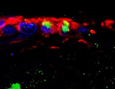
The researchers at the Karolinska Institute and MIT found that neural stem cells in the adult spinal cord are limited to a layer of cube- or column-shaped, cilia-covered cells called ependymal cells. These cells make up the thin membrane lining the inner-brain ventricles and the connecting central column of the spinal cord.
"We have been able to genetically mark this neural stem cell population and then follow their behavior," Konstantinos Meletis said, the first author of the study and presently a postdoctoral fellow at the Picower Institute at MIT in Cambridge, Massachusetts, USA.
"We find that these cells proliferate upon spinal cord injury, migrate toward the injury site and differentiate over several months."
 Ependymal cells in the adult spinal cord self-renew in vivo. Stack of 14 images depicting vimentin expressing ependymal cells (purple) of the central canal that have incorporated BrdU (have divided, orange). Spinal cord neurons are labelled with NeuN (white). Credit: Image/Design by Marie Carlén; confocal image by Fanie Barnabé-Heider.
The study uncovers the molecular mechanism underlying the tantalizing results of the rodent and primate and goes one step further: By identifying for the first time where this subpopulation of cells is found, they pave a path toward manipulating them with drugs to boost their inborn ability to repair damaged nerve cells. "The ependymal cells' ability to turn into several different cell types upon injury makes them very interesting from an intervention aspect: Imagine if we could regulate the behaviour of this stem cell population to repair damaged nerve cells," Meletis said. Upon injury, ependymal cells proliferate and migrate to the injured area, producing a mass of scar-forming cells, plus fewer cells called oligodendrocytes. The oligodendrocytes restore the myelin, or coating, on nerve cells' long, slender, electrical impulse-carrying projections called axons. Myelin is like the layer of plastic insulation on an electrical wire; without it, nerve cells don't function properly. "The limited functional recovery typically associated with central nervous system injuries is in part due to the failure of severed axons to regrow and reconnect with their target cells in the peripheral nervous system that extends to our arms, hands, legs and feet," Meletis said. "The function of axons that remain intact after injury in humans is often compromised without insulating sheaths of myelin." If scientists could genetically manipulate ependymal cells to produce more myelin and less scar tissue after a spinal cord injury, they could potentially avoid or reverse many of the debilitating effects of this type of injury, the researchers said. This study was supported by grants from the Swedish Research Council, the Swedish Cancer Society, the Foundation for Strategic Research, the Karolinska Institute, EuroStemCell and the Christopher and Dana Reeve Foundation. Reference: Spinal Cord Injury Reveals Multilineage Differentiation of Ependymal Cells Konstantinos Meletis, Fanie Barnabé-Heider, Marie Carlén, Emma Evergren, Nikolay Tomilin, Oleg Shupliakov, Jonas Frisén PLoS Biol 6(7): e182 doi:10.1371/journal.pbio.0060182 ......... ZenMaster
For more on stem cells and cloning, go to CellNEWS at http://cellnews-blog.blogspot.com/ and http://www.geocities.com/giantfideli/index.html
Sunday, 20 July 2008
The Genetics of the White Horse Unravelled
The Genetics of the White Horse Unravelled
Sunday, 20 July 2008
The white horse is an icon for dignity which has had a huge impact on human culture across the world. An international team led by researchers at Uppsala University has now identified the mutation causing this spectacular trait and show that white horses carry an identical mutation that can be traced back to a common ancestor that lived thousands of years ago. The study is interesting for medical research since this mutation also enhance the risk for melanoma. The paper is published on July 20 on the website of Nature Genetics.
Exactly when the mutation for ‘Greying with age’ was appearing is not known, but it was first described in western literature 2.500 years ago by the Greek historian Herodotus, telling that the Persian King Xerxes had a whole stable with Holy White Horses.
The great majority of white horses carry the dominant mutation ‘Greying with age’. A Grey horse is born coloured (black, brown or chestnut) but the greying process starts already during its first year and they are normally completely white by six to eight years of age but the skin remains pigmented. Thus, the process resembles greying in humans but the process is ultrafast in these horses. The research presented now demonstrates that all Grey horses carry exactly the same mutation which must have been inherited from a common ancestor that lived thousands of years ago.
“It is a fascinating thought that once upon a time a horse was born that turned grey and subsequently white and the people that observed it were so fascinated by its spectacular appearance that they used the horse for breeding so that the mutation could be transmitted from generation to generation,” says Leif Andersson, who led the study, and professor in functional genomic at the Department of Medical Biochemistry and Microbiology (IMBIM), Uppsala Biomedical Centre, at Uppsala University. Today about one horse in ten carries the mutation for Greying with age.
It is obvious that humans across the world have greatly valued these white horses as documented by the rich collection of stories and paintings featuring white horses. In the paper the white horse as an icon for dignity is illustrated by reproducing a painting from the late 17th century of the Swedish king Karl XI on his white horse Brilliant.
“The discovery was quite unexpected, since the mutation for ‘Greying with age’ was found in a DNA segment never before associated with colour pigmentation,” says Leif Andersson.
 Lipizzaner horses.
DNA from more than 900 horses was analysed, several of them from the white Lipizzaner horses from the Spanish Riding School in Vienna. In all white horses, but not in any black, brown or chestnut coloured horse, the exact same DNA change was found, a large segment of DNA duplication. This mutation does not change any protein coding gene, but regulate how active two other genes will be. The Grey horse is also very interesting from a medical point of view since the mutation also predisposes for development of melanoma. About 75% of Grey horses older than 15 years of age have a benign form of melanoma that in some cases develops into a malignant melanoma. Thus, the study reported today has also given new insight in a molecular pathway that may lead to tumour development. “We propose that the Grey mutation stimulates growth of melanocytes and that this leads to a premature loss of the melanocyte stem cells needed for hair pigmentation whereas the mutation promotes an expansion of some of the melanocytes causing skin pigmentation,” says Leif Andersson. Domestic animals constitute extraordinary models for evolution of biological diversity as already recognized by Charles Darwin. The white horse is a beautiful illustration of the importance of regulatory mutations as a major underlying mechanism for phenotypic diversity within and between species. The Grey mutation does not change any protein structure but it affects the genetic regulation of two genes. The researchers found that the white horses carry an extra copy of a DNA segment located in one of these genes. “It is very likely that regulatory mutations like the one we found in these white horses constitute the dominating class of mutations explaining differences between breeds of domestic animals as well as between species like humans and chimpanzee,” concludes Leif Andersson. Referens: A cis-acting regulatory mutation causes premature hair greying and susceptibility to melanoma in the horse Gerli Rosengren Pielberg, Anna Golovko, Elisabeth Sundström, Ino Curik, Johan Lennartsson, Monika H Seltenhammer, Thomas Druml, Matthew Binns, Carolyn Fitzsimmons, Gabriella Lindgren, Kaj Sandberg, Roswitha Baumung, Monika Vetterlein, Sara Strömberg, Manfred Grabherr, Claire Wade, Kerstin Lindblad-Toh, Fredrik Pontén, Carl-Henrik Heldin, Johann Sölkner & Leif Andersson Nature Genetics Published online: 20 July 2008, doi:10.1038/ng.185 ......... ZenMaster
For more on stem cells and cloning, go to CellNEWS at http://cellnews-blog.blogspot.com/ and http://www.geocities.com/giantfideli/index.html
Friday, 18 July 2008
Standards in Stem Cell Research Needed
Standards in Stem Cell Research Needed
Friday, 18 July 2008
Standards in stem cell research help both scientists and regulators to manage uncertainty and the unknown, according to new research funded by the Economic and Social Research Council, UK. Efforts to standardise practices across different labs is, however, a balancing act where the autonomy of scientists and fragility of living material need to be weighed against the need for comparable data.
The ambition in many quarters to scale up the production of human embryonic stem cells and move towards clinical trials requires that different laboratories are able to produce to a standard quality of cells. Developing common standards in stem cell production is not straightforward as so much is still unknown in this new science.
Professor Andrew Webster and Dr Lena Eriksson of York University interviewed and observed a range of scientists and technicians working in stem cell laboratories in the UK, USA and Sweden.
Accurately describing human embryonic stem cell lines is one way to begin setting standards. A stem cell line is a family of constantly-dividing cells, the product of a single parent group of stem cells. Embryonic stem cells are unique in that they have yet to 'decide' which developmental path to choose: they have the ability to turn into almost all human cell types. However each human embryonic stem cell holds the genetic signature of the donor which differs between donors just as people themselves differ. Further the state of a stem cell is by its very nature temporary as it is defined by its ability to develop into many different cell types.
Some scientists argued that as the stem cell cannot be standardised, the process and materials used should be standardised. Currently differences in laboratory practices are thought to result in differences in stem cell lines reflecting the way they are treated rather than an innate quality of the lines themselves. The skills of the laboratory technician also play a key role. But pinpointing all the factors that contribute to producing successful stem cell lines remains elusive.
"Scientists often explain that their laboratory produces successful human embryonic stem cell lines because their laboratory uses the factor X when they grow them or its lab technicians have green fingers," says Dr Lena Eriksson of the research team.
Some researchers prefer not to develop standards as these will constrain the science and may close off promising areas of research.
"Others argue that it is simply futile," explains Lena Eriksson.
"Can you standardise how all children sleep by giving them the same bed, sheet and blanket? Of course not. So why bother standardising the materials of stem cell production when other differences such as donor history and derivation methods are so complex, manifold and, to date, largely unknown?"
However, the research shows most stem cell scientists are keen to collaborate on the technical side as they feel this is necessary in order to move the field as a whole forward. The research team followed one particularly successful effort – the International Stem Cell Initiative – that adopted a multi-sited experimental approach in which a large number of stem cell lines were analysed and compared.
Because of the imprecise nature of the manual laboratory work, standardisation opens a potential market for automation technologies to be introduced into human embryonic stem cell laboratories. Yet the research shows this also brings tensions. By attracting businesses keen to become suppliers of laboratory material for this emerging market, the expense of such equipment as well as the skills and staff needed to operate it may exclude small laboratories. Even those that can afford to meet the costs have reservations about the robustness of stem cells to withstand the automation process. The relationship between standards, automation and stem cell quality will be key to the future scale-up of the field and so its clinical application.
The research project 'Quality Assured Science: The Role of Standards in Stem Cell Research' was funded by the Economic and Social Research Council. It was carried out by Professor Andrew Webster and Dr Lena Eriksson of York University together with Professor Peter Andrews and Professor Harry Moore of Sheffield University. The project is part of the ESRC's Social Science Stem Cell Initiative set up in 2005.
See also:
Scientist Work Out Stem Cell Guidelines
CellNEWS - Saturday, 14 June 2008
.........
ZenMaster
For more on stem cells and cloning, go to CellNEWS at http://cellnews-blog.blogspot.com/ and http://www.geocities.com/giantfideli/index.html
For more on stem cells and cloning, go to CellNEWS at http://cellnews-blog.blogspot.com/ and http://www.geocities.com/giantfideli/index.html
Sunday, 13 July 2008
Stem Cell Tourism Seen as ‘Medical Hoax’ by Specialists
Stem Cell Treatment for Children with Eye Nerve Disease Called ‘Medical Hoax’
Sunday, 13 July 2008
Two paediatric eye surgeons at Washington University School of Medicine in St. Louis expressed alarm over what they label a “21st century snake oil” scam.
Recent newspaper stories — including several from Missouri — have reported parents flying their children to main land China for umbilical cord stem cell (CSC) infusions. The cost of these treatments, paid for entirely out-of-pocket by the parents, can be $50,000 or more. CSCs are extracted from the umbilical cords of Chinese mothers and their newborns and injected into the fluid around the spinal cord of the American children. The parents are led to believe by Chinese doctors that these CSCs are an effective treatment for optic nerve hypoplasia (ONH), a disease causing partial blindness at birth.
ONH is growth failure of one or both optic nerves during the first trimesters of pregnancy. The nerve is the “cable” connecting the eye to the brain and each nerve should have one million fibres; in ONH the number of fibres ranges from 200,000-800,000. ONH, which affects about 1 in 5,000 newborns, is not hereditary and the exact cause is unknown.
Lawrence Tychsen, M.D., and Gregg Lueder, M.D., professors of ophthalmology and visual sciences at Washington University School of Medicine and paediatric ophthalmologists at St Louis Children’s Hospital, diagnose and treat dozens of children each year with ONH. They are concerned that the CSC reports will mislead many parents of children with ONH, who may bankrupt savings, go deeply into debt or organize fundraisers to pay for sham treatment.
Although some parents claim improvement in their child’s vision after returning from China, Tychsen and Lueder caution that no objective visual gains after CSC treatment have been demonstrated in any child with ONH. They can measure visual improvements objectively in infants and toddlers using non-invasive nerve and brain imaging and electronic measures of visual brain activity.
They add that one would expect “a powerful placebo effect after these purported treatments. The temptation to believe vision had improved, after the expenditure of so much time and money, would be difficult to resist.”
Aside from grave ethical concerns, they say that the injections could be dangerous, introducing infection or toxic matter into the brain fluids.
Tychsen, who is also a neurobiologist studying visual brain development in infant monkeys, listed a number of reasons to disbelieve reports of improvement. First, CSCs placed in human spinal fluid would not be transported into the fibres of the optic nerve. Second, CSCs have never been shown to transform into optic fibres, even in fish or rodent experiments. In a monkey or human, the task would be “several orders of magnitude more complex,” Tychsen said. Third, to improve vision, 100,000 or so fibres would need to grow, not just a few, and each of the fibres would need to connect precisely in the brain.
“CSCs are used legitimately throughout the United States to treat blood diseases (such as leukaemia) when the donor and recipient are genetically matched,” Tychsen said.
“But CSCs from an unrelated person are rejected and destroyed. Even if an unmatched CSC survived, found its way inside the optic nerve and transformed itself into a new fibre, the fibre would need to find the correct connection among more than 500,000 connections in the visual brain. “
“Such a series of events would be so improbable as to qualify as miraculous, the equivalent of a chimpanzee typing the five acts of King Lear at one sitting,” Tychsen said. He said he believes that experiments by neuroscientists devoted to the discovery of nerve growth molecules may hold the best hope for future cures.
Lueder pointed out that parents of ONH children should not despair.
“Many babies born with ONH will have some improvement as they mature, because they learn to exploit more effectively the optic fibres that remain,” he said. Children with ONH can also achieve some improvements with surgery for eye crossing and nystagmus (roving movements of the eyes). He added that many ONH children function reasonably well in school using enlarged print, magnifiers and other aids for the visually impaired.
About Washington University School of Medicine:
Washington University School of Medicine's 2,100 employed and volunteer faculty physicians also are the medical staff of Barnes-Jewish and St. Louis Children's hospitals. The School of Medicine is one of the leading medical research, teaching and patient care institutions in the nation, currently ranked third in the nation by U.S. News & World Report. Through its affiliations with Barnes-Jewish and St. Louis Children's hospitals, the School of Medicine is linked to BJC HealthCare.
See also:
Stem-Cell Tourism Troubles Experts
The Philadelphia Inquirer - Sunday, 13 July 2008
.........
ZenMaster
For more on stem cells and cloning, go to CellNEWS at http://cellnews-blog.blogspot.com/ and http://www.geocities.com/giantfideli/index.html
For more on stem cells and cloning, go to CellNEWS at http://cellnews-blog.blogspot.com/ and http://www.geocities.com/giantfideli/index.html
Friday, 11 July 2008
Genetic Recombination gets More Precise
Scientists generate the most precise map of genetic recombination ever
Friday, 11 July 2008
Genetic recombination, the process by which sexually reproducing organisms shuffle their genetic material when producing germ cells, leads to offspring with a new genetic make-up and influences the course of evolution. In the current issue of Nature, researchers at the European Molecular Biology Laboratory (EMBL) in Heidelberg, Germany, and the EMBL-European Bioinformatics Institute (EMBL-EBI) in Hinxton, UK, present the most precise map of genetic recombination yet. The study sheds light on fundamental questions about genetic shuffling and has implications for the tracking of disease genes and their inheritance.
In order to generate germ cells, sexually reproducing organisms undergo a complex series of cell divisions (meiosis) that includes the shuffling of genetic material inherited from the two parents. Equivalent chromosomes from mother and father pair up and exchange sections of DNA in a process called crossover. In a different type of recombination, called non-crossover, a small piece of DNA is copied from one chromosome onto the other without reciprocal exchange leading to gene conversion. Non-crossovers are minute events with a subtler effect than the exchange of larger fragments, but both types of recombination can increase genetic diversity and explain why organisms of the same species differ in many ways. Both types of recombination can also act to separate the transmission of neighbouring genes, which are normally inherited together.
The groups of Lars Steinmetz at EMBL and Wolfgang Huber at EMBL-EBI have produced the most detailed map to date of recombination events in the yeast genome.
"Our map has the highest resolution of recombination events that currently exists for any organism. We can locate crossovers and even hard-to-trace non-crossovers, typically with a precision of about 80 bases. This resolution is 20 times higher than in any existing yeast map and more than 360 times higher than a recent human map," says Steinmetz.
The map revealed many new insights into the organisation of recombination in yeast. On average over 150 recombination events were observed during a typical meiosis. These events did not occur uniformly across the genome. The recombination rate varied according to location, with events concentrated at so-called hotspots, some of which favoured either crossovers or non-crossovers. The researchers also found evidence for interference between crossovers and non-crossovers - a phenomenon previously only known to occur between crossovers - that makes it unlikely for two recombination events to happen in close proximity.
The fundamental principles of recombination are likely to be shared between yeast and humans.
"Our map expands our understanding of crossover and provides a wealth of new information about non-crossovers and gene conversion. It will act as a reference for future research into recombination," says Richard Bourgon from Huber's group, who developed the statistical methodology for this new type of data.
The insights gained will not only help tackle questions about the basic mechanisms of recombination; they will also have practical implications for the tracking of disease genes in humans.
Reference:
High-resolution mapping of meiotic crossovers and non-crossovers in yeast
Eugenio Mancera, Richard Bourgon, Alessandro Brozzi, Wolfgang Huber & Lars M. Steinmetz
Nature advance online publication 9 July 2008 doi:10.1038/nature07135
.........
ZenMaster
For more on stem cells and cloning, go to CellNEWS at http://cellnews-blog.blogspot.com/ and http://www.geocities.com/giantfideli/index.html
For more on stem cells and cloning, go to CellNEWS at http://cellnews-blog.blogspot.com/ and http://www.geocities.com/giantfideli/index.html
Genes That Control Embryonic Stem Cell Fate Identified
Genes That Control ESC Fate Identified
Friday, 11 July 2008
Scientists have identified about two dozen genes that control embryonic stem cell fate. The genes may either prod or restrain stem cells from drifting into a kind of limbo, they suspect. The limbo lies between the embryonic stage and fully differentiated, or specialized, cells, such as bone, muscle or fat.
By knowing the genes and proteins that control a cell's progress toward the differentiated form, researchers may be able to accelerate the process – a potential boon for the use of stem cells in therapy or the study of some degenerative diseases, the scientists say.
Their finding comes from the first large-scale search for genes crucial to embryonic stem cells. The research was carried out by a team at the University of California, San Francisco and is reported in a paper in the July 11, 2008 issue of "Cell."
"The genes we identified are necessary for embryonic stem cells to maintain a memory of who they are," says Barbara Panning, PhD, associate professor of biochemistry and biophysics at UCSF, and senior author on the paper.
"Without them the cell doesn't know whether it should remain a stem cell or differentiate into a specialized cell."
The scientists used a powerful technique known as RNA interference, or RNAi, to screen more than 1,000 genes for their role in mouse embryonic stem cells. The technique allows researchers to "knock down" individual genes, reducing their abundance in order to determine the gene's normal role.
The research focused on proteins that help package DNA. In the nucleus, DNA normally wraps around protein complexes called nucleosomes, forming a structure known as chromatin. This is what makes up chromosomes.
They found 22 proteins, each of which is essential for embryonic stem cells to maintain their consistent shape, growth properties, and pattern of gene expression.
Most of the genes code for multi-protein complexes that physically rearrange, or "remodel" nucleosomes, changing the likelihood that the underlying genes will be expressed to make proteins.
The main player they identified is a 17-protein complex called Tip60-p400. This complex is necessary for the cellular memory that maintains embryonic stem cell identity, Panning explains. Without it, the embryonic stem cells turned into a different cell type, which had some features of a stem cell but many features of a differentiated cell.
The scientists believe that Tip60-p400 is necessary for embryonic stem cells to correctly read the signals that determine cell type. These findings are not only important for understanding cellular memory in embryonic stem cells, but will also likely be relevant to other cell types, they say.
Inactivation of other genes disrupted embryonic stem cell proliferation. These genes were already known to have only slight influence on viability of mature cells in the body. This suggests that embryonic stem cells are "uniquely sensitive to certain perturbations of chromatin structure," the scientists report.
If other types of stem cells are also found to be sensitive to these chromatin perturbations, this could lead to novel cancer therapies in the future, Panning says.
Reference:
An RNAi Screen of Chromatin Proteins Identifies Tip60-p400 as a Regulator of Embryonic Stem Cell Identity
Thomas G. Fazzio, Jason T. Huff, and Barbara Panning
Cell, Vol 134, 162-174, 11 July 2008
.........
ZenMaster
For more on stem cells and cloning, go to CellNEWS at http://cellnews-blog.blogspot.com/ and http://www.geocities.com/giantfideli/index.html
For more on stem cells and cloning, go to CellNEWS at http://cellnews-blog.blogspot.com/ and http://www.geocities.com/giantfideli/index.html
Stem Cells Restore Muscle in Muscular Dystrophy
Muscle stem cell transplant boosts diseased muscle function and replenishes stem cell pool
Friday, 11 July 2008
 Researchers at the Joslin Diabetes Center have demonstrated for the first time that transplanted muscle stem cells can both improve muscle function in animals with a form of muscular dystrophy and replenish the stem cell population for use in the repair of future muscle injuries.
By injecting purified stem cells isolated from adult skeletal muscle, researchers have shown they can restore healthy muscle and improve muscle function in mice with a form of muscular dystrophy. Those muscle-building stem cells were derived from a larger pool of so-called satellite cells that normally associate with mature muscle fibers and play a role in muscle growth and repair.
In addition to their contributions to mature muscle, the injected cells also replenished the pool of regenerative cells normally found in muscle. Those stem cells allowed the treated muscle to undergo subsequent rounds of injury repair, they found.
"I'm very excited about this," said lead author Amy J. Wagers, Ph.D., Principal Investigator in the Joslin Section on Developmental and Stem Cell Biology, principal faculty member at the Harvard Stem Cell Institute and Assistant Professor of Stem Cell and Regenerative Biology at Harvard University.
"This study indicates the presence of renewing muscle stem cells in adult skeletal muscle and demonstrates the potential benefit of stem cell therapy for the treatment of muscle degenerative diseases such as muscular dystrophy."
"Our work shows proof-of-concept that purified muscle stem cells can be used in therapy," said Wagers, noting that in some cases the stem cells replaced more than 90 percent of the muscle fibers. Such an advance would require isolation of stem cells equivalent to those in the mouse from human muscle, something Wagers said her team is now working on.
The study was designed to test the concept that skeletal muscle precursor cells could function as adult stem cells and that transplantation of these cells could both repair muscle tissue and regenerate the stem cell pool in a model of Duchenne muscular dystrophy, she said.
Duchenne muscular dystrophy is the most common form of the disease and is characterized by rapidly progressing muscle degeneration. The disease is caused by a genetic mutation and there is currently no cure.
The data from this new study demonstrate that regenerative muscle stem cells can be distinguished from other cells in the muscle by unique protein markers present on their surfaces. The authors used these markers to select stem cells from normal adult muscle and transferred the cells to diseased muscle of mice carrying a mutation in the same gene affected in human Duchenne muscular dystrophy.
Satellite cells were first described decades ago and have since generally been considered as a homogeneous group, Wagers said. While anatomically they look similar under a microscope, they nonetheless show considerable variation in their physiology and function. In a previous study, Wagers' identified a set of five markers that characterize the only subset of satellite cells responsible for forming muscle, which they also refer to as skeletal muscle precursors or SMPs.
In the new study, the researchers analyzed the stem cell and regenerative properties of those SMPs. When engrafted into muscle of mice lacking dystrophin, purified SMPs contributed to up to 94 percent of muscle fibers, restoring dystrophin expression and significantly improving muscle structure and contractile function, they report. (The dystrophin gene encodes a protein important for muscle integrity. Mice lacking dystrophin, also known as mdx mice, are a model for Duchenne muscular dystrophy, the most prevalent form of muscular dystrophy.)
"Importantly, high-level engraftment of transplanted SMPs in mdx animals shows therapeutic value — restoring defective dystrophin gene expression, improving muscle histology, and rescuing physiological muscle function," the researchers said.
"Moreover, in addition to generating mature muscle fibers, transplanted SMPs also re-seed the satellite cell niche and are maintained there such that they can be recruited to participate in future rounds of muscle regeneration.”
"Taken together, these data indicate that SMPs act as renewable, transplantable stem cells for adult skeletal muscle. The level of myofiber reconstitution achieved by these myogenic stem cells exceeds that reported for most other myogenic cell populations and leads to a striking improvement of muscle contraction function in SMP-treated muscles. These data thus provide direct evidence that prospectively isolatable, lineage-specific skeletal muscle stem cells provide a robust source of muscle replacement cells and a viable therapeutic option for the treatment of muscle degenerative disorders."
Wagers noted however that there may be complications in the delivery of cell therapy in humans, particularly for those with conditions influencing skeletal muscle throughout the body. Even so, the new findings present an "opportunity to understand what happens [to these regenerative cells] in disease and identify factors and pathways that may boost their activity," she said.
"We may get a handle on drugs that could target muscle impairment" not only in those with muscular dystrophies, but also in elderly people suffering from the muscle wasting that comes with age.
"Once the healthy stem cells were transplanted into the muscles of the mice with muscular dystrophy, they generated cells that incorporated into the diseased muscle and substantially improved the ability of the treated muscles to contract," said Wagers.
"At the same time, the transplantation of the healthy stem cells replenished the formerly diseased stem cell pool, providing a reservoir of healthy stem cells that could be re-activated to repair the muscle again during a second injury."
According to the paper, these cells provide an effective source of immediately available muscle regenerative cells as well as a reserve pool that can maintain muscle regenerative activity in response to future challenges.
"This work demonstrates, in concept, that stem cell therapy could be beneficial for degenerative muscle diseases," Wagers said.
Wagers also said the study will lead to other studies in the near-term that will identify pathways that regulate these muscle stem cells in order to figure out ways to boost the normal regenerative potential of these cells. These could include drug therapies or genomic approaches, she said. In the long-term, the idea will be to replicate these findings in humans.
"This is still very basic science, but I think we're going to be able to move forward in a lot of directions. It opens up many exciting avenues," she said.
The Wagers Lab at Joslin studies both hematopoietic stem cells, which constantly maintain and can fully regenerate the entire blood system, as well as skeletal muscle stem cells, involved in skeletal muscle growth and repair. The work is aimed particularly at defining novel mechanisms that regulate the migration, expansion, and regenerative potential of these two distinct adult stem cells.
About Joslin Diabetes Center
Joslin Diabetes Center is the world's largest diabetes clinic, diabetes research center and provider of diabetes education. Joslin is dedicated to ensuring people with diabetes live long, healthy lives and offers real hope and progress toward diabetes prevention and a cure for the disease. Founded in 1898 by Elliott P. Joslin, M.D., Joslin is an independent non-profit institution affiliated with Harvard Medical School.
Reference:
Highly Efficient, Functional Engraftment of Skeletal Muscle Stem Cells in Dystrophic Muscles
Massimiliano Cerletti, Sara Jurga, Carol A. Witczak, Michael F. Hirshman, Jennifer L. Shadrach, Laurie J. Goodyear, and Amy J. Wagers
Cell, Vol 134, 37-47, 11 July 2008
.........
ZenMaster
Researchers at the Joslin Diabetes Center have demonstrated for the first time that transplanted muscle stem cells can both improve muscle function in animals with a form of muscular dystrophy and replenish the stem cell population for use in the repair of future muscle injuries.
By injecting purified stem cells isolated from adult skeletal muscle, researchers have shown they can restore healthy muscle and improve muscle function in mice with a form of muscular dystrophy. Those muscle-building stem cells were derived from a larger pool of so-called satellite cells that normally associate with mature muscle fibers and play a role in muscle growth and repair.
In addition to their contributions to mature muscle, the injected cells also replenished the pool of regenerative cells normally found in muscle. Those stem cells allowed the treated muscle to undergo subsequent rounds of injury repair, they found.
"I'm very excited about this," said lead author Amy J. Wagers, Ph.D., Principal Investigator in the Joslin Section on Developmental and Stem Cell Biology, principal faculty member at the Harvard Stem Cell Institute and Assistant Professor of Stem Cell and Regenerative Biology at Harvard University.
"This study indicates the presence of renewing muscle stem cells in adult skeletal muscle and demonstrates the potential benefit of stem cell therapy for the treatment of muscle degenerative diseases such as muscular dystrophy."
"Our work shows proof-of-concept that purified muscle stem cells can be used in therapy," said Wagers, noting that in some cases the stem cells replaced more than 90 percent of the muscle fibers. Such an advance would require isolation of stem cells equivalent to those in the mouse from human muscle, something Wagers said her team is now working on.
The study was designed to test the concept that skeletal muscle precursor cells could function as adult stem cells and that transplantation of these cells could both repair muscle tissue and regenerate the stem cell pool in a model of Duchenne muscular dystrophy, she said.
Duchenne muscular dystrophy is the most common form of the disease and is characterized by rapidly progressing muscle degeneration. The disease is caused by a genetic mutation and there is currently no cure.
The data from this new study demonstrate that regenerative muscle stem cells can be distinguished from other cells in the muscle by unique protein markers present on their surfaces. The authors used these markers to select stem cells from normal adult muscle and transferred the cells to diseased muscle of mice carrying a mutation in the same gene affected in human Duchenne muscular dystrophy.
Satellite cells were first described decades ago and have since generally been considered as a homogeneous group, Wagers said. While anatomically they look similar under a microscope, they nonetheless show considerable variation in their physiology and function. In a previous study, Wagers' identified a set of five markers that characterize the only subset of satellite cells responsible for forming muscle, which they also refer to as skeletal muscle precursors or SMPs.
In the new study, the researchers analyzed the stem cell and regenerative properties of those SMPs. When engrafted into muscle of mice lacking dystrophin, purified SMPs contributed to up to 94 percent of muscle fibers, restoring dystrophin expression and significantly improving muscle structure and contractile function, they report. (The dystrophin gene encodes a protein important for muscle integrity. Mice lacking dystrophin, also known as mdx mice, are a model for Duchenne muscular dystrophy, the most prevalent form of muscular dystrophy.)
"Importantly, high-level engraftment of transplanted SMPs in mdx animals shows therapeutic value — restoring defective dystrophin gene expression, improving muscle histology, and rescuing physiological muscle function," the researchers said.
"Moreover, in addition to generating mature muscle fibers, transplanted SMPs also re-seed the satellite cell niche and are maintained there such that they can be recruited to participate in future rounds of muscle regeneration.”
"Taken together, these data indicate that SMPs act as renewable, transplantable stem cells for adult skeletal muscle. The level of myofiber reconstitution achieved by these myogenic stem cells exceeds that reported for most other myogenic cell populations and leads to a striking improvement of muscle contraction function in SMP-treated muscles. These data thus provide direct evidence that prospectively isolatable, lineage-specific skeletal muscle stem cells provide a robust source of muscle replacement cells and a viable therapeutic option for the treatment of muscle degenerative disorders."
Wagers noted however that there may be complications in the delivery of cell therapy in humans, particularly for those with conditions influencing skeletal muscle throughout the body. Even so, the new findings present an "opportunity to understand what happens [to these regenerative cells] in disease and identify factors and pathways that may boost their activity," she said.
"We may get a handle on drugs that could target muscle impairment" not only in those with muscular dystrophies, but also in elderly people suffering from the muscle wasting that comes with age.
"Once the healthy stem cells were transplanted into the muscles of the mice with muscular dystrophy, they generated cells that incorporated into the diseased muscle and substantially improved the ability of the treated muscles to contract," said Wagers.
"At the same time, the transplantation of the healthy stem cells replenished the formerly diseased stem cell pool, providing a reservoir of healthy stem cells that could be re-activated to repair the muscle again during a second injury."
According to the paper, these cells provide an effective source of immediately available muscle regenerative cells as well as a reserve pool that can maintain muscle regenerative activity in response to future challenges.
"This work demonstrates, in concept, that stem cell therapy could be beneficial for degenerative muscle diseases," Wagers said.
Wagers also said the study will lead to other studies in the near-term that will identify pathways that regulate these muscle stem cells in order to figure out ways to boost the normal regenerative potential of these cells. These could include drug therapies or genomic approaches, she said. In the long-term, the idea will be to replicate these findings in humans.
"This is still very basic science, but I think we're going to be able to move forward in a lot of directions. It opens up many exciting avenues," she said.
The Wagers Lab at Joslin studies both hematopoietic stem cells, which constantly maintain and can fully regenerate the entire blood system, as well as skeletal muscle stem cells, involved in skeletal muscle growth and repair. The work is aimed particularly at defining novel mechanisms that regulate the migration, expansion, and regenerative potential of these two distinct adult stem cells.
About Joslin Diabetes Center
Joslin Diabetes Center is the world's largest diabetes clinic, diabetes research center and provider of diabetes education. Joslin is dedicated to ensuring people with diabetes live long, healthy lives and offers real hope and progress toward diabetes prevention and a cure for the disease. Founded in 1898 by Elliott P. Joslin, M.D., Joslin is an independent non-profit institution affiliated with Harvard Medical School.
Reference:
Highly Efficient, Functional Engraftment of Skeletal Muscle Stem Cells in Dystrophic Muscles
Massimiliano Cerletti, Sara Jurga, Carol A. Witczak, Michael F. Hirshman, Jennifer L. Shadrach, Laurie J. Goodyear, and Amy J. Wagers
Cell, Vol 134, 37-47, 11 July 2008
.........
ZenMaster
For more on stem cells and cloning, go to CellNEWS at http://cellnews-blog.blogspot.com/ and http://www.geocities.com/giantfideli/index.html
For more on stem cells and cloning, go to CellNEWS at http://cellnews-blog.blogspot.com/ and http://www.geocities.com/giantfideli/index.html
Primer on Mitochondria
Primer on Mitochondria

1. What are mitochondria and where are they found?
Mitochondria are bean-shaped compartments within cells that supply energy. These compartments, a type of membrane-bound organelle, are found in eukaryotes — organisms whose cells have nuclei, the home of the genome. Multicellular organisms (humans, mice, fish, etc.) as well as some unicellular ones, like yeast, are counted as eukaryotes. Bacteria, though, are not: They are considered prokaryotes for their lack of organelles, including mitochondria and nuclei. Intriguingly, mitochondria vary widely across organisms and even within an organism. Drastic differences can exist in the number of mitochondria per cell, their size and morphology, and even their biochemical capabilities. For example, fatty acids readily broken down by mitochondria in muscle, but not brain tissue. Because of a lack of molecular knowledge about mitochondria and their resident proteins, the basis for such differences is largely unclear.
2. What do mitochondria do? Although mitochondria are perhaps best known for their roles in energy metabolism, they also participate in a plethora of other key biological processes. These include critical functions such as programmed cell death (or “apoptosis”), a normal mechanism through which old or damaged cells can be eliminated. Defects in mitochondria are associated with more than 50 human diseases, ranging from in-born errors of metabolism in infants to neurodegeneration in adults. Moreover, several common diseases, such as cancer and type 2 diabetes, have been associated with mitochondrial dysfunction. Prescription drugs can also disrupt mitochondria. Such drug-induced toxicity is a reason why some drugs are pulled from the market and why some potential drugs fail the clinical trial process.
3. Where do mitochondria come from? Mitochondria, it turns out, have their own tiny genome. And in humans, this mitochondrial DNA is inherited solely from the mother. Such maternal inheritance arises because mitochondria from sperm are lost following fertilization, while those contributed by the egg persist. Because it is maternally inherited, mitochondrial DNA can provide clues about human history, including the most recent common matrilineal ancestor of living humans (so-called “Mitochondrial Eve”.) But there are, in fact, paternal contributions to mitochondria. The parts of the mitochondria that are derived from nuclear genes actually come from both parents (see below). This follows a core principle of human genetics: of the 23 pairs of chromosomes that make up your nuclear genome, roughly half come from Mom and the other half from Dad. Evolutionarily speaking, mitochondria have a very interesting history. They are descendants of an ancient bacterium — a relative of the modern bacterial species, Rickettsia prowazekii — that some 2 billion years ago was enveloped by another cell. That moment marked the beginning of a long and mutually beneficial relationship with eukaryotic cells, known as endosymbiosis. As a result of such “co-habitation”, eukaryotic cells and mitochondria have evolved and adapted to life together, such that now, neither can survive alone.
4. Where do the proteins in mitochondria come from? Because of the organelle’s unusual past, the molecular pieces that make up mitochondria have undergone some shuffling of their own. Mitochondria carry a small circular genome, a vestige of their days as free-living bacteria that has been winnowed during evolution to just a few protein-coding genes. The human mitochondrial genome was decoded in 1981, a full 20 years before the human genome itself was decoded. The organelle’s genome consists of roughly 16,000 chemical units called base pairs, much smaller than the nuclear genome’s 3 billion base pairs. The mitochondrial genome includes just 37 genes: 13 genes that encode proteins and 24 additional non-protein coding genes. The rest of the genes required for a functioning mitochondrion, roughly 1,200 to 1,500 in total, now reside in the nucleus. Identifying these genes from DNA sequence data alone has proven immensely difficult, which is why other large-scale approaches — namely proteomics and computational methods — are required to pinpoint them.
See also:
MitoCarta - Protein Catalogue for Mitochondria
CellNEWS - Friday, 11 July 2008
.........
ZenMaster
For more on stem cells and cloning, go to CellNEWS
at http://cellnews-blog.blogspot.com/